0951
High-performance portable spin-exchange optical pumping polarizer for hyperpolarized 129Xe MRI1University of Sheffield, Sheffield, United Kingdom
Synopsis
There is increasing interest in hyperpolarized (HP) 129Xe MRI for research and clinical imaging questions in the lungs and other organs where dissolved xenon can be used to assess tissue perfusion. With the motivation of increasing the availability of HP 129Xe technology we undertook to design and build a portable 129Xe polarizer. We demonstrate here a compact portable 129Xe polarizer capable of generating 129Xe polarized to ~35% at a production rate of 1.8 L/h. The polarizer is entirely self-contained, requiring only mains electricity at the site of interest.
Introduction
Hyperpolarized (HP) 129Xe MRI is an emerging research and clinical imaging modality with interesting applications in lung diseases1 and in other organs where dissolved 129Xe MRI can be used to study tissue perfusion and gas exchange.2,3 Critical to the success of this modality is the availability and cost of 129Xe polarization equipment. Spin-exchange optical pumping (SEOP) polarizer equipment typically resides in close proximity to the MR scanner to minimize polarization losses during 129Xe transit. Of particular advantage is a portable 129Xe polarizer, i.e. a self-contained, stand-alone transportable device. The portable polarizer could be positioned on-site or be taken temporarily to any facility where there is interest in using hyperpolarized 129Xe, removing the need for costly stationary equipment and not interfering with existing infrastructure required for operation. In this study we demonstrate a new custom-built 129Xe polarizer optimized for portability and high-volume Xe production. The design constraints of the system are as follows: (i) readily transportable to a novice clinical MRI facility; (ii) no need for additional site infrastructure (i.e. it should run on mains electricity without the need for compressed air supplies); and (iii) should provide doses of 129Xe for high- quality lung MRI in <15 minutes.Materials and Methods
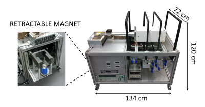
129Xe polarizer components: Four square coils (60cm width) with geometry based on the design of Merrit et al.2 for optimized B0 homogeneity at 3mT; 1767cm3 (dimensions 7.5cm diameter, 40cm length) cylindrical borosilicate cell filled with <1g rubidium located within a calcium silicate oven; on-board air compressor in-line with heating element to heat the oven; 75W diode (QPC) in combination with a beam expander to produce a 7.5cm diameter circular beam; retractable storage magnet (~0.3T NdBFe, see Fig. 1) and spiral glassware (6 rings, 4.5cm diameter, 1cm thickness) for collection of frozen Xe; and an on-board NMR spectrometer for 129Xe polarization monitoring. Polarizer footprint: 134cm length (163cm with magnet out), 120cm height, 72cm depth – see Fig. 1.
129Xe SEOP flow parameters: A gas mixture of 3% Xe, 10% N2, 87% He was flowed through the cell at a volumetric flow rate of 1000sccm (equivalent to a 1.8 L/h pure Xe production rate) and collected in its solid state within a cryostat (spiral submerged in liquid nitrogen) for 8min. The recovered gaseous Xe volume after submerging the spiral in hot water was 250mL, where the Xe was collected in a pre-evacuated Tedlar bag and topped up to 1L with medical grade N2. The oven was regulated to T = 125°C and the SEOP cell pressure was 1.25 bar. These parameters were based on optimized 129Xe polarization from previously reported volume/flow rate production maps.5 The 129Xe polarization was measured after 10 min of cryogenic collection at Xe production rate of 1.8 L/h using a polarization method described previously.6
Imaging parameters: Enriched Xe (86% 129Xe) lung imaging parameters: 3D SSFP sequence at 1.5T (GE, HDx), TE/TR = 2.2ms/6.7ms; FA = 10°; BW = ±8.0kHz; FOV = 400×320×240mm; voxel size = 4.2×4.2×10mm. Inhaled Xe volume = 250mL by a healthy adult male volunteer.
Results and Discussion
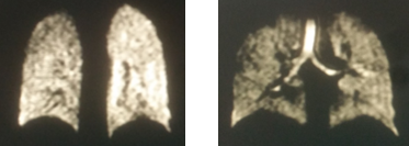
The 129Xe polarization was measured to be 35% at a Xe production rate of 1800mL/h. A performance metric called dose equivalence rate has been defined in a previous study7 as DErate = f129PXeQXe, where f129 = isotopic fraction of 129Xe (assumed to be 1 in the calculation), PXe = 129Xe polarization and QXe = Xe production rate. Using this formula with QXe = 1800mL/h and PXe = 35% yields DErate = 630mL/h, considerably higher than those recorded previously in a recent tabulation of 129Xe polarizer performance.7 This high 129Xe polarization value and fast production rate has enabled collection of 250mL Xe in 8 minutes for good-quality 129Xe lung imaging with enriched 129Xe (Fig. 2). From an installation and running perspective, the system fits easily in the back of a transit van, runs reliably on 240V mains electricity and the on-board air compressor was able to heat the oven to 125°C in ~20min with <1°C fluctuation once thermal stability is reached.Conclusion
By optimizing the polarizer running parameters from previous models we have demonstrated the generation of highly polarized 129Xe with fast production rates on a portable polarizer. We have demonstrated high-SNR lung imaging, and the system has successfully been transported and tested at novice clinical imaging centers where no specialist equipment is needed for polarizer installation. The system could form the basis of a cost effective platform for wider dissemination and multi-center clinical evaluation of 129Xe MRI.Acknowledgements
This work was supported by NIHR grant NIHR-RP-R3-12-027 and MRC grant MR/M008894/1. The views expressed in this work are those of the author(s) and not necessarily those of the NHS, the National Institute for Health Research or the Department of Health.References
1. Mugler J et al. Hyperpolarized 129Xe MRI of the human lung. Journal of magnetic resonance imaging. 2013; 37(2):313-331.
2. Stewart NJ, et al. Experimental validation of the hyperpolarized 129Xe chemical shift saturation recovery technique in healthy volunteers and subjects with interstitial lung disease. 2015; 74(1):196-207
3. Rao M et al. Imaging human brain perfusion with inhaled hyperpolarized 129Xe MR Imaging. Radiology. 2017 (early view); doi:162881.
4. Merritt R, et al. Uniform magnetic field produced by three, four, and five square coils. Review of Scientific Instruments. 1983; 54(7):879-882.
5. Norquay G, et al. Large-scale production of highly-polarized 129Xe. Proc. ISMRM 2017; p2140.
6. Norquay G, et al. Optimized production of hyperpolarized 129Xe at 2 bars for in vivo lung magnetic resonance imaging. Journal of Applied Physics. 2013; 113(4):044908.
7. He M, et al. Dose and pulse sequence considerations for hyperpolarized 129Xe ventilation MRI. Magnetic Resonance Imaging. 2015; 33(7):877-885.
Figures

