0949
Ex vivo continuous Overhauser nuclear dynamic polarization in a SQUID-based ultralow field magnetic resonance imaging system1High-field Magnetic Resonance, Max Planck Institute for Biological Cybernetics, Tübingen, Germany, 2Physikalisches Institut and Center for Quantum Science (CQ) in LISA+, Eberhard Karls Universität, Tübingen, Germany
Synopsis
Overhauser Dynamic Nuclear Polarization (ODNP) is a method to achieve continuous hyperpolarization in MR measurements. Here, the polarization of free radicals is transferred to 1H using High Frequency (HF) pulses at the electron Larmor frequency. At UltraLow Fields (ULF) the frequency of the HF pulse lies in the range of several 100 MHz and is able to penetrate large sample volumes, making continuous in vivo ODNP measurements possible. Since conventional Faraday coils are not sensitive enough at ULF, a Superconducting QUantum Interference Device (SQUID) based detector is employed. First ex vivo images using ODNP enhanced MR have been acquired successfully.
Introduction
Conventional hyperpolarizers 1 produce hyperpolarized contrast agents, which can be injected into a sample. The hyperpolarized state decays with time and lasts for minutes or even only seconds, 2 limiting the usability of this technique. Here, we demonstrate a method which allows for continuous hyperpolarization of a sample using Overhauser Dynamic Nuclear Polarization (ODNP) at UltraLow magnetic Fields (ULF) below 10 mT, 3 Continuous-ODNP requires free electrons within the sample, which are provided by free radicals.2 However, there are two major problems with continuous ODNP. First, the free radicals have to be biocompatible in order to perform in vivo measurements.4,5 Secondly, the hyperpolarization is induced by a High Frequency (HF) pulse, with frequencies 660 times higher than the corresponding nuclear Larmor frequency. In conventional high field MRI-scanners, the penetration depth of such HF pulses is too short to hyperpolarize a sufficiently large sample volume.6 A simple approach to increase the penetration depth is to employ ULF MRI where the HF pulse can penetrate several centimeters into the sample. To counteract the inherent low sensitivity of conventional Faraday coils at ULF, a Superconducting QUantum Interference Device (SQUID)-based detector is used. Here, we present a SQUID-detector based ULF MRI scanner. This is an optimal tool for the investigation of different free radicals intended for ODNP, an important step towards the development of new biocompatible free radicals.
With continuous ODNP, signal enhancement factors of up to two orders of magnitude can be achieved.7 Using a hyperpolarization field strength of about 20 mT would enhance the MR signal to a level comparable to the thermal equilibrium magnetization of 1T. This fact alone would allow one to perform unique experiments at ULF, such as the detection of functional MRI signals.
Methods
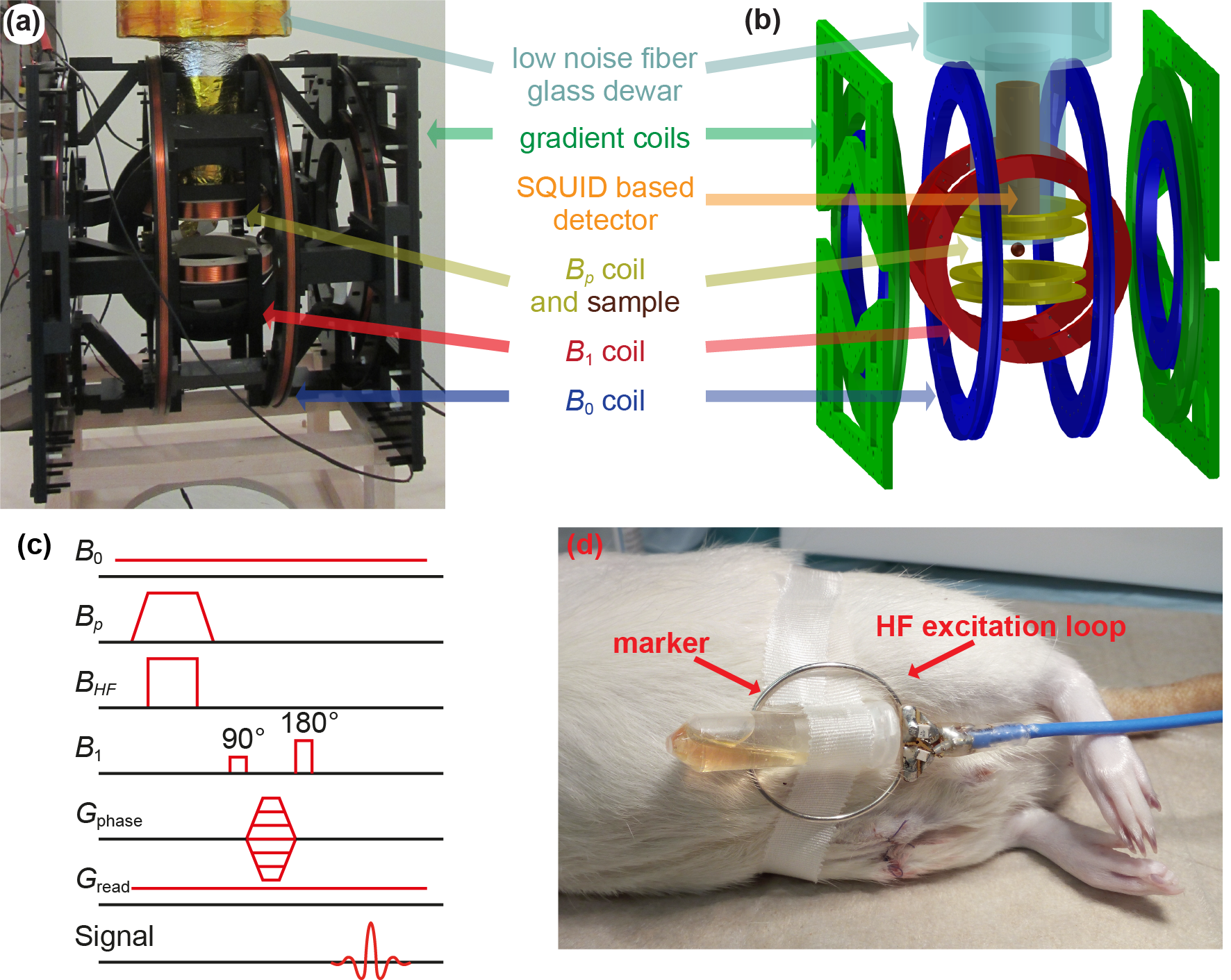
The SQUID-based ULF MRI setup contains a tetracoil for B0 field generation, a Helmholtz B1 coil, three gradient coils for spatial encoding (Gphase and Gread), as well as a Helmholtz Bp coil for prepolarization of the sample.8 The SQUID-based magnetic field detector is immersed inside a low-noise liquid helium fiber glass dewar [Fig. 1 (a) and (b)].9 A second order gradiometric pickup coil with 40 mm diameter is used for signal detection. The sample is positioned 12 mm below the lowest loop of the pickup coil. The HF pulse BHF, used for ODNP enhancement, is transmitted by a loop antenna (30mm diameter). Various loop configurations can be tuned to frequencies of 80 – 250 MHz.
Due to the high sensitivity of the SQUID to signals from the DC up to the GHz range, the whole system is placed inside a multilayer shielding chamber, which shields static magnetic fields as well as electric HF fields.10
For first proof-of-principle measurements inside a biological environment, carboxy-proxyl free radicals 11 were injected into the lower belly of a rat carcass ex vivo. The HF excitation coil together with a marker tube was placed on the outside of the belly as shown in Fig. 1 (d).
The spin-echo pulse sequence used to acquire the ULF MR images is shown in Fig. 1 (c). For comparison, the rat was also scanned at a clinical 3 T scanner, using a standard gradient echo sequence.
Results
Hyperpolarized images utilizing continuous ODNP inside a biological environment were obtained with a SQUID-based ULF-MRI system. With up to 50-fold enhanced signals, the images show the same anatomic tissue structures as comparison measurements made at 3 T, as indicated in Fig. 2 by the red line. The free radicals remained stable and ODNP enhancement could be induced for 60 – 90 minutes after injection into the body of the rat.Discussion
We successfully demonstrated the use of continuous ODNP in combination with a SQUID-based ULF MRI setup for ex vivo measurements on rats. The free radicals showed sufficient stability and good hyperpolarization properties. The enhanced MR signal makes ULF functional MRI feasible. Since magnetoencephalography (MEG) uses similar SQUID based detectors, this is an important step into the direction of a combined ULF functional MRI and MEG system.12Acknowledgements
No acknowledgement found.References
1. Bruker. Available from: https://www.bruker.com/products/mr/nmr/accessories/hyperpolarization.html.
2. Lingwood, M.D., et al., Continuous flow Overhauser dynamic nuclear polarization of water in the fringe field of a clinical magnetic resonance imaging system for authentic image contrast. Journal of Magnetic Resonance, 2010. 205(2): p. 247-254.
3. Zotev, V.S., et al., Microtesla MRI with dynamic nuclear polarization. Journal of Magnetic Resonance, 2010. 207(1): p. 78-88.
4. Golman, K., et al., Molecular imaging with endogenous substances. Proceedings of the National Academy of Sciences of the United States of America, 2003. 100(18): p. 10435-10439.
5. Ito, S. and F. Hyodo, Dynamic nuclear polarization-magnetic resonance imaging at low ESR irradiation frequency for ascorbyl free radicals. Scientific Reports, 2016. 6.
6. Wu, T., T.S. Rappaport, and C.M. Collins, The Human Body and Millimeter-Wave Wireless Communication Systems: Interactions and Implications. 2015 Ieee International Conference on Communications (Icc), 2015: p. 2423-2429.
7. Franck, J.M., et al., Quantitative cw Overhauser effect dynamic nuclear polarization for the analysis of local water dynamics. Progress in Nuclear Magnetic Resonance Spectroscopy, 2013. 74: p. 33-56.
8. Buckenmaier, K., et al., SQUID-based detection of ultralow-field multinuclear NMR of substances hyperpolarized using signal amplification by reversible exchange. Scientific Reports, 2017. 7: p. 13431.
9. Seton, H.C., J.M.S. Hutchison, and D.M. Bussell, Liquid helium cryostat for SQUID-based MRI receivers. Cryogenics, 2005. 45(5): p. 348-355.
10. Vacuumschmelze. Available from: http://www.vacuumschmelze.de/.
11. SigmaAldrich. Available from: http://www.sigmaaldrich.com/catalog/product/aldrich/253324?lang=de®ion=DE.
12. Vesanen, P.T., et al., Hybrid ultra-low-field MRI and magnetoencephalography system based on a commercial whole-head neuromagnetometer. Magnetic Resonance in Medicine, 2013. 69(6): p. 1795-1804.
Figures