0946
Joint dynamic shimming using the scanner’s spherical harmonic shim combined with a local multi-coil shim array1Max Planck Institute for Biological Cybernetics, Tuebingen, Germany, 2IMPRS for Cognitive and Systems Neuroscience, University of Tuebingen, Tuebingen, Germany, 3Skope Magnetic Resonance Technologies AG, Zurich, Switzerland, 4Department of Biomedical Magnetic Resonance, University of Tuebingen, Tuebingen, Germany
Synopsis
In this work, we combined scanner's spherical harmonic shim coils with a local multi-coil shim array to work in parallel for dynamic shimming of the human brain at 9.4 T. Performance of the combined method is compared with global shimming with scanner's built-in shim setup, global shimming with multi-coil and dynamic shimming with multi-coil.
INTRODUCTION
Magnetic field variations due to different susceptibilities in tissue and air cavities lead to distorted and quality degraded images. A set of dedicated shim coils can be used to compensate these magnetic field variations. To this aim, the most efficient basis functions are spherical harmonic (SH) terms1,2 which are orthogonal to each other. Conventional shim coils that generate spherical harmonics are integrated within the large-volume gradient coil system, and they are usually not actively shielded. Therefore, they are prone to interact and generate eddy-current for the higher orders. As an alternative, a local multi-coil approach has been proposed3–5 which is constructed of the small local coils. Each technique has its own cons and pros, but the eddy-currents issue and basis functions orthogonality are the most important ones. The aim of this work is to demonstrate and apply dynamic slice-wise shimming, jointly with SH fields and a local multi-coil setup that combines the advantages of both methods.METHODS
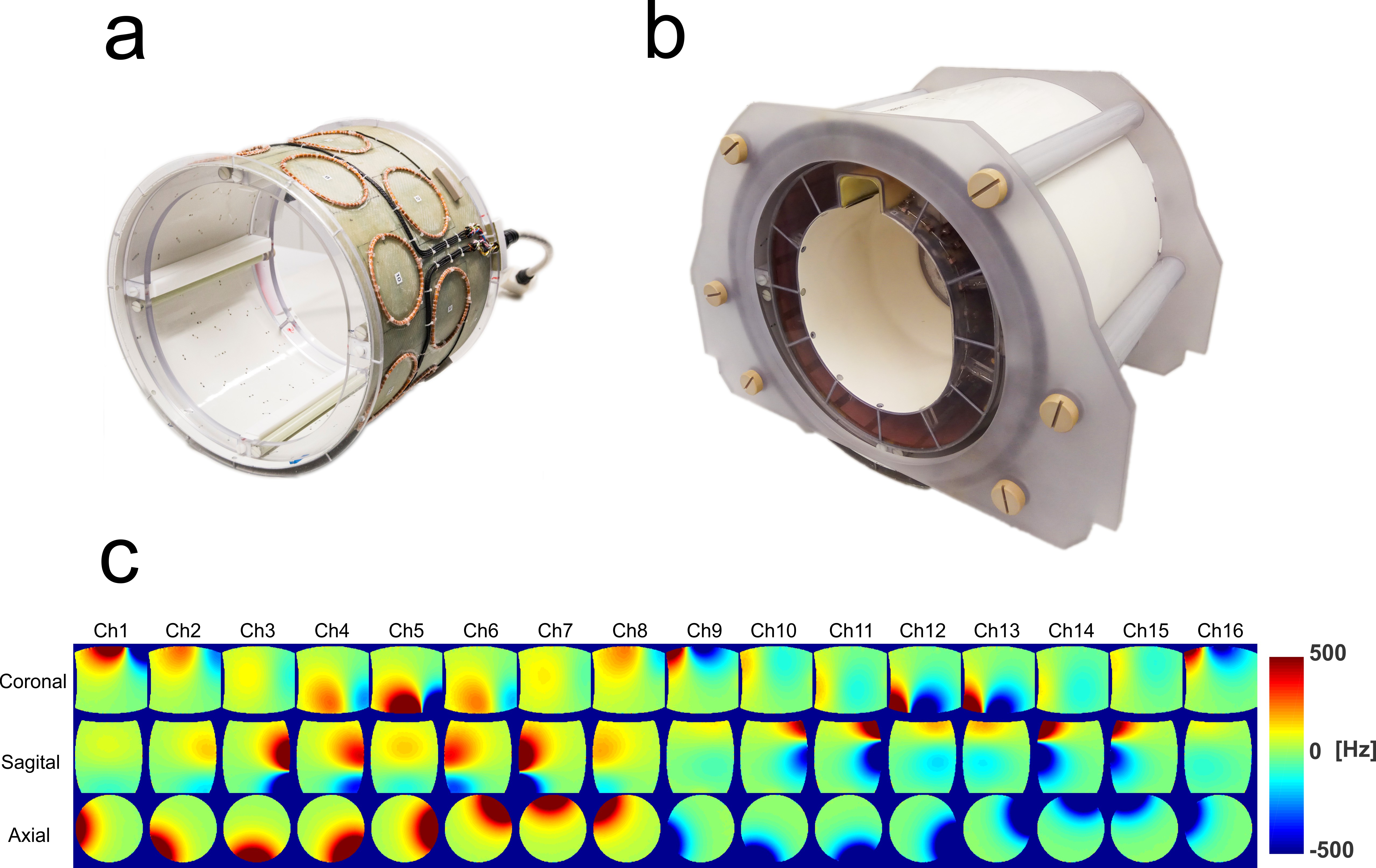
The multi-coil setup used in the study6,7 consisted of 16 identical circular coils with a diameter of 100 mm which were placed in two rows on a fiber-glass cylinder with a diameter of 370 mm (Fig. 1). As SH terms, we used zeroth and first order built-in scanner’s SH shim setup. The scanner’s vendor provides interface for rapid switching and update of its zeroth (i.e. RF frequency) and first order (i.e. X, Y, and Z gradient) SH shim. These are the low orders SH components which have the highest impact on globally distributed inhomogeneities and are actively shielded to compensate induced eddy currents. Instead of using ideal analytical values, the spatial profile of all linear gradients was measured and used to calculate SH shim currents.
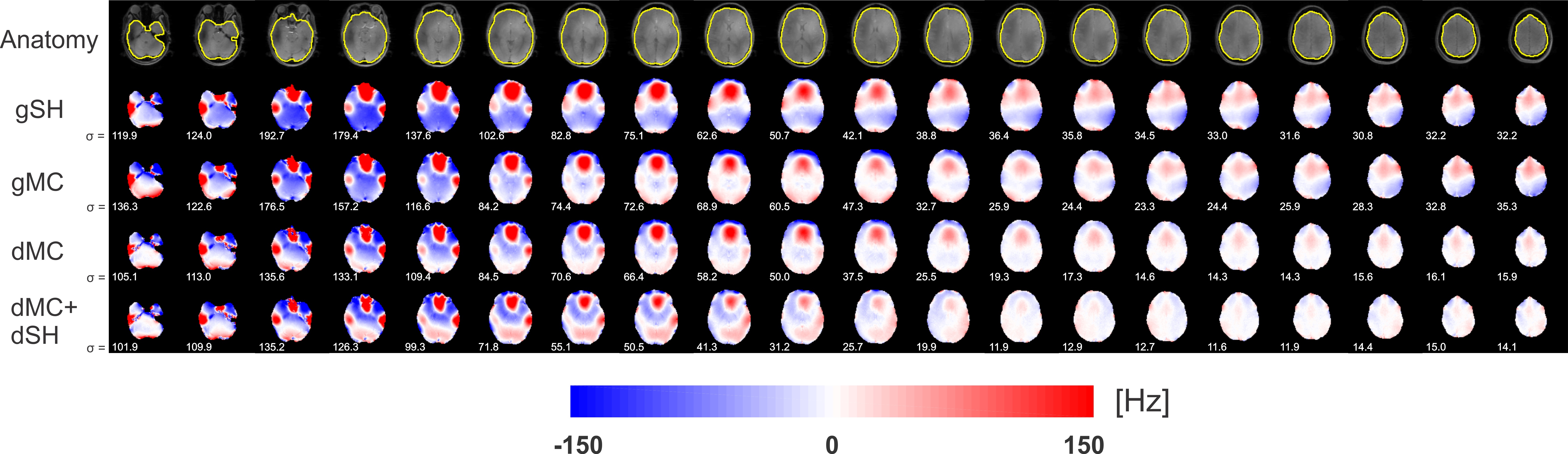
A dual-echo 2D GRE sequence was used for B0 mapping at 9.4T. (TE1/TE2/TR= 3.03/4.56/11 ms and 2 mm isotropic). The measured B0 map at the beginning of the study was used as a reference for the later processing with the assumption that subject doesn’t move during the whole study. The resulting reduction of distortion artifacts was evaluated using an EPI sequence (TE/TR = 25/1830 ms, BW = 1894 Hz/pixel, 2 mm isotropic and echo-spacing = 0.59 ms). The study includes four shimming scenarios: global shimming with the scanner’s shim setup up to 2nd order, global shimming with multi-coil, dynamic shimming with multi-coil and a combined dynamic shimming with multi-coil and scanner SH shim up to 1st order.
RESULTS
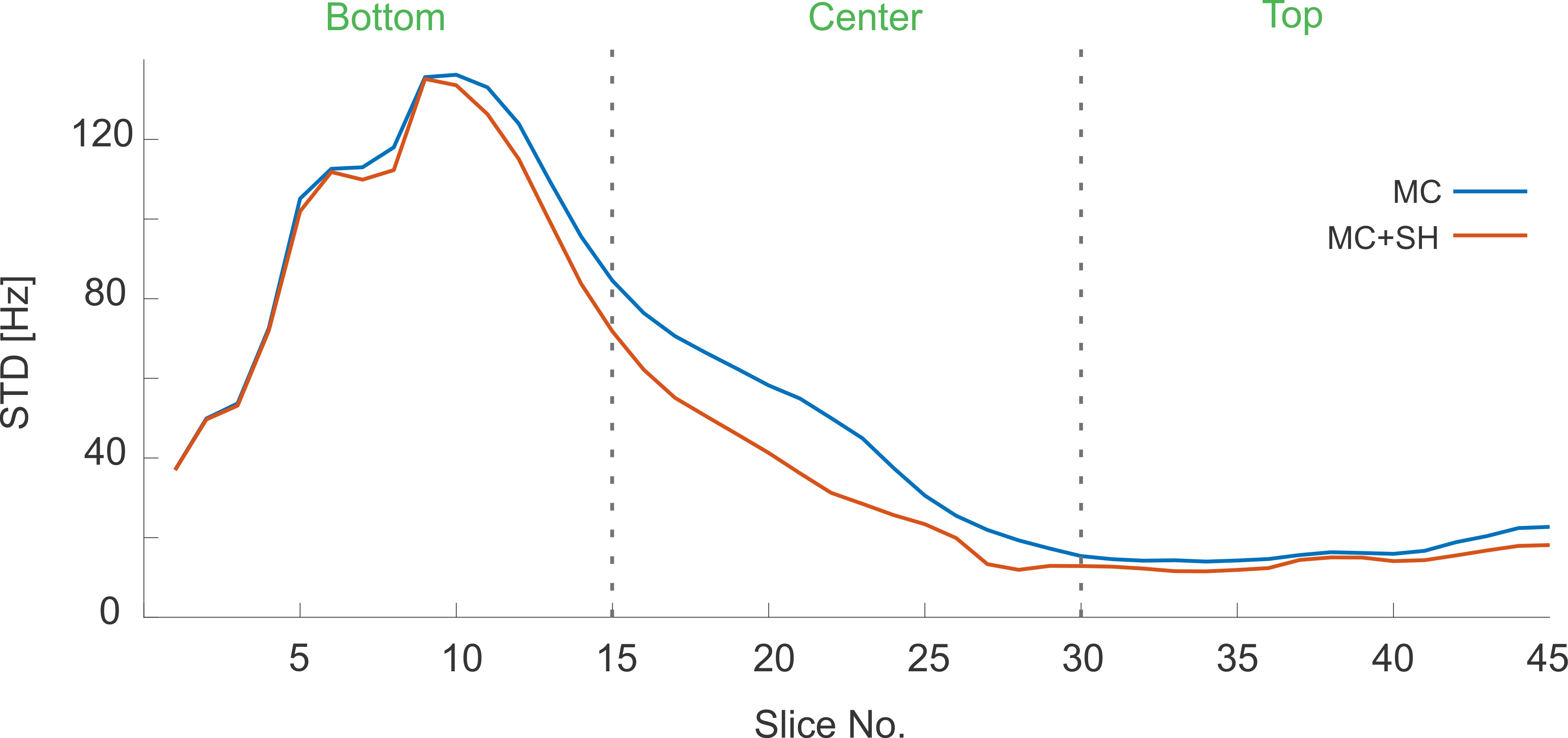
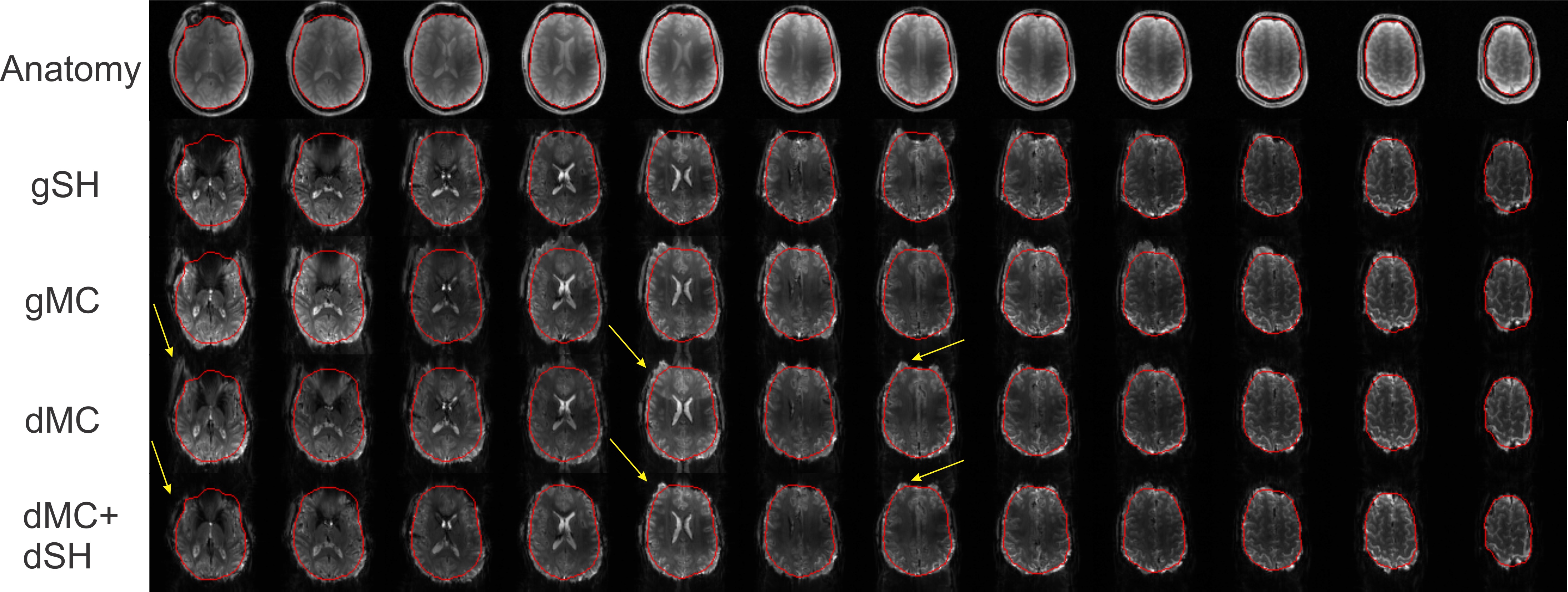
Figure 2 shows a comparison between the four shimming strategies used in this study. All shimming with multi-coil were conducted while the ROI was initially shimmed with the scanner’s 2nd order SH. Standard deviation of B0 inhomogeneity for the whole brain was 96, 86, 67 and 61 Hz at 9.4T for shimming with scanner’s 2nd order SH, global multi-coil, dynamic multi-coil and combined dynamic multi-coil and SH, respectively. Figure 3 shows the impact of SH terms on shimming of different slices of brain. The plot compares field homogeneity when dynamic multi-coil shimming is conducted with and without employing SH terms. The average improvement in B0 standard deviation for bottom, center and top slices are 4.5%, 35.5%, and 17.8% respectively. Figure 4 demonstrates EPI images while different shimming strategies are used to mitigate the geometric distortion. Brain boundary calculated from anatomical images is highlighted by red color to determine the amount of distortion.DISCUSSION AND CONCLUSION
MR scanners are equipped with dedicated gradient coils for imaging which can be employed for dynamic shimming in combination with dynamic local multi-coil shimming. Not only these linear gradient coils bring more degrees of freedom in shimming process, but also they generate orthogonal spatial fields which are the most efficient for nulling B0 inhomogeneity. Results from B0 shimming of the whole brain show that combination of multi-coil and SH up to 1st order works better in the center slices where field inhomogeneity is globally distributed, whereas in bottom slices it is very local and abrupt changes in B0 requires higher orders SH. However, the combined method for dynamic shimming is limited to acquisitions for which the whole volume can be split into independent sub-regions (e.g. 2D sequences). Neither single multi-coil nor hybrid dynamic shimming could substantially mitigate inhomogeneity in bottom slices (including the temporal lobe). Not only B0 perturbation is very strong in this region, but also weak B1+ coverage at our ultra-high field scanner leads to slightly wrong automatic brain mask calculation which is later used as shimming ROI.Acknowledgements
No acknowledgement found.References
1. Stockmann JP, Wald LL. In vivo B0 field shimming methods for MRI at 7 T. Neuroimage. June 2017. doi:10.1016/j.neuroimage.2017.06.013.
2. De Graaf RA, Juchem C. B0 Shimming Technology. In: Magnetic Resonance Technology : Hardware and System Component Design. The Royal Society of Chemistry; 2016:166-207. doi:10.1039/9781782623878.
3. Juchem C, Nixon TW, McIntyre S, Rothman DL, Graaf RA De. Magnetic field modeling with a set of individual localized coils. J Magn Reson. 2010;204(2):281-289. doi:10.1016/j.jmr.2010.03.008.
4. Stockmann JP, Witzel T, Keil B, et al. A 32-channel combined RF and B0 shim array for 3T brain imaging. Magn Reson Med. 2016;75(1):441-451. doi:10.1002/mrm.25587.
5. Juchem C, Nixon TW, McIntyre S, Boer VO, Rothman DL, De Graaf RA. Dynamic multi-coil shimming of the human brain at 7 T. J Magn Reson. 2011;212(2):280-288. doi:10.1016/j.jmr.2011.07.005.
6. Aghaeifar A, Zivkovic I, Mirkes C, Steffen T, Scheffler K. Global and Dynamic Shimming with the Scanner’s Inbuilt Shim System and a Custom-Made Multi-Coil Setup at 9.4 T. In: 25th Annual Meeting and Exhibition of the International Society for Magnetic Resonance in Medicine. Honolulu, Hawaii, USA; 2017:4330.
7. Aghaeifar A, Zivkovic I, Steffen T, Mirkes C, Scheffler K. Flexible gradient driver system for a multi-coil setup; design considerations and implementation. In: 34th Annual Scientific Meeting of the European Society for Magnetic Resonance in Medicine and Biology. Barcelona, Spain; 2017:50.
Figures