0919
Automated Coil Ranking using a Neural Network for Image Quality Assessment: An Explorative Study in Coronary MRI1Department of Radiology, University Hospital (CHUV) and University of Lausanne, Lausanne, Switzerland, 2Advanced Clinical Imaging Technology, Siemens Healthcare AG, Lausanne, Switzerland, 3Institute of Bioengineering/Center for Neuroprosthetics, École Polytechnique Fédérale de Lausanne (EPFL), Lausanne, Switzerland, 4Center for Biomedical Imaging (CIBM), Lausanne, Switzerland, 5LTS5, École Polytechnique Fédérale de Lausanne (EPFL), Lausanne, Switzerland
Synopsis
A novel method that uses a neural network to rank individual coil elements of phased arrays based on their image quality is proposed. With a ranking of the coil elements, the specific subset of coils that leads to the best image reconstruction can be selected. Alternatively, the contribution of coils with high levels of artifacts and noise to the final image can be reduced. We show that both selection and weighting of coil elements can reduce the level of image artifacts while maintaining a high signal intensity in the region of the examined organ.
Introduction
Phased array coils are nowadays standard in clinical MRI and the number of coil elements continuously increases to enable higher acceleration factors and improve signal-to-noise. However, the localized sensitivity of small receivers is not only beneficial since peripheral elements “seeing” strong unwanted signal sources may amplify undersampling artifacts, that typically appear as streaking or aliasing in radial and Cartesian images, respectively. Hence, the choice of how many and which coil elements to use for reconstruction is a trade-off between signal and artifact level. Coil selection relying on mathematical metrics has been suggested 1, 2. As an alternative, we propose an automated method for coil ranking using a recently presented neural network for image quality (IQ) assessment 3. Firstly, we investigate whether this automated ranking of coil elements correlates with human perception. Secondly, we evaluate the impact that the number of coil elements has on IQ in gridding reconstructions. Lastly, we test if weighting coil elements in compressed sensing reconstructions can suppress artifacts to a higher degree than what is already accomplished by regularization.Methods
Our method exploits a convolutional neural network 3 trained to assess IQ in whole-heart coronary MRI scans. The network was trained on 324 coil-combined patient scans, acquired with a prototype respiratory self-navigated radial sequence 4, and graded on a standard 0-4 IQ scale 5 by an expert reader. Ranking of the coil elements according to IQ is here obtained by using this neural network to grade the volumetric reconstructions from individual coil elements. The coil ranking was tested on healthy volunteer datasets (N=11, 27.9±5.4 years, 9M) acquired with the same sequence 4 (FoV=210x210x210 mm3, Matrix=1923) using a 1.5T clinical scanner (MAGNETOM Aera, Siemens Healthcare, Erlangen, Germany). First, the coil ranking was computed on conventional gridding reconstructions. Thereafter, two reviewers (J.H. and D.P.) visually selected the five coil elements with best IQ for each subject. Their selection was compared to the network’s ranking and the differences in the number of matching coil elements among reviewers and network compared with Wilcoxon signed rank tests (p<0.05 was considered statistically significant). To evaluate the impact of the number of coil elements on final IQ, respiratory motion-corrected 4 gridding reconstructions were completed with the full range of coil elements added in decreasing quality order (from only the best ranked element to the full set). The IQ obtained with the best number and all coils for each subject was compared with a paired t-test. Finally, respiratory-resolved 6 compressed sensing 7 reconstructions were performed using all coils, both with a uniform weighting and by weighting the elements by multiplying the k-space data with their individual IQ grades. Visible length and sharpness of the right coronary artery were measured 8 and compared with paired sample t-tests and the artifact level visually examined.Results
The neural network assigned high IQ to coil elements with high signal from the heart (Figure 1) and the agreement in the selection of the five best coils was similar between the network and human readers (Figure 2) with no significant differences in the number of matching coils. In the reconstructions with increasing number of ranked coil elements (Figures 3-4), the highest IQ was obtained using on average the best 61.6±26.9% of all coil elements. Significantly higher IQ was seen with the best number of coils compared to all coils (mean IQ grade difference: 0.17±0.25, p=0.044). In the compressed sensing reconstructions, non-significant improvements of visible vessel length and sharpness resulted from weighting the coils (Figure 5). Visually, coil weighting decreased streaking at the expense of signal inhomogeneity across the field of view.Discussion
The coil ranking obtained from the neural network correlates well with human perception and even though the network was trained on coil-combined reconstructions, it also provides meaningful results for individual coils. Maximum IQ was obtained with a subset of the coil elements which confirms that blindly including all elements in reconstructions is sub-optimal. Moreover, excluding coil elements reduces computational complexity which may be advantageous for demanding iterative reconstructions. Both selection and weighting of coil elements showed potential for improving the reconstructed images. However, the risk of increasing signal inhomogeneity across the field of view should be considered. Therefore, future developments may take the physical position of the coil elements into account when computing the ranking. Alternatively, training a network specifically for coil ranking could improve performance, but that would be a less general approach than exploiting image quality.Conclusion
Coil ranking based on automated IQ assessment is a flexible and subject-specific method that enables reduction of image artifacts while maintaining high signal intensity in the region of the examined organ.Acknowledgements
No acknowledgement found.References
[1] Y. Xue, J. Yu, H. S. Kang, S. Englander, M. A. Rosen, and H. K. Song, “Automatic coil selection for streak artifact reduction in radial MRI,” Magn. Reson. Med., vol. 67, no. 2, pp. 470–476, 2012.
[2] M. Doneva and P. Börnert, “Automatic coil selection for channel reduction in SENSE-based parallel imaging,” Magn. Reson. Mater. Physics, Biol. Med., vol. 21, no. 3, pp. 187–196, 2008.
[3] R. Demesmaeker et al., “Image quality through the eyes of deep learning: A first implementation in coronary MRI”, ISMRM Workshop on Magnetic Resonance Imaging of Cardiac Function, NYU Langone Medical Center, New York City, NY, USA, 2017.
[4] D. Piccini, A. Littmann, S. Nielles-Vallespin, and M. O. Zenge, “Respiratory self-navigation for whole-heart bright-blood coronary MRI: Methods for robust isolation and automatic segmentation of the blood pool,” Magn. Reson. Med., vol. 68, no. 2, pp. 571–579, 2012.
[5] P. Monney et al., “Single centre experience of the application of self navigated 3D whole heart cardiovascular magnetic resonance for the assessment of cardiac anatomy in congenital heart disease.,” J. Cardiovasc. Magn. Reson., vol. 17, p. 55, 2015.
[6] D. Piccini et al., “Four-dimensional respiratory motion-resolved whole heart coronary MR angiography,” Magn. Reson. Med., vol. 77, no. 4, pp. 1473–1484, 2017.
[7] L. Feng, L. Axel, H. Chandarana, K. T. Block, D. K. Sodickson, and R. Otazo, “XD-GRASP: Golden-angle radial MRI with reconstruction of extra motion-state dimensions using compressed sensing,” Magn. Reson. Med., vol. 75, no. 2, pp. 775–788, 2016.
[8] A. Etienne, R. M. Botnar, A. M. C. Van Muiswinkel, P. Boesiger, W. J. Manning, and M. Stuber, “‘Soap-Bubble’ visualization and quantitative analysis of 3D coronary magnetic resonance angiograms,” Magn. Reson. Med., vol. 48, no. 4, pp. 658–666, 2002.
Figures
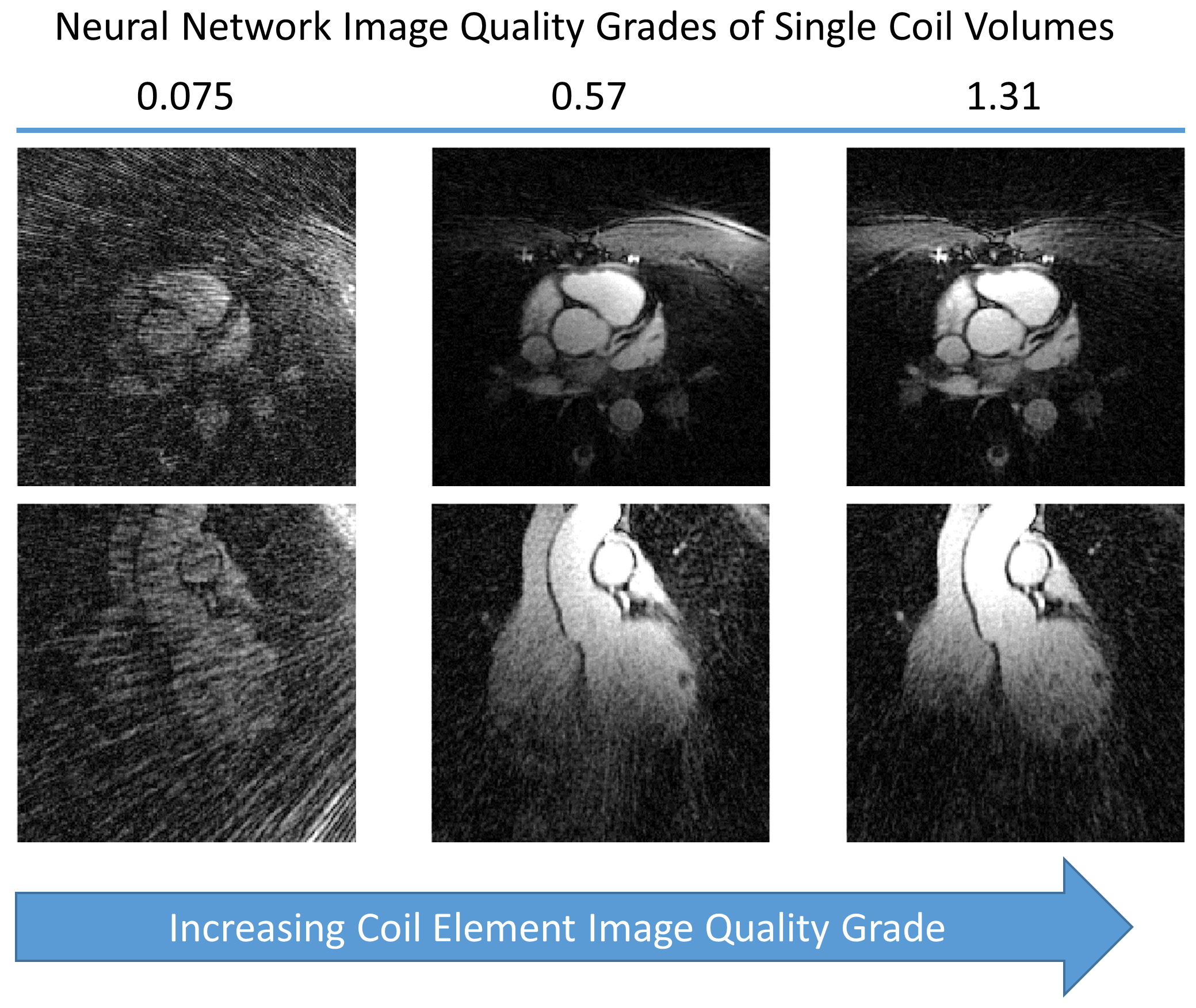
Figure 1. Example of Coil Images and Corresponding Grades
The network consistently assigned high IQ grades to coil elements with high signal from the heart and low general noise and artifact levels as depicted in the figure comprising single-coil volumes from one example subject. The grade difference between the single-coil volumes with grades 0.57 and 1.31 respectively, indicates that the network seems to assign better IQ when high signal is received from a larger part of the heart.
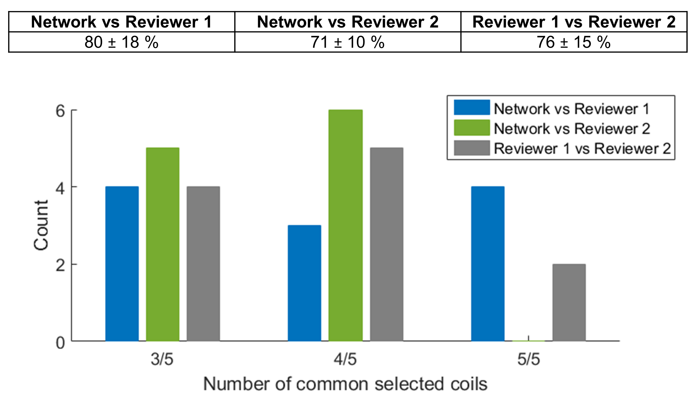
Figure 2. Agreement in Coil Selection between the Neural Network and Human Reviewers
For each subject, the five coil elements with highest image quality according to the network and the human reviewers were compared in order to study the agreement and assess the inter-observer variability. To express the agreement as a percentage (cf. table), the ratio of common selected coils was computed on a subject by subject basis and then averaged. The variability between the human readers and the network was found to be similar with no significant differences in the number of common selected coils.
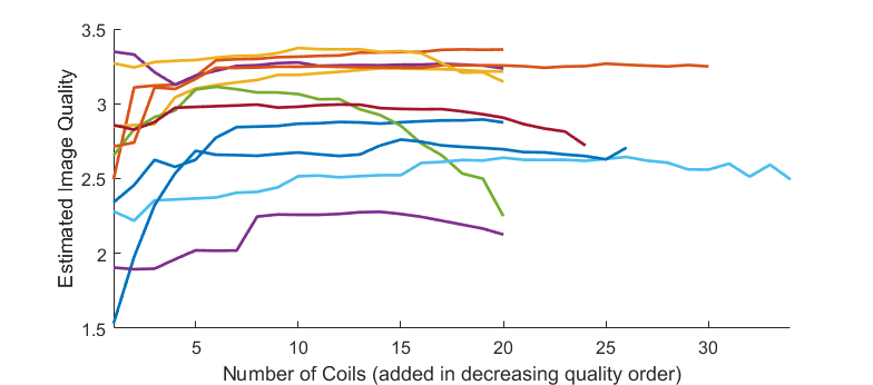
Figure 3. Image Quality as a Function of the Number of Coil Elements
Respiratory self-navigated radial reconstructions were performed with the full range of coil elements beginning with only the one with highest IQ to all coil elements. The inclusion of coil elements was done in decreasing quality order. In general, the network’s assessed image quality of the coil-combined reconstructions either reached a plateau or started decaying after reaching its maximum value. In one subject (topmost purple curve), the reconstruction with a single coil obtained the highest grade.
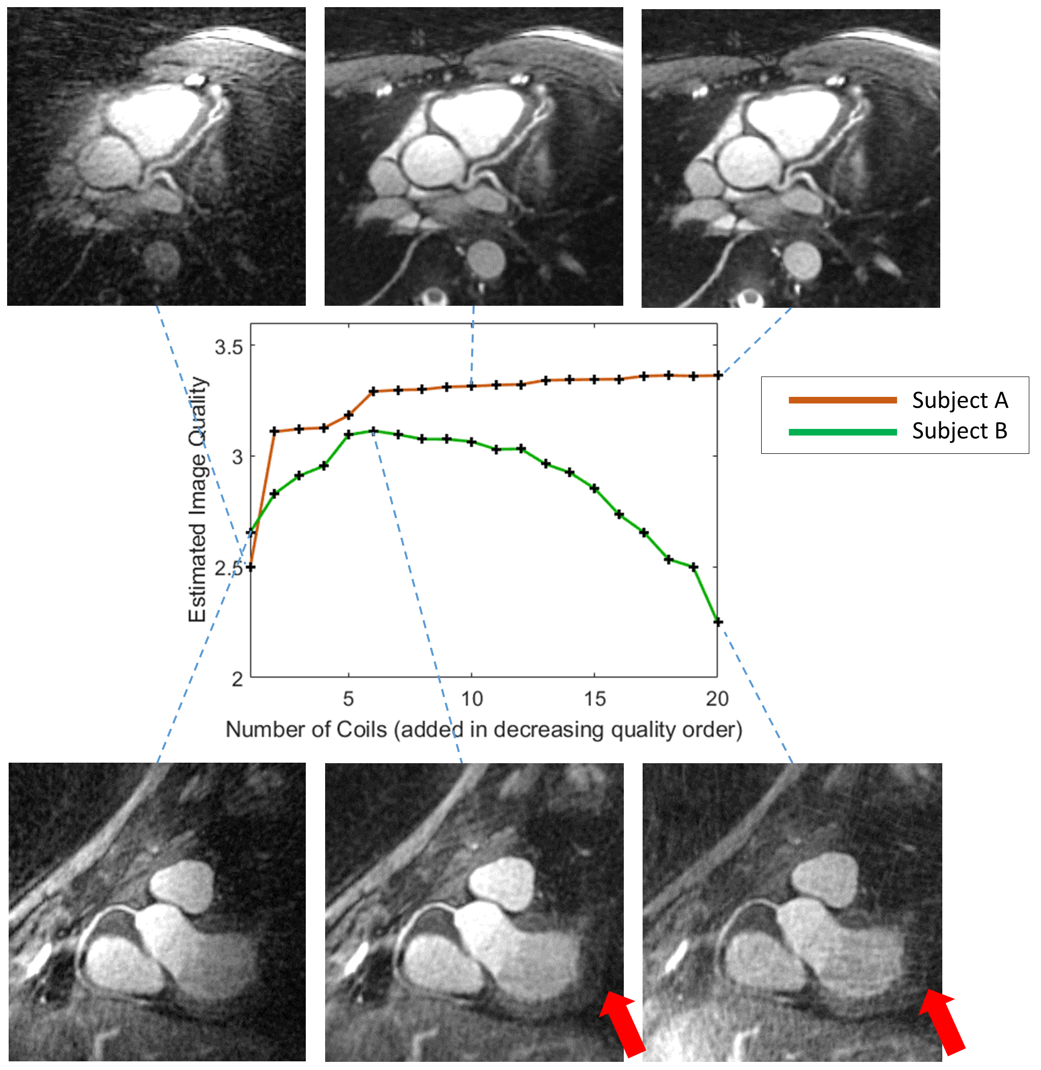
Figure 4. Examples of Reformats from Reconstructions with Different Number of Coils
In subject A, optimum IQ according to the neural network was obtained using almost all coils, although only small improvements of the IQ was seen from six coils upwards. However, in subject B a considerable decrease in IQ resulted from using a majority of the coils for reconstruction. Visually, a larger degree of streaking can be seen in the reformat to the bottom right using 20 coil elements compared to the middle reformat with maximum IQ using 6 coils (red arrows).
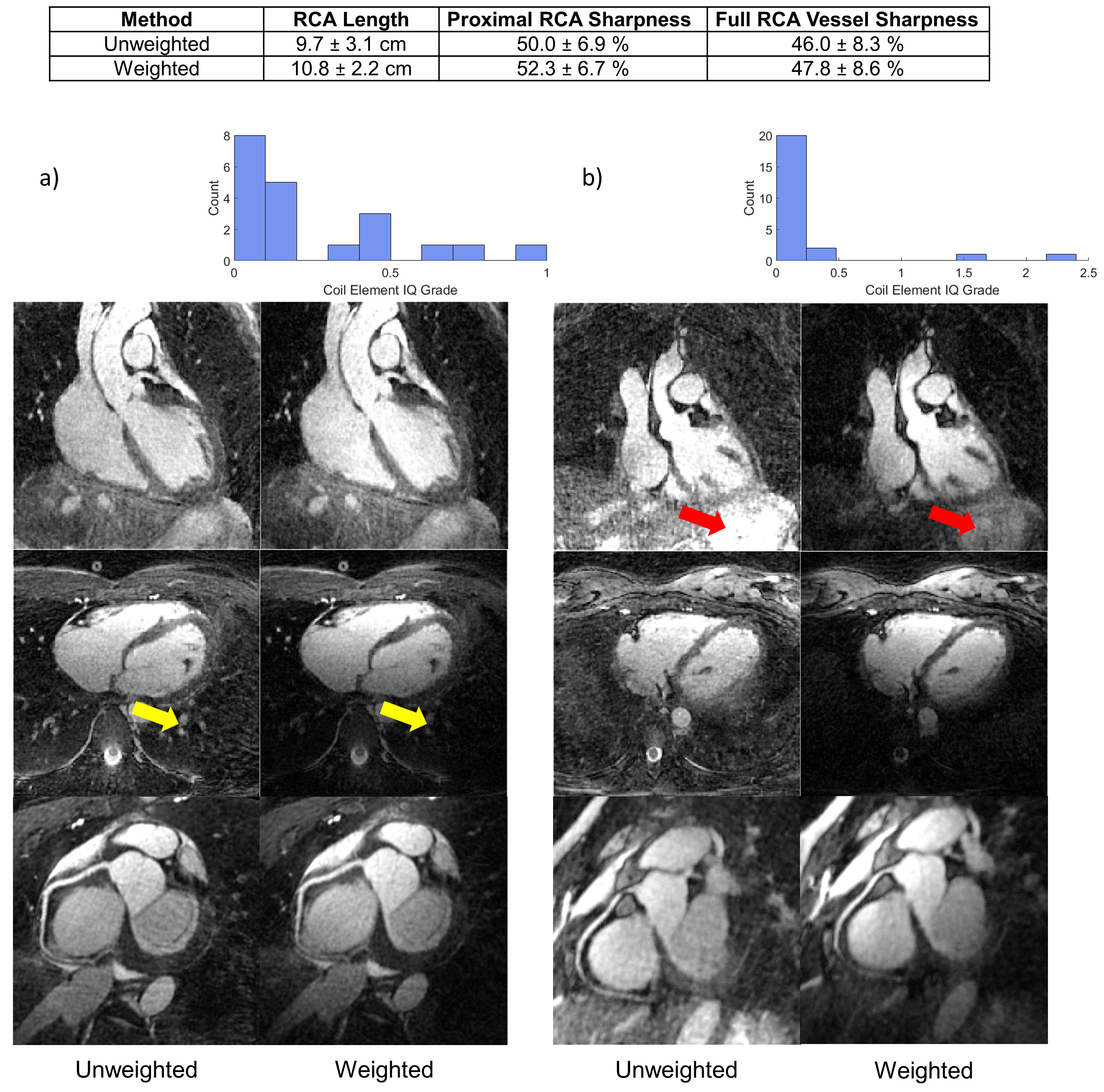
Figure 5. Compressed Sensing Reconstructions with and without Coil Weighting
Coil weighting non-significantly improved the sharpness and visible length (cf. table) of the right coronary artery (RCA). In a), where the coil grades (cf. histogram) were more uniformly distributed, streaking was reduced but with some signal loss posterior to the heart (yellow arrows). In b), weighting the coils attenuated the bright structure by the liver (red arrows). However, the axial view and reformats show that too much weight was given to the two coil elements with the highest IQ, resulting in spatial imbalance in the signal level across the field of view.