0853
Simultaneous measurements of global cerebral blood flow with 2D pseudo-continuous multi-TI arterial spin labeling and 15O-H2O PET in a hybrid PET/MR system1Department of Clinical Physiology, Nuclear Medicine and PET, Rigshospitalet, University of Copenhagen, Copenhagen, Denmark
Synopsis
Arterial spin labeling (ASL) provides easy-access non-invasive quantification of regional cerebral blood (CBF) but its accuracy is not established. The aim of the study was to compare simultaneous measurements of absolute CBF obtained by ASL MRI and 15O-H2O PET CBF measurements in healthy subjects using a hybrid PET/MR system. Simultaneous 15O-H2O PET and ASL MRI were performed in resting state, hypoperfusion and hyperperfusion. Overall highly significant positive and linear correlation of ASL MRI and H2O PET in gCBF was observed across all perfusion states, confirming the ability of ASL MRI to quantify gCBF in good agreement with PET.
Introduction and Objective
Arterial spin labeling (ASL) provides easy-access non-invasive quantification of regional cerebral blood (CBF). By obtaining measurements at multiple post-labeling delays and acquiring a separate equilibrium magnetization map (M0), absolute quantitation of CBF is feasible even when the arterial transit time is unknown or variable across brain regions (1). However, the accuracy of absolute ASL CBF measurements is not established. The aim of the study is to compare simultaneous measurements of absolute CBF obtained by ASL MRI and 15O-H2O PET CBF measurements in healthy subjects using a hybrid PET/MR system.Methods
A total of 14 healthy male volunteers were studied on a hybrid PET/MR Siemens mMR system. In each volunteer duplicate simultaneous measurements by each method were performed during rest, hypoperfusion (hyperventilation) and hyperperfusion (post-acetazolamide). ASL was obtained using 2D pseudo-continuous ASL sequence (TR: 3400 ms, TE 12 ms, labeling duration 1800 ms, Post labeling delay 800, 1100, 1400, 1700 and 2000 ms, 9 pairs of measurements at each post labeling delay, scan time: 7 minutes. Resolution: 4 x 4x 6 mm). Absolute CBF maps were generated using model-based analysis (FSL BASIL) (2). Dynamic PET scans were performed with arterial blood sampling after inj. of approx. 500 MBq of 15O-H2O. Kinetic modelling was performed using a 1-tissue 2 compartment model (3). A T1 MRI derived brain mask was applied to parametric ASL and PET CBF images to obtain global CBF (gCBF) in ml/100g/min. Methods agreement and correlations were investigated by Bland-Altman analysis and random effects models.Results
Overall highly significant positive and linear correlation of ASL MRI and H2O PET in CBF was observed across all perfusion states (Figure 1, R2 = 0.80, p < 0.001). Also within the individual states significant, but weaker, positive correlations were demonstrated in rest (R2 = 0.15, p = 0.007), post-acetazolamide (R2 = 0.32, p < 0.001 and during hyperventilation R2 = 0.44, p = 0.001).Conclusions
This study confirms the ability of ASL MRI to quantify gCBF in good agreement with PET, but also shows a slight relative difference of gCBF values obtained by the two methods. The study also demonstrates the potential of hybrid PET/MR systems for simultaneous validation of quantitative MRI CBF measurements.Acknowledgements
The authors would like to thank the hard work and dedication of the nuclear medicine technologists and radiographers Nadia Azizi, Marianne Federspiel, Jakup Poulsen and Karin Stahr; the staff of Cyclotron Unit and Radiochemistry, especially Jesper Jørgensen and Anne Sørensen and the secretaries of the PET department Gudrun L. Semitoje and Tina Vikman. We also thank Anne Marie Sørensen from the Trauma Center for her assistance with the arterial blood gases analysis and Annette Ulrich from the thorax anestesiology department for the placing of the arterial lines. We would also like to thank The John and Birthe Meyer Foundation, who generously donated the PET/MRI scanner and the cyclotron to Copenhagen University Hospital Rigshospitalet. Oriol Puig is supported by a training grant from the Fundación Alfonso Martín Escudero and Mark B. Vestergaard is supported by a post-doc grant from the Danish Council for Independent Research (reference number: 6110-00692A).References
1. Petersen ET, Zimine I, Ho YC, Golay X. Non-invasive measurement of perfusion: a critical review of arterial spin labelling techniques. Br J Radiol. 2006 Aug;79(944):688-701
2. Chappell MA, Groves AR, Whitcher B, Woolrich MW. Variational Bayesian inference for a non-linear forward model. IEEE Transactions on Signal Processing 57(1):223-236, 2009.
3. Ohta S, Meyer E, Fujita H, Reutens D, Evans A, Gjedde A. Cerebral [15O] water clearance in humans determined by PET: I. Theory and normal values. J Cereb Blood Flow Metab. 1996;16:765-780.
Figures
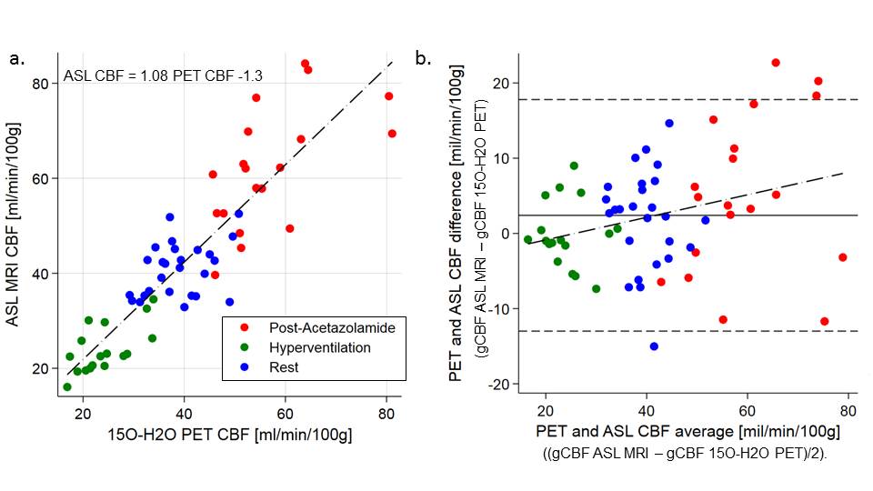
Figure 1 Agreement of ASL MRI and 15O-H2O PET gCBF measurements.
1a. Correlation between global cerebral blood flow (gCBF) measured by 15O-H2O PET and ASL MRI. 1b. Bland-Altman plot showing the difference between the two methods against mean of the methods. Dot-dashed lines show the linear fit. In the Bland-Altman plot, the horizontal solid line and dashed lines show bias +/- 2 SD. The slope of the regression line in the Bland-Altman plot was not significant from zero.
