0848
Long term Brain Functional Connectivity changes after Neonatal Arterial Ischemic Stroke (NAIS): A resting state fMRI study1Institute Joliot/DRF/CEA, U1129/UNIACT, Neurospin, CEA-Saclay, Gif sur Yvette, France, 2Département de SSR pédiatrique, CHU Angers, Angers, France, 3Institute Joliot/DRF/CEA, UNATI, Neurospin, CEA-Saclay, Gif sur Yvette, France, 4Departement de Rééducation Fonctionnelle, CHU Saint Etienne, Saint Etienne, France, 5University Hospital Necker-Enfants Malades, Paris, France, 6CHU Lille, Lille, France
Synopsis
Resting state fMRI enables the study of plastic inter- and intra- hemispheric connectivity changes after early brain lesions. We studied 38 7yo children having suffered an arterial ischemic stroke in the neonatal period (NAIS) and 29 age-matched controls, with rs-fMRI, and language fMRI. Preprocessing took into account various sources of spurious signals (motion, lesion, interdependency of correlation measures, etc…). Tangent metric appeared the most accurate to classify groups of subjects and highlighted mostly a reduced inter-hemispheric connectivity in the auditory, language, and attentional networks, especially in patients with atypical clinical or fMRI language profiles, but with little evidence for intra-hemispheric changes.
INTRODUCTION
Neonatal Arterial Ischemic Stroke (NAIS) is an exquisite model to study long-term brain plasticity after early focal lesions. Studies have shown inconstant cerebral palsy, good language outcome with contralesional development of language network, and frequent executive functions and visuo-spatial impairments. Resting state fMRI enables the study of inter- and intra- hemispheric connectivity without requiring cognitive engagement. We aimed to study rs-fMRI functional connectivity in school-age children having suffered a NAIS, to i- explore dysconnectivity, as compared to age-matched controls, and ii. understand its clinical correlates with a focus on language outcome.METHODS
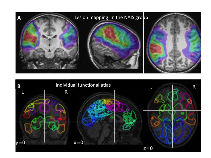
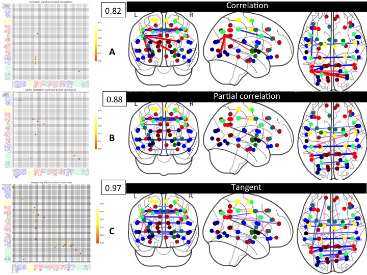
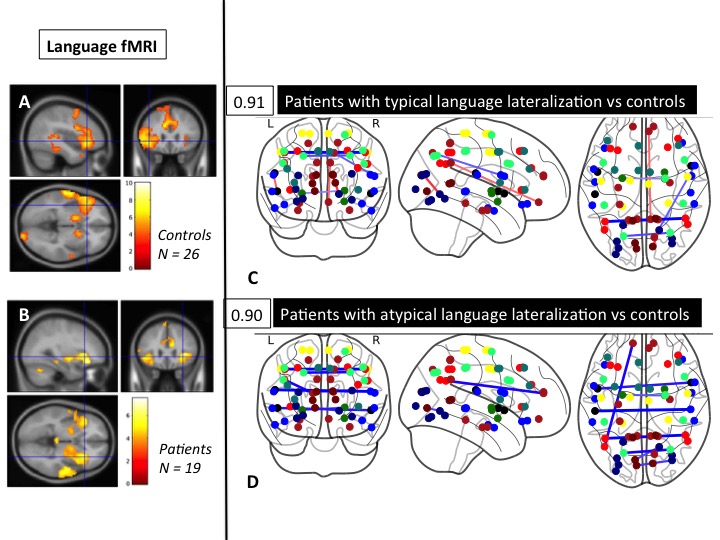
From 100 newborns with NAIS (AVCnn cohort, 1), 72 were followed at age 7 (2/3 boys, 85% in the MCA territory, 2/3 on the left, Fig 1A). 30% had unilateral Cerebral Palsy. Global language performance was good (mean ICV = 93), but 49 % of children had atypical language profiles (N-EEL battery). Among them, 38 patients (along with 29 age-matched controls) underwent 3T MRI protocol including rs-fMRI (TR=2.4 sec, 3x3x3 mm3 voxel, 3x90 volumes). In addition, block-designed sentence production fMRI was exploitable in 19 patients/26 controls. Data preprocessing included simultaneous slice timing and motion correction (Nipy), image registration (DARTEL, SPM12), 4mm kernel smoothing, and removal of motion-corrupted images using ART (volumes > 4mm SD on the movement norm and datasets with >13 volumes). In addition to movement parameters and derivatives, we regressed out signals with highest variances (CompCor 2) and spurious signals in white matter, CSF and the pontine cistern. Rs-fMRI was exploitable in 32 patients/26 controls. For connectivity analyses, a dedicated functional atlas was built up from the control population with a dictionary learning algorithm 3 to learn sparse spatial maps resulting in 65 nodes. Corresponding ROIs were labeled according to the anatomical position of their centroids, and grouped into 10 functional networks 4. We built individual atlases to account for the presence of the lesion and to avoid including extra-cerebral signals: ROIs were excluded when the intersection of the subject brain mask and the corresponding atlas ROIs was over 0,5. (With this criterion, only two ROIs in one patient were excluded (Fig 1B)). In all other instances, ROI was reduced according to the resulting intersection of the ROI and the subject brain mask. We then extracted standardized and detrended time series over the regions with a least square algorithm (Nilearn python). We estimated connectivity matrices with three metrics: correlation, partial correlation and tangent (Nilearn python 5). For the three metrics, we used a SVM classifier with a linear kernel and L2 regularization. We computed a mean accuracy score under a random split scheme in test set and train set repeated 100 times. Then, two samples t-tests on all pairs of connectivity coefficients were used for group comparisons (significant at a type I error rate of 0.05, using False Discovery Rate correction for multiple comparisons). Various group comparisons were tested according to lesion side, sex, clinical language assessment, language lateralization at fMRI....RESULTS
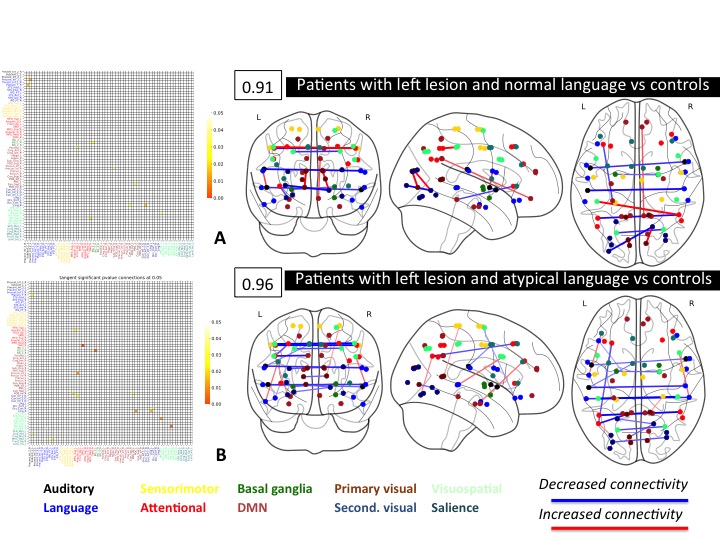
While correlation showed numerous versatile positive and negative links, partial correlation and tangent metrics showed mostly reduced inter-hemispheric connectivity in the patient groups as compared with controls (mainly in auditory, language and attentional/visuospatial networks). In the classification process, the tangent metric systematically outperformed correlation and was most often largely better than partial correlation, often reaching 90-95% (fig 2). Significantly reduced inter-hemispheric connectivity was constantlyfound in the superior parietal lobules. We found no significant differences in direct comparisons of patients groups according to age, lesion side, and clinical language status and lateralization (typical or atypical). However, when patients were compared to controls, both language status and language lateralization did impact on the connectivity matrices, with stronger inter-hemispheric dysconnectivity in the language homotopic regions (supramarginal gyrus, and planum temporale, Fig 3 and 4).DISCUSSION
Rs-fMRI can capture connectivity changes reflecting networks plasticity long after a neonatal lesion, providing careful assessment of data quality is done. Tangent metric seems most robust to classify subjects and enabled to unravel involved networks. While correlation and partial correlation matrices are inherently interdependent 6, the tangent metric strongly reduces the statistical dependency of connectivity coefficients and outperforms correlation measures in binary classifications 6,7. Inter-hemispheric dysconnectivity is the leading abnormality after an early lesion, but surprisingly, not clear intra-hemispheric change pattern could be revealed. This might reflect the limits in sensitivity of the methods in clinical groups.CONCLUSION
Rs-fMRI is useful to show the plasticity of functional connectivity after NAIS, showing mostly widespread reduction of inter-hemispheric connectivity, especially in the auditory, language, and attentional networks. The clinical correlates of this complex pattern remain to be understood.Acknowledgements
The Research was supported by the University Hospital of Angers (eudract number 2010-A00976-33), the University Hospital of Saint Etienne (eudract number 2010-A00329-30 and the Fondation de l'Avenir ( ET0-571).
References
(1) Chabrier et al, Multimodal Outcome at 7 Years of Age after Neonatal Arterial Ischemic Stroke. J of Ped 2016 [2] Behzadi, A component based noise correction method (CompCor), Neuroimage, 2007. [3] Mensch et al, Compressed online dictionary learning for fast resting-state fMRI decomposition, ISBI, 2016 (4) Shirer et al: Decoding subject-driven cognitive states with whole-brain connectivity patterns. Cereb Cortex (2012) [5] Abraham et al, Machine learning for neuroimaging with scikit-learn, Front. Neuroinform 2014. [6] Ng et al, Transport On Riemannian Manifold for Connectivity-Based Brain Decoding, MICCAI 2014 [7] Varoquaux et al, Detection of brain functional-connectivity difference in post-stroke patients using group level covariance modeling, MICCAI 2010.Figures