0847
Connectome of the Extremely Preterm Brain at Adolescence and Measures of Cognitive Functioning: Early Experience with White Matter Fibrography (WMF)1Boston University, Boston, MA, United States, 2University of North Carolina Children’s Hospital, Chapel Hill, NC, United States
Synopsis
Purpose: To analyze the fiber bundle architecture via white matter connectomes of extremely preterm (EP) born adolescents generated by Synthetic-MRI based white matter fibrography, and to study associations with summary measures of intelligence quotient (IQ) and executive function (EF). Methods: Eleven EP adolescents were scanned the tri-turbo spin echo pulse sequence, which is the concatenation of dual-echo TSE and single-echo TSE pulse sequences, both acquired with same geometry and scanner settings. Results: Connectome were successfully created for all subjects. Conclusion: White matter fibrography is a direct and reliable method for generating whole-brain white matter connectomes in adolescents born EP, and appears to have clinical relevance.
Purpose
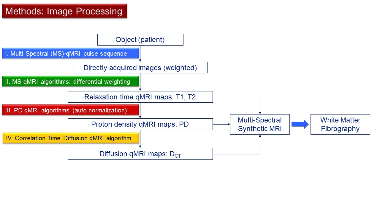
Although survival rates for children born extremely preterm (EP) (gestational age <28 weeks) have greatly increased over the last 40 years, EP children remain at risk of developing a broad range of neurodevelopmental impairments including motor and sensory impairment, cognitive and learning disabilities, and psychiatric and behavioral disorders. The increased risk for development of neurological deficits in children born EP are probably linked to perturbations of critical maturational processes of the central nervous system (CNS) that occur during the first two trimesters of pregnancy and in the postnatal period. These preterm birth associated perturbations can also have long-term architectural CNS sequelae. The purpose of this early proof-of-concept work is to examine qualitatively the architecture of whole-brain connectomes of 11 EP adolescents directly generated by Synthetic-MRI based white matter fibrography (WMF) (Figure 1) (1), and to study possible associations with their profiles on measures of neurocognitive impairment as previously classified by level of cognition over the ELGAN cohort (n=873) at age 10 (2).Methods
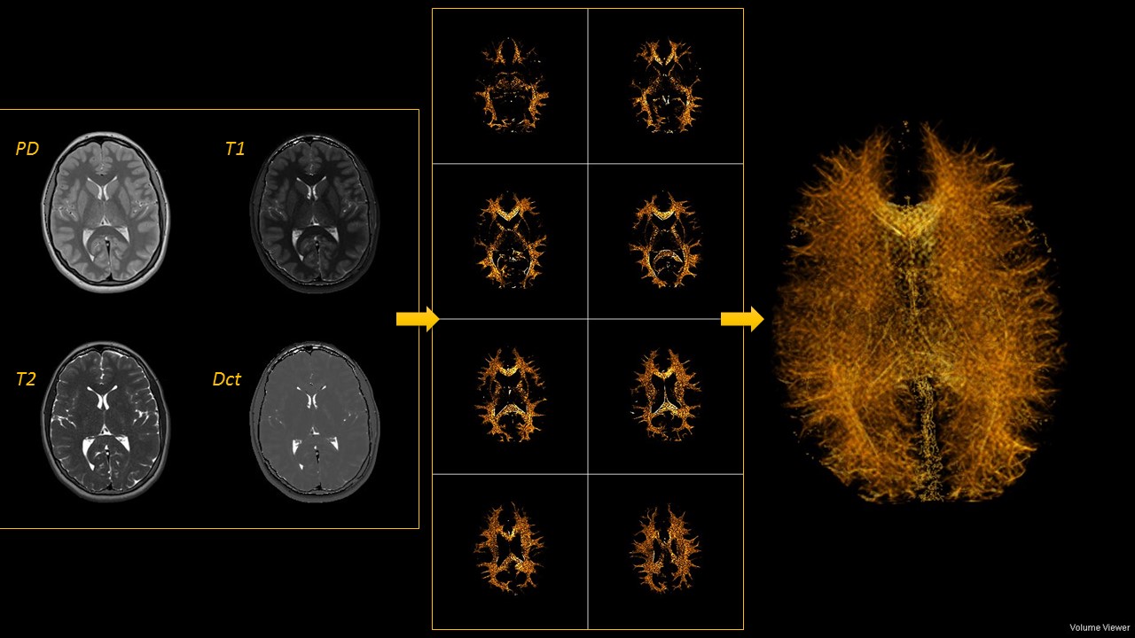
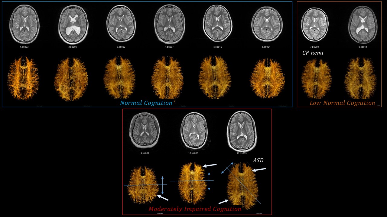
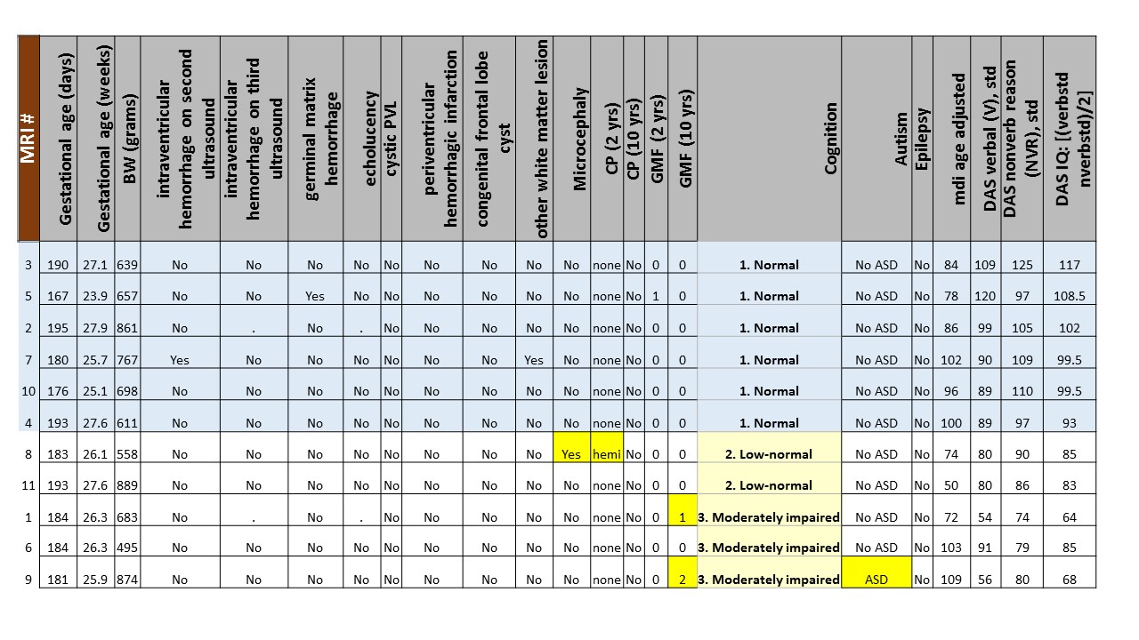
This study was approved by the Institutional Review Boards of the 12 participating institutions of the Extremely Low Gestational Age Newborn (ELGAN) study (3). Eleven EP adolescents were scanned with a 3T MRI protocol that included the tri-turbo spin echo (Tri-TSE) pulse sequence, which is the concatenation of dual-echo TSE (TE1&2eff=12ms & 101ms, TR=10s) and single-echo TSE (TEeff=12ms, TR=0.5s) pulse sequences, both acquired with identical geometry (80 contiguous axial slices and voxel = 0.5 x 0.5 x 2 mm3) and scanner settings. The three directly-acquired images per slice were used to create maps of the relaxation times (T1 and T2), the normalized proton density (PD), and correlation time diffusion coefficient (Dct) using in-house developed qMRI algorithms. Longitudinal relaxation rate (R1=1/T1) heavily weighted images of the intracranium were generated with a synthetic MRI engine (all programs coded in Mathcad, PTC, Needham, MA). The R1-weighted synthetic images, which show well-defined white matter structure, were processed with ImageJ (https://imagej.nih.gov/ij/): first sharpened and then 3D-to-2D projected using the Volume Viewer plugin (Figure 2). Connectomes were grouped by their cognitive classification (Figure 3): normal (n=6), low normal (n=2), and moderately impaired (n=3), and were further organized by IQ.Results
Successful connectome renderings in eleven adolescents (Figure 3) yielded high spatial resolution connectomes with high levels of left-right symmetry and well-organized fiber bundle patterns in the posterior as well as the anterior aspects of the brain among the six children with normal cognition. One exception in this normal group, is a subject with ventriculomegaly, in whom fiber bundles are shorter and the overall order appears diminished. The two subjects in the low-normal cognition group show lower degree of L/R symmetry and diminished connectome organization. The three subjects in the moderately impaired cognition group have disorganized fiber bundles in one case and asymmetric, simplified and disorganized connectomes in the other two.Discussion
This proof of concept study points to the potential utility of WMF via Synthetic MRI as an informative tool for the evaluation of white matter and for establishing associations with independently generated measures of intelligence and cognition. Further work is needed for establishing connectome reference standards in the form of quantitative connectome-descriptive parameters representative of fiber bundle order, state of development, and overall health.Conclusion
White matter fibrography has been successfully used with eleven extremely preterm adolescents showing its reliability and clinical relevance. This work could have implications for developing quantitative connectome metrics and for constructing and assessing ultra-high spatial resolution connectomes in routine clinical practice.Acknowledgements
Supported by the National Institute of Neurological Disorders and Stroke (5U01NS040069-05 and 2R01NS040069-09), National Institutes of Health Office of the Director (1UG3OD022348-01), and the National Institute of Child Health and Human Development (5P30HD018655-28).
List of additional ELGAN Study Investigators is available at www.jpeds.com (Appendix).
References
1. Jara H, Sakai O, Anderson SW, Soto JA. MR Fibrography: an application of correlation time diffusion synthetic MRI (1.5T and 3.0T). Proceeding RSNA (Chicago) 2016.
2. Heeren T, Joseph RM, Allred EN, O'Shea TM, Leviton A, Kuban KC. Cognitive functioning at age 10 years among children born extremely preterm: A latent profile approach. Pediatric research 2017.
3. O'Shea TM, Allred EN, Dammann O, et al. The ELGAN study of the brain and related disorders in extremely low gestational age newborns. Early Human Development 2009;85(11):719-725.
Figures