0845
Early postnatal music intervention in preterm birth reorganizes salience network interactions1Division of Development and Growth, Department of Pediatrics, University of Geneva, Geneva, Switzerland, 2Institute of mathematics, Ecole Polytechnique Fédérale de Lausanne, Lausanne, Switzerland, 3Neuroscience of Emotion and Affective Dynamics Lab, Department of psychology and educational sciences, University of Geneva, Geneva, Switzerland, 44 Swiss center for affective neurosciences, University of Geneva, Geneva, Switzerland, 5Department of Radiology and Medical Informatics, University of Geneva, Geneva, Switzerland, 6Institute of Bioengineering, Ecole Polytechnique Fédérale de Lausanne, Lausanne, Switzerland
Synopsis
The aim of this study is to characterize the preterm brain functional connectivity and to test the effectiveness of an early postnatal music intervention on preterm newborn’s functional brain development. We explored functional connectivity by resting state fMRI in 16 full-term 1 to 4 days old newborns and 29 preterm infants (born < 32weeks) at term equivalent age (TEA) randomly assigned to either music intervention (14 newborns) or control group. We identified a network of interest that represents resting state functional connectivity decrease in preterm control compared to full-term newborns. We found salience network functional connectivity to be especially altered by a premature birth but music intervention significantly increased its connectivity. To the best of our knowledge, this study is the first one to observe music effects on brain development in the preterm newborns.
Introduction
In recent studies alterations of networks connectivity in preterm compared to full-term newborns have been linked to the stressful environment in neonatal intensive care units (NICUs). NICUs are thus willing to propose enrichment of the NICUs’ environment using music, but to date, no evidence of a music effect on preterm newborns’ brain development has been provided. The aim of this study is to characterize the preterm brain functional connectivity and to test the effectiveness of an early postnatal music intervention on preterm newborn’s functional brain development.Methods
Subjects
We explored resting state fMRI functional connectivity of 16 full-term 1 to 4 days old newborns and 29 preterm infants (born < 32weeks) at term equivalent age (TEA) randomly assigned to either music intervention (14 newborns) or control group.
Infants Preparation
All infants were fed before MRI, acquisition was done without sedation with babies swaddled in a blanket and set up in a vacuum pillow, during natural sleep or while resting quietly in the scanner. Infants were monitored (heart rate and oxygen level) during the MRI. MR-compatible headphones were used to protect newborns from noise in the MRI.
Acquisition
8 minutes of RS-fMRI (300 volumes) were acquired (EPI sequences: RT=1600ms, ET=30 ms, slice thickness =3mm, flip angle=90°, matrix=64x52) on Siemens 3T scanners (Siemens, Erlangen, Germany): Siemens Trio using a 32 channel head coil (7PM, 8PC, 11T) and Siemens Prisma using 64 channel head coil. No significant difference due to the different coils or scanners was observed. A T2*-weighted structural image was also acquired (113 coronal slices, RT= 4990ms, TE=151ms, flip angle=150°, Matrix size=256x164; voxel size =0.78x0.78x1.2mm3).
Analysis
We conducted a group independent component analysis to determine robust brain components. By considering these components as regions of interest, we built functional connectivity networks based on a bivariate non-parametric estimator that reflects coupling and anti-coupling between regions of interest1. We identified a network of interest (NOI) that represents resting state functional connectivity decrease in preterm control compared to full-term newborns using statistical testing at each connection of the connectivity matrices. Finally, we performed a t-test comparing PM and PC in each connection that belongs to the NOI using adaptive local correction2.
Results and discussion
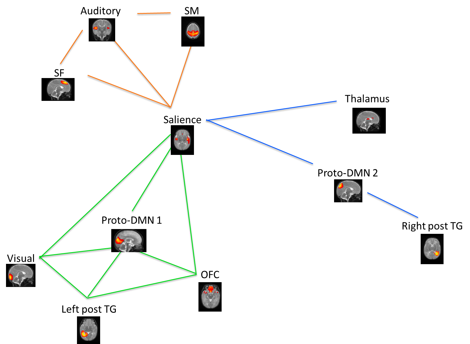
The difference between preterm control and full-term subjects resulted in a network of interest (NOI) that comprises 11 nodes and 16 connections that represent lower functional connectivity in preterm infants (Bonferroni correction for multiple comparison p<0.05). Three subnetworks can be identified including two modules with dense intra-connectivity as modules (Figure 1). A first module (orange module) consists of medial superior frontal (SF1), auditory and sensorimotor (SM) networks. The second (green module) contains the orbitofrontal, posterior cingulate cortex and precuneus (proto-DMN1), visual and left posterior temporal networks (LpTG). The third subnetwork consists of the remaining connections and consists of the thalamus, the precuneus (proto-DMN 2) and right posterior temporal networks (RpTG).
When compared to preterm control infants, the preterm music group had significantly higher functional connectivity between salience, thalamus, medial superior frontal (SF 1), auditory and sensorimotor (SM) networks and a higher connectivity between salience, precuneus (proto-DMN2) and posterior middle and superior temporal networks (RpTG). After performing the adaptive local correction, only one connection survived the strong Bonferroni procedure (Auditory - Sensorimotor). However, five additional connection survived FDR control (Salience - Sensorimotor; Salience - SF1; Auditory - SF1; Salience - Thalamus; Salience - Proto-DMN 2). See Figure 1.
We found salience network functional connectivity to be especially altered by a premature birth but music intervention significantly increased its connectivity. However, an effect of music intervention was observed only in the second module composed of salience, thalamus, precuneus (proto-DMN2) and right posterior temporal gyri.
Conclusion
The Salience network has clearly been identified in this study as an articulation point between three modules and we observed altered connectivity between salience network and numerous networks including sensory networks, networks composed of regions known to be part of the default mode network and regions implicated in executive functions and emotional regulation. We also showed that an early music intervention can increase functional connectivity in these impaired networks and thus, that auditory enrichment of the NICUs environment can have long lasting effects on brain maturation.Acknowledgements
No acknowledgement found.References
1. Meskaldji, Djalel Eddine et al. 2016. “Prediction of Long-Term Memory Scores in MCI Based on Resting-State fMRI.” NeuroImage: Clinical 12:785–95.
2. Meskaldji, Djalel Eddine et al. 2015. “Improved Statistical Evaluation of Group Differences in Connectomes by Screening-Filtering Strategy with Application to Study Maturation of Brain Connections between Childhood and Adolescence.” NeuroImage 108:251–64.
Figures