0843
Fixel-based analysis of white matter fibre density and morphology in the preterm brain.1Centre for the Developing Brain, King's College London, London, United Kingdom, 2Centre for Medical Image Computing, University College London, London, United Kingdom
Synopsis
Fixel-based analysis (FBA) provides measures of fibre cross-section (FC) and fibre density (FD) for individual fibre populations within voxels containing crossing fibres, overcoming the limitations of the diffusion tensor model. FBA was applied to a preterm neonatal cohort to assess the effects of perinatal risk factors on white matter, identifying decreases in FD and FC in distinct fibre populations associated with increased prematurity, low birthweight, respiratory support and parenteral nutrition. We show that aberrant white matter development previously attributed to microstructural changes may be related to alterations in the cross-sectional area of specific fibre bundles at the macroscopic scale.
Background
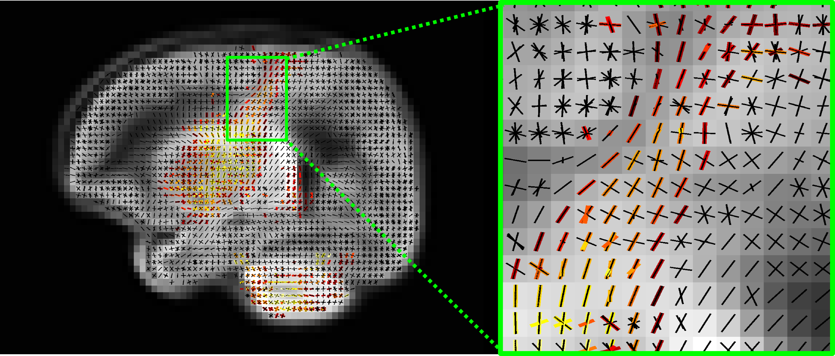
The developing preterm white matter is vulnerable to injury and diffusion MRI (dMRI) studies using the tensor model have identified abnormal white matter development associated with perinatal risk factors in preterm infants studied at term equivalent age. However, this model is an over simplification of the underlying neuroanatomy. Fixel-based analysis (FBA)1 is a novel quantitative framework, which discriminates between alterations in white matter due to microstructural abnormalities (alterations in fixel-specific fibre density, FD) and morphological changes (alterations in fibre cross-section, FC), and combined microstructural and morphological changes (alterations in fibre density and cross-section, FDC).Aims
The aims of this study were to (i) assess the feasibility of applying FBA to assess preterm white matter in the neonatal period, and (ii) investigate the relationship between fixel-based measures of white matter at term equivalent age and perinatal risk factors.Methods
We studied 50 infants (28 male) born at 24.0–32.9 (median 30.4) weeks gestational age (GA) and imaged at 38.6–47.1 (median 42.1) weeks postmenstrual age (PMA). The perinatal characteristics of the cohort were as follows; median birthweight, 1202.5 grams (range 645–1990 grams); mean birthweight z-score, -0.71±0.876; median number of days on mechanical ventilation, 0 (range 0-40); median number of days of total parenteral nutrition (TPN), 6.5 (range 0-89); median index of multiple deprivation, 14.9 (range 6.2 - 46.8). One infant underwent surgery for necrotising enterocolitis, two infants had chorioamnionitis and one infant was treated for patent ductus arteriosus.
High angular resolution, high b-value dMRI (HARDI) (64 non-collinear directions b-value 2500 s/mm2 and 4 non-diffusion-weighted images, TR = 9000 ms, TE = 62 ms, voxel size: 2mm isotropic, SENSE factor of 2) were acquired on a 3 Tesla system sited on the neonatal intensive care unit. Fixel-based analysis was performed to assess the relationship between FD, FC and FDC (given by FD multiplied by FC) and the following;
- PMA at scan (GA and sex as covariates),
- GA at birth (PMA and sex as covariates),
- days on mechanical ventilation (PMA, GA and sex as covariates),
- days of TPN (PMA, GA and sex as covariates),
- birth weight z-score (PMA, GA and sex as covariates).
The results were corrected for multiple comparisons using connectivity-based fixel enhancement2.
Results
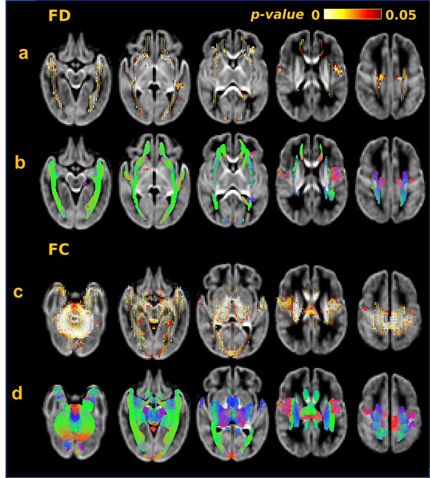
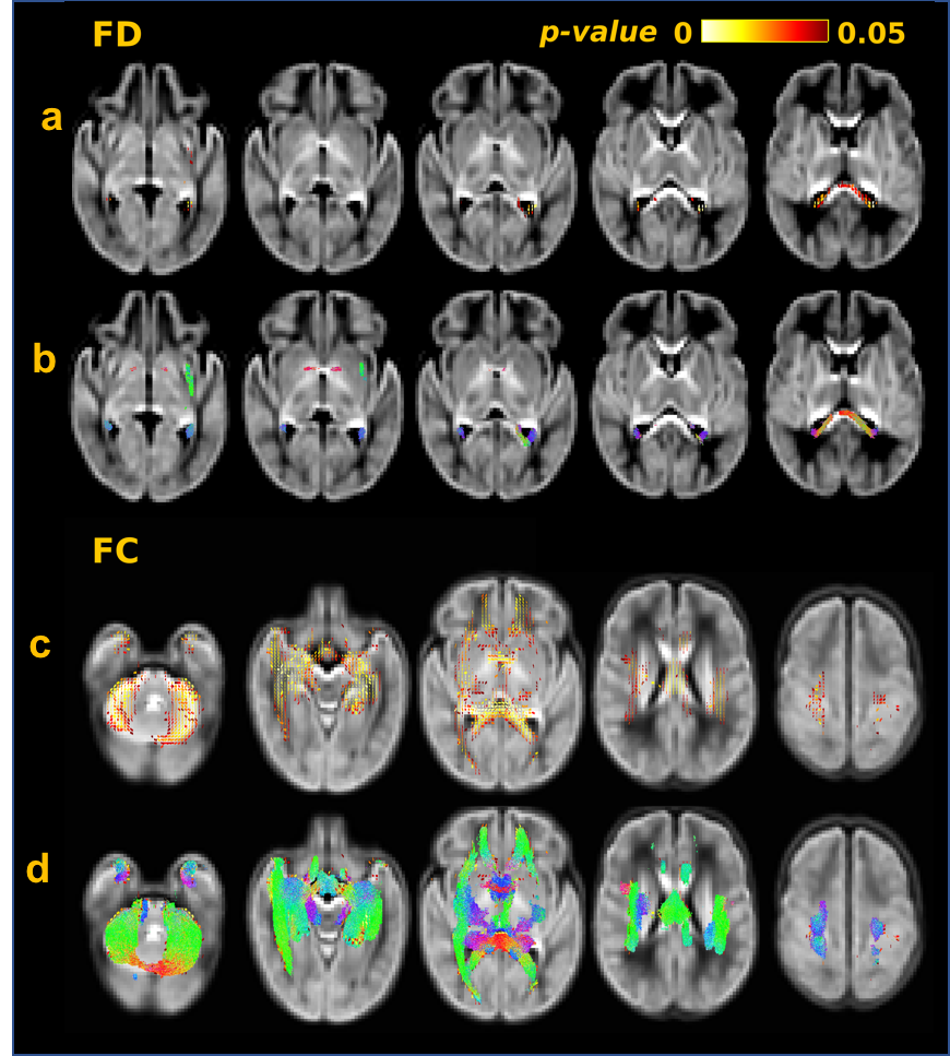
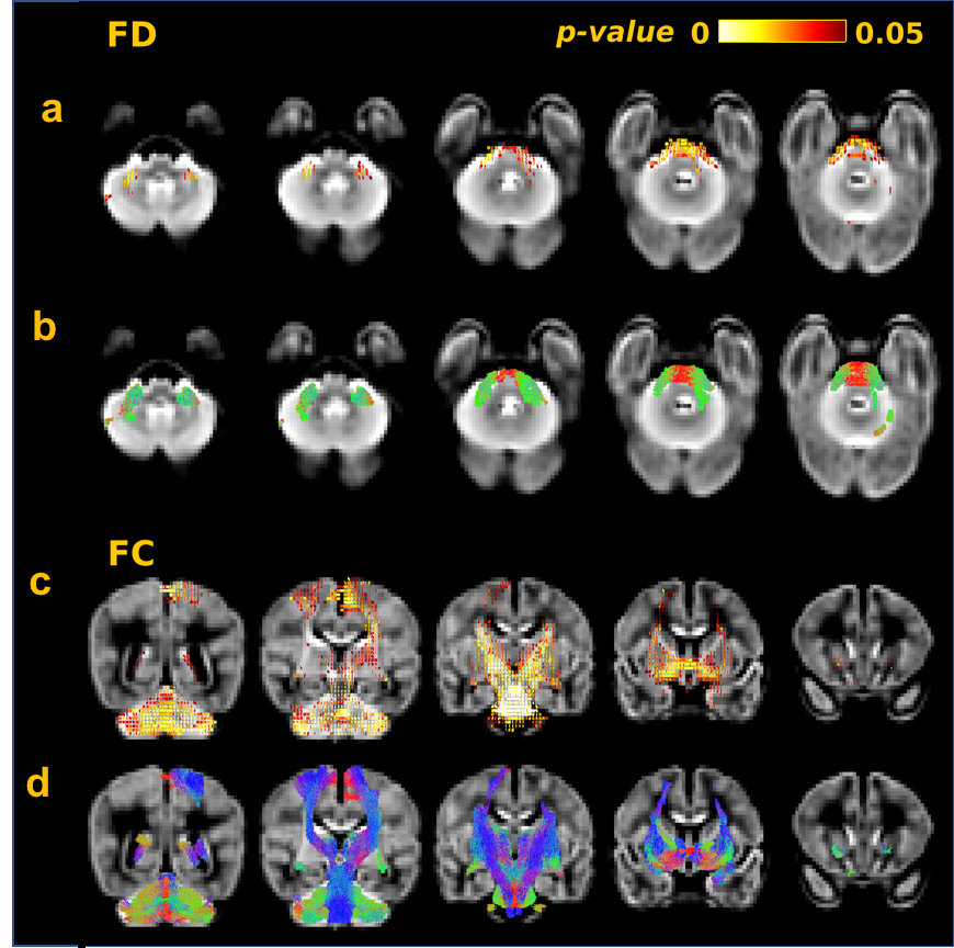
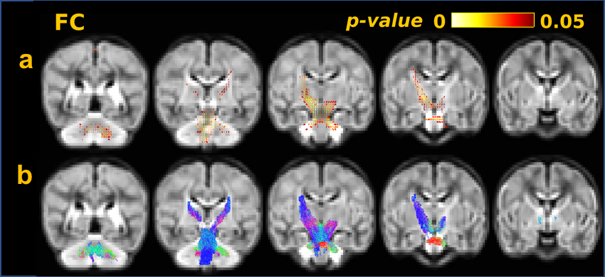
FD was positively correlated with PMA at scan (Figure 1a,b ), GA at birth (Figure 2a,b), and was negatively correlated with number of days requiring ventilation (Figure 3a,b) across a number of white matter fasciculi. FC was positively correlated with PMA at scan (Figure 1c,d), GA at birth (Figure 2c,d) and birthweight z-score. FC was negatively correlated with number of days on ventilation (Figures 3c,d) and days on TPN (Figure 5). FDC was positively correlated with PMA at scan, GA at birth and birthweight z-scores, and negatively correlated with days on ventilation and days of TPN.
Using fixel-based measures it was possible to identify whether morphological changes associated with increased exposure to perinatal risk factors are localised to specific fibre bundles or were related to changes across the whole of the white matter. This is of particular importance in regions of crossing fibres. For example, in the centrum semiovale, we observed that FC in the corticospinal tract was diminished with an increased requirement for mechanical ventilation, however there was no statistically significant relationship between duration of mechanical ventilation and FC in the association fibres traversing the region (Figure 4).
Discussion and Conclusion
This study demonstrates the feasibility of applying FBA to investigate white matter properties in the developing preterm brain, revealing the relationship between fibre population-specific measures of microstructural fibre density and macroscopic morphology of white matter fasciculi in the developing preterm brain and perinatal risk factors. We show that aberrant white matter development previously attributed to microstructural changes may be related to alterations in the size (fibre cross-sectional area) of specific fibre bundles at the macroscopic scale.Acknowledgements
This work was supported by the Wellcome EPSRC Centre for Medical Engineering at Kings College London (WT 203148/Z/16/Z), MRC strategic grant MR/K006355/1 and by the National Institute for Health Research (NIHR) Biomedical Research Centre based at Guy’s and St Thomas’ NHS Foundation Trust and King’s College London. The views expressed are those of the authors and not necessarily those of the NHS, the NIHR or the Department of Health.References
- David A. Raffelt, J.-Donald Tournier, Robert E. Smith, David N. Vaughan, Graeme Jackson, Gerard R. Ridgway, Alan Connelly, Investigating white matter fibre density and morphology using fixel-based analysis, In NeuroImage, Volume 144, Part A, 2017, Pages 58-73.
- David A. Raffelt, Robert E. Smith, Gerard R. Ridgway, J-Donald Tournier, David N. Vaughan, Stephen Rose, Robert Henderson, Alan Connelly, Connectivity-based fixel enhancement: Whole-brain statistical analysis of diffusion MRI measures in the presence of crossing fibres, In NeuroImage, Volume 117, 2015, Pages 40-55.
Figures