0842
Integrated Parallel Reception, Excitation, and Shimming (iPRES) Breast Coil Array for Simultaneous MR Image Acquisition and Localized B0 Shimming1Brain Imaging and Analysis Center, Duke University, Durham, NC, United States, 2Medical Physics Graduate Program, Duke University, Durham, NC, United States, 3Medical Physics Graduate Program, Duke Kunshan University, Kunshan, China, 4GE Healthcare, Aurora, OH, United States
Synopsis
In breast imaging, the image quality is severely degraded by susceptibility-induced B0 inhomogeneities. Here, we apply the novel integrated parallel reception, excitation, and shimming (iPRES) technology to enable simultaneous image acquisition and localized B0 shimming of the breasts with the same coil array. Proof-of-concept in vivo experiments with only four iPRES(1) breast coil elements already show up to 43.3% reduction in B0 root-mean-square error with no SNR compromise. Simulations show that the shimming can be further improved by implementing more complex iPRES(N) geometries with N smaller shim loops per RF coil element.
Introduction
In breast imaging, susceptibility-induced B0 inhomogeneities caused by the air-breast interfaces, heart, lungs, and liver severely degrade the image quality (incomplete fat suppression, distortions, signal loss) in applications like MR spectroscopy or diffusion-weighted imaging, which is increasingly used for breast cancer screening1,2. These localized B0 inhomogeneities cannot be effectively shimmed with traditional spherical harmonic shim coils. Localized shimming methods have been proposed, but require additional shim coils and are limited to one or two coils3,4.
A novel coil design, termed integrated parallel reception, excitation, and shimming (iPRES), allows RF and DC currents to flow in the same coil elements, enabling image acquisition and localized B0 shimming with the same coil array without reducing the SNR or taking additional space, and resulting in a significant reduction in B0 inhomogeneities in the human brain5,6. An improved design, termed iPRES(N), which divides each RF coil element into N smaller shim loops with N independent DC currents, enables the shimming of even more localized B0 inhomogeneities, as demonstrated in body imaging7.
Here, we apply the iPRES technology to enable localized B0 shimming for breast imaging. As a proof-of-concept, we implement four iPRES(1) coil elements into a breast coil array to assess its RF and shimming performance in vivo. We further use simulations to evaluate the improvement in shimming achieved by more complex iPRES(N) geometries.
Methods
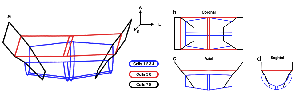
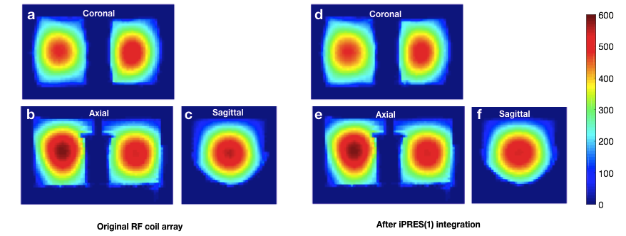
iPRES(1) implementation. Four iPRES(1) coil elements (1234 in Fig. 1) were implemented into an existing 8-channel 3T breast coil array (GE Healthcare). Inductors and chokes were added to bypass capacitors in the RF loops and to provide RF isolation between the coil elements and the DC power supply, allowing DC currents to flow in the coil elements for shimming5,7. S-parameters and SNR maps from these coil elements were acquired before and after modification to ensure that their RF performance was maintained.
In vivo shimming experiment. Basis B0 maps were acquired on a water phantom with a DC current of 1 A separately applied in each iPRES(1) coil element. B0 maps were then acquired on a healthy volunteer before and after shimming with the breast coil array. The optimal DC currents to shim each axial slice were determined by minimizing the root-mean-square error (RMSE) between the baseline B0 map and a weighted combination of the basis B0 maps, computed in an ROI covering both breasts. Dynamic shimming was implemented to update the optimal DC currents for each slice acquisition within a scan.
iPRES(N) simulations. Based on the geometry of the 8 breast coil elements, we used the Biot-Savart law to simulate the basis B0 maps corresponding to more complex iPRES(N) geometries. We then simulated the B0 maps resulting from shimming the baseline B0 map acquired in vivo with the simulated basis B0 maps.
Results
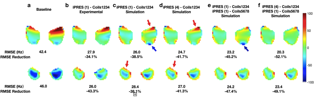
SNR maps are virtually identical before and after iPRES(1) integration (Fig. 2, average over the phantom = 194.9 vs. 194.6), showing that the coil modifications did not impact the SNR. Preliminary in vivo results show that implementing only four iPRES(1) coil elements can already reduce the B0 RMSE in the breasts by up to 43.3% (Fig. 3a-b). Simulations performed with the same coil geometry provided similar, but not identical, results due to slight differences between the simulated and experimental coil geometries (Fig. 3b-c). Nevertheless, the simulations show that the shimming can be further improved by implementing more complex iPRES(N) geometries with more degrees of freedom, smaller shim loops, and/or a better location of the coil elements, resulting in an RMSE reduction of up to 52.1% (Fig. 3c-f). For example, implementing iPRES(4) in coils 1234 improves the shimming in the anterior regions (blue arrows), whereas implementing iPRES(1) in coils 5678, which are more posterior to the breasts, improves the shimming in the posterior regions (red arrows).Discussion and Conclusion
The in vivo shimming experiment shows that our proof-of-concept breast coil array with only four iPRES(1) coil elements can already achieve a very promising reduction of localized B0 inhomogeneities in the breasts, which we plan to further improve by implementing more complex iPRES(N) designs, as shown in the simulations. Combined with dynamic shimming, this novel technology can enable a highly effective and efficient localized B0 shimming of the breasts, without compromising the SNR or requiring additional shim coils, which will greatly improve the image quality, spatial fidelity, and diagnostic accuracy of many clinical applications in breast imaging.Acknowledgements
This work was in part supported by grants R21 EB018951, R24 MH106048, R21 EB024121 from the National Institutes of Health, by GE Healthcare, and by the Duke-Coulter Translational Partnership.References
[1] Lee SK, Tan ET, Govenkar A, Hancu I. Dynamic slice-dependent shim and center frequency update in 3T breast diffusion weighted imaging. Magn Reson Med. 2014;71(5):1813-1818
[2] Lin C, Rogers CD, Majidi S. Fat suppression techniques in breast magnetic resonance imaging: a critical comparison and state of the art. Reports in Medical Imaging 2015; 8:37-49
[3] Lee SK et al. B0 shimming in 3T Bilateral Breast Imaging with Local Shim Coils. Proc ISMRM 2011, p 715
[4] Boer VO et al. Bilateral shimming of the breast at 7T. Proc ISMRM 2014, p 1617
[5] Truong T-K, Darnell D, Song AW. Integrated RF/shim coil array for parallel reception and localized B0 shimming in the human brain. NeuroImage 2014;103:235-240
[6] Stockmann JP, Witzle T, Keil B, Polimeni JR, Mareyam A, LaPierre C, Setsompop K, Wald LL. A 32-channel combined RF and B0 shim array for 3T brain imaging. Magn Reson Med. 2016;75(1):441-451.
[7] Darnell D, Truong TK, Song AW. Integrated parallel reception, excitation, and shimming(iPRES) with multiple shim loops per radio-frequency coil element for improved B0 shimming. Magn Reson Med. 2017;77(5):2077-2086.
Figures