0840
Constrained Optimization for Static and Dynamic B0 Shimming1MRRC, Dept. of Radiology, University of Pittsburgh, Pittsburgh, PA, United States, 2Medical Imaging Physics (INM-4), Institute of Neuroscience and Medicine, Jülich, Germany
Synopsis
Dynamic shimming compared to global shimming improves B0 homogeneity over whole brain. However, typical shim optimization could result in high currents, particularly for small volume, e.g. small slab or slice. To address this we investigated the extent to which large reductions in aggregate shim current could be achieved through constrained optimization at 7T. We also investigated the extent to which alternate imaging orientation could improve homogeneity. The results show constrained optimization can provide robust shimming at substantially reduced aggregate current values, and angulated imaging improved shimming condition. The proposed methods enhanced multi-slice imaging by reducing both susceptibility-induced signal losses and distortions.
Introduction
Typically optimization of B0 homogeneity within a 3D volume is achieved through least square minimization of the residual B0 field constrained only by the maximum output of the shim current supplies. Unfortunately, with higher order shim systems (e.g., 1st-4th) this can result in high currents for volumes of limited size, e.g., small slab or slice. The higher currents used place greater demands on the accuracy of the calibration of the shims, including any imperfections, thereby requiring longer iterative approaches. Further, eddy current effects are increased when the high valued shims are switched dynamically. To overcome this effect we investigated the extent to which large reductions (>60%) in aggregate shim current could be achieved through constrained optimization without significant loss in B0 homogeneity. We also investigated the extent to which alternate imaging plane orientations could improve homogeneity.Methods
All data were acquired on a 7T Siemens whole body system (with 1st - 2nd shims embedded) using a high degree head shim insert coil (RRI, Resonance Research Inc., MA) providing 3rd and 4th degree shims1. Dynamic updating of 2nd-4th degree shims was achieved using an RRI power supply. Offsets for 1st degree shims (gradient offset) were controlled via imaging sequence in MR console. The shim unit and scanner were synchronized through trigger signal which generated in the scanner sequence. Data processing and shim controls were done by a PC with home-made MATLAB program (MathWorks, MA). The B0 field was mapped using BOLERO with incremental B0 encoding intervals (TE = 1,2,4 and 8 ms)2. Data was acquired using an 8×2 transceiver array in pTx mode. B0 maps, 33-41 contiguous slices, 3mm thickness, matrix size 64×64 over a FOV of 192×192mm2 were acquired by using multi-echo GRE sequence. The shim optimization was applied for 3D volume in static shimming and 2D multi-slices in dynamic shimming. In multi-slice 2D imaging, a constrained optimization (15A total aggregate current) was used to reduce the optimal shim currents. For dynamic shim update, eddy currents due to dynamic updating of the shims was reduced further by using a 2ms ramp to switch the shims. For the feasibility test to real MR application, the developed shim methods were applied to multi-slice 2D GE EPI with identical spatial parameters as B0 mapping sequence.Results
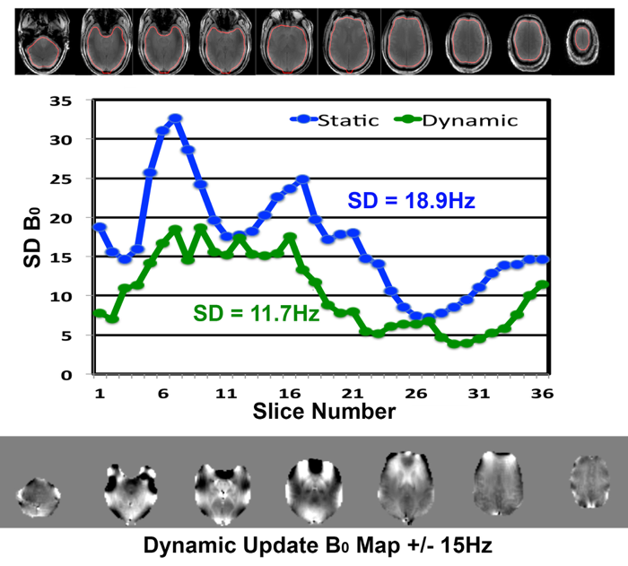
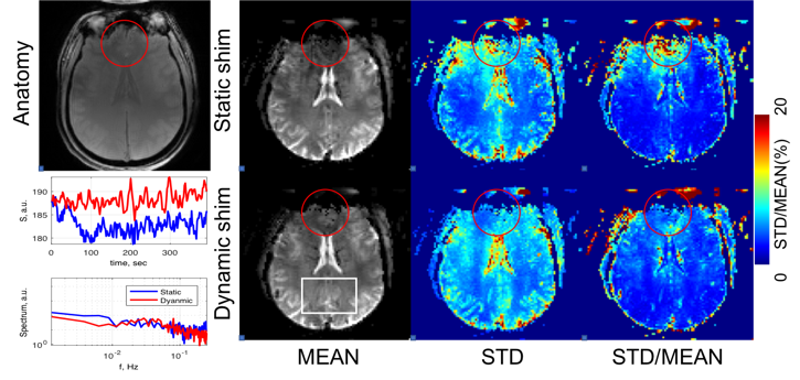
Fig. 1 shows the dependence of the achieved B0 inhomogeneity (SD) to the aggregate current applied for all 2nd, 3rd and 4th degree shims using static shimming; the unconstrained optimizations required 42.9, 46.5, 24.1 and 8.4A of aggregate current for TL, SCN, CBL, SMA, respectively. However, constraint of the aggregate current to 15A, results in increases of 0% - 8% for the four loci. Thus a >60% reduction in aggregate current can be achieved with only minimal loss in B0 homogeneity. For multi-slice studies the B0 homogeneity can be optimized by updating the values of the shims for each slab (3 adjacent 3mm slices) per slice, allowing each slice(s) to have different values. For axial slices, B0 shimming over the temporal and inferior frontal lobe is often complicated due to the need to simultaneously correct for different susceptibility effects arising from both the frontal sinuses and lateral ear canals. These effects can be decoupled from each other by angulating the slice orientation along the temporal pole. In addition to providing an optimal view of the hippocampus imaging, this approach places the frontal susceptibility artifact in more superior slices. Fig. 2 shows the B0 map acquired for 36 3mm slices acquired parallel to the planum temporale. The overall inhomogeneity across all brain pixels was measured as 11.7Hz with dynamic shim, while it was18.9Hz under static shimming condition. Fig. 3 shows resting state EPI data acquired by using dynamically updated and static 1st-4th degree shims. The improvement in B0 homogeneity from the anterior region above the sinuses reduces dropout, distortion and thus increases SNR and temporal stability.Conclusions
Constrained optimization of high order B0 shimming can provide robust shim optimization at substantially reduced aggregate current values (up to 70%) with minimal loss (<10%) in B0 homogeneity. Angulation of the imaging plane along the planum temporale spatially segregates the frontal and temporal susceptibility effects enabling improved shimming condition for the temporal lobe and inferior frontal regions when dynamically updating the shims. Combined these methods enhance the quality of multi-slice imaging by reducing both susceptibility induced signal losses and distortions in difficult brain regions.Acknowledgements
The study is supported by NIH EB011639, EB009871, NS090417, NS081772.References
1. Pan JW, Lo KM, Hetherington HP. Magnetic resonance in medicine. 2012;68:1007-1017.
2. Hetherington H, Chu WJ, Gonen O, Pan JW, Magnetic resonance in medicine, 2006;56:26-33.
Figures