0838
Application of Integrated Parallel Reception, Excitation, and Shimming (iPRES) for Signal Loss Recovery in fMRI1Brain Imaging and Analysis Center, Duke University, Durham, NC, United States, 2Medical Physics Graduate Program, Duke University, Durham, NC, United States
Synopsis
Integrated parallel reception, excitation, and shimming (iPRES) coil arrays enable simultaneous image acquisition and localized B0 shimming with a single coil array. We propose to apply this novel technology to recover signal loss in fMRI by using a new shim optimization algorithm. Simulations and in vivo experiments performed with a 32-channel iPRES head coil array demonstrate that the proposed method can reduce both the B0 inhomogeneity and the signal loss in gradient-echo EPI images, particularly in the inferior frontal brain region, resulting in more activation in that region in a breath-holding fMRI experiment.
Introduction
Magnetic susceptibility differences at air/tissue interfaces induce static magnetic field (B0) inhomogeneities, which cause distortions and signal loss in gradient-echo EPI images. This effect is most significant in the inferior frontal brain region, because of the air cavities in the sinuses, and hampers our ability to functionally image this region using BOLD fMRI. Various methods have been proposed to correct for these artifacts, but with some limitations. For example, post-processing techniques cannot recover signal loss, z-shimming techniques1 have a trade-off between shimming effectiveness and scan time, and hardware methods like intra-oral coils2 or passive shims3 have limited subject comfort or flexibility. Integrated parallel reception, excitation, and shimming (iPRES) is a novel technology that allows RF and DC currents to flow in the same coil elements, thereby enabling simultaneous image acquisition and localized B0 shimming with a single coil array, without compromising the SNR or requiring additional space in the scanner. Previously, it has been used to correct for distortions in EPI images4-6. Here, we propose to use it to recover signal loss in fMRI without the limitations of previous methods.Methods
In previous work, the DC currents used for shimming were optimized by minimizing a cost function defined as the root-mean-square error (RMSE) between the in vivo B0 map to shim and a weighted combination of basis B0 maps acquired on a phantom with a DC current of 1 A separately applied in each iPRES coil element4,5:
$$\small C_1 = \sqrt{\text{mean}\left(B_{0,\text{shimmed}}^2\right)} \text{ where } B_{0,\text{shimmed}} = B_{0, \text{to shim}}+\sum_k{\text{DC}_k B_{0,\text{basis,}k}}$$
This method minimizes the overall B0 inhomogeneity, but, when applied to a thin slab, tends to minimize the in-plane B0 gradients (causing distortions) at the expense of the through-plane B0 gradient (causing signal loss). To more effectively minimize the signal loss for fMRI applications, we propose a new cost function:
$$\small C_2 = C_1 + w \sqrt{\text{mean}\left(\text{signal loss}^2\right)}$$
where w is a weight and:
$$\small \text{signal loss} = 1-\mid\text{sinc}\left(\frac{\phi}{2}\right)\mid$$
with
$$\small \phi = \gamma \frac{dB_{0,\text{shimmed}}}{dz} \Delta z \text{TE}$$
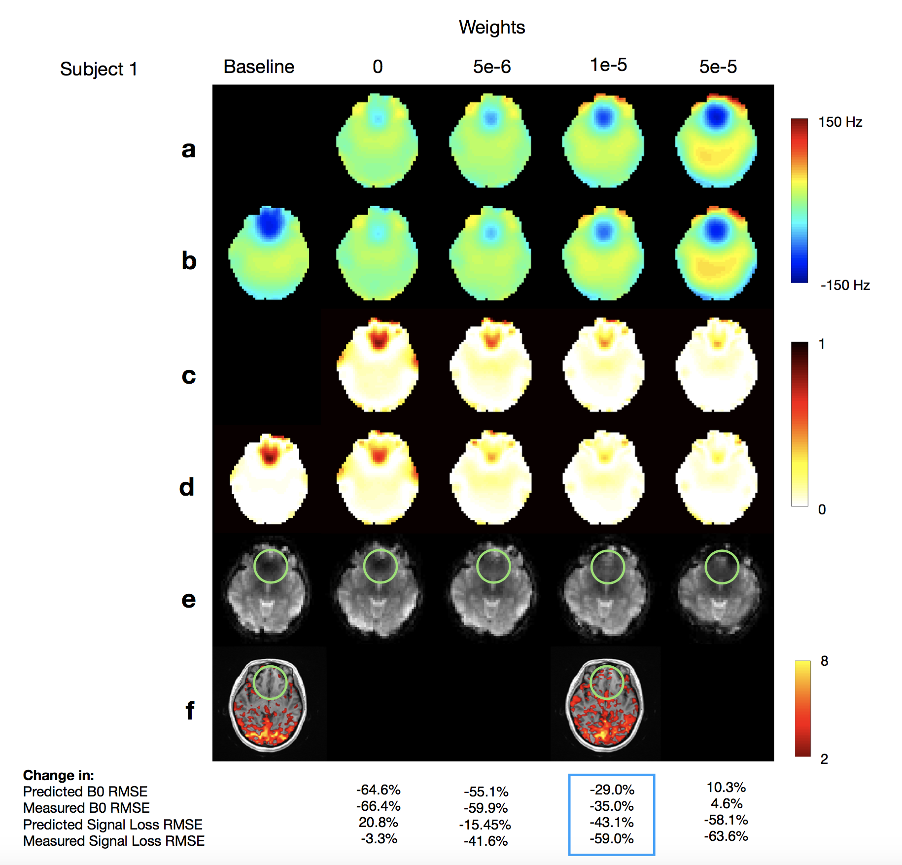
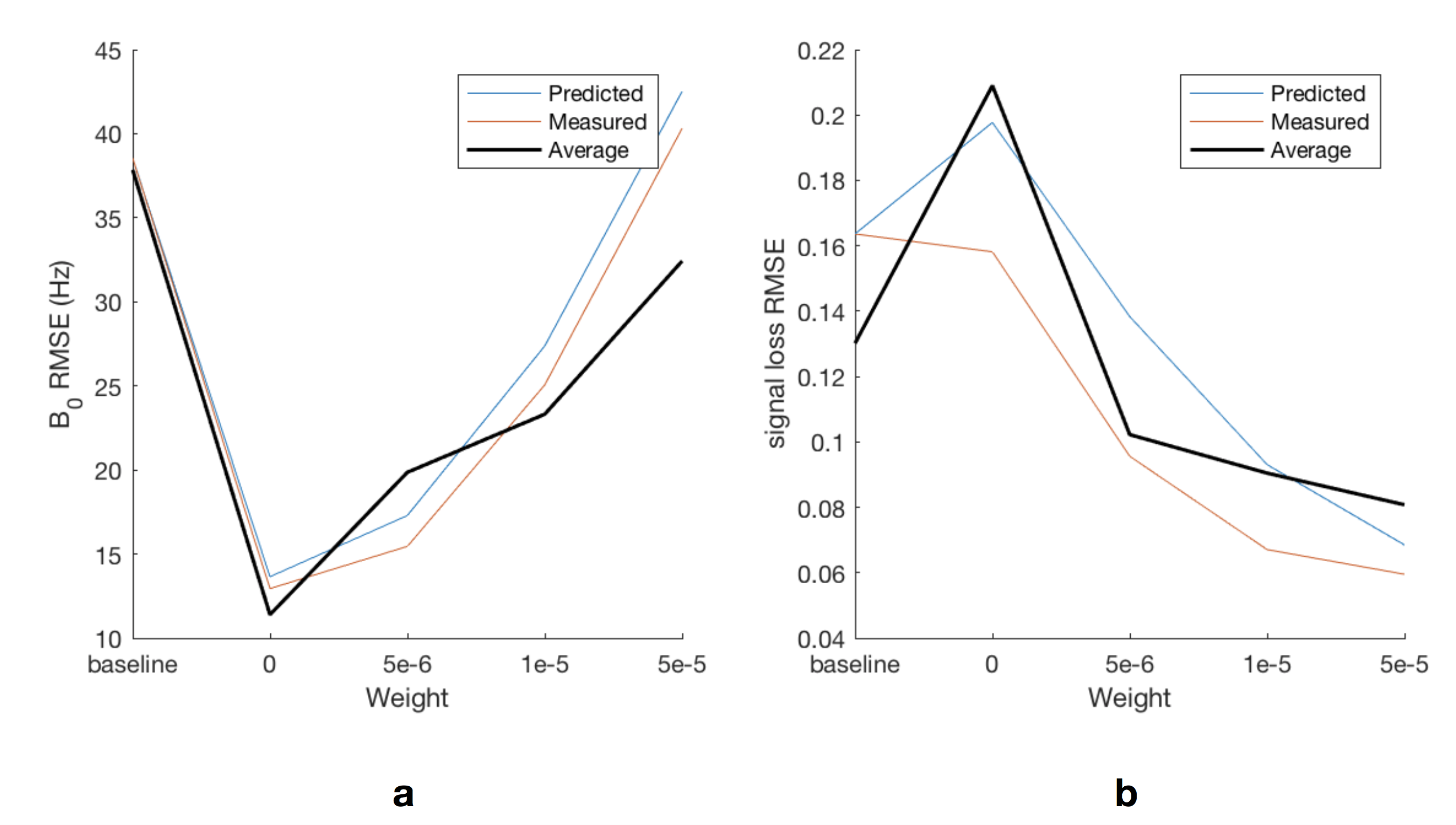
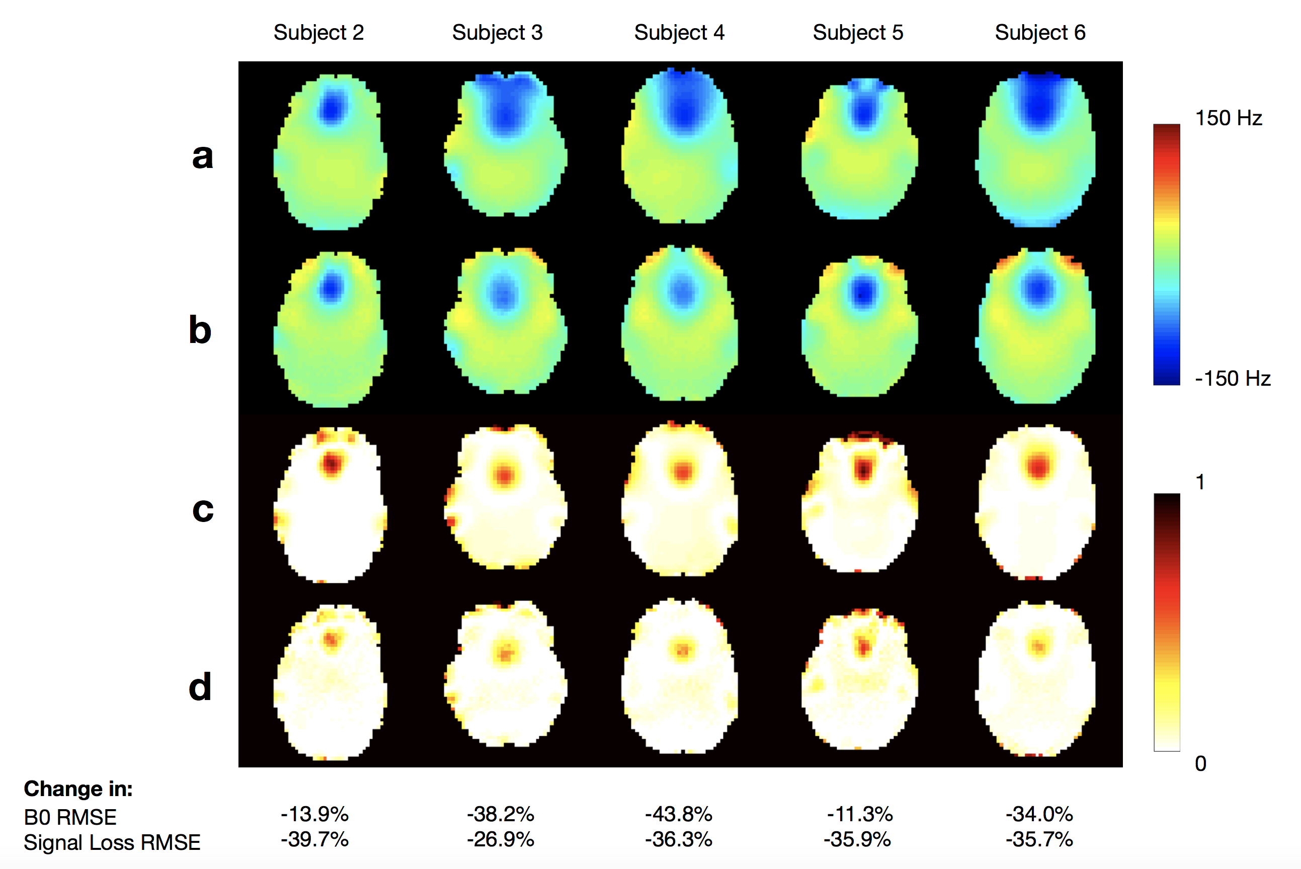
where $$$\gamma$$$ is the gyromagnetic ratio and $$$\Delta z$$$ the slice thickness7. To determine the optimal weight, simulations were performed by using different weights to shim axial B0 maps acquired through the inferior frontal brain region of 15 healthy subjects. To assess the shimming performance in vivo, experiments were also performed by acquiring B0 maps and gradient-echo EPI images (TR = 2 s, TE = 30 ms, voxel size = 4x4x4 mm) on a healthy subject before and after shimming with different weights. Furthermore, to evaluate the viability of this method for signal recovery in fMRI, EPI time series were acquired with a block design alternating between normal breathing and breath-holding, before and after shimming with the optimal weight. All scans were performed on a 3T scanner with a 32-channel iPRES head coil array using linear shimming, but no high-order spherical harmonic shimming.
Results and Discussion
As expected, the simulation results show that as the weight increases from 0 (old cost function) to 5e-5, the shimming becomes less effective at reducing the B0 inhomogeneity, but more effective at reducing the signal loss, particularly in the frontal brain region (Figs. 1a,c and 2). A weight of 1e-5 was found to provide an optimal tradeoff between the reduction in B0 inhomogeneity and the reduction in signal loss. Virtually identical results were obtained in the experiment on the same subject (Figs. 1b,d and 2) and similar results were obtained in the simulations on all other subjects (Figs. 2 and 3). The B0 and signal loss RMSEs were reduced by up to 43.8% and 59.0%, respectively, but the optimal weight can be adjusted to achieve an even more effective signal recovery at the expense of the B0 homogeneity, in applications where this is more important (e.g., the distortions could be corrected with post-processing). The EPI images (Fig. 1e) follow the same trend as the signal loss maps, but with some differences since the latter assumed a uniform spin density and did not take into account the distortions. Importantly, the signal loss in the frontal brain region (green circle) was largely recovered after shimming with the optimal weight. Consequently, the fMRI results show more activation in this region after shimming (Fig. 1f).Conclusion
While further optimization and in vivo validation in additional subjects is ongoing, our promising preliminary results demonstrate that the iPRES technology can effectively recover the signal loss in fMRI without compromising the SNR, scan time, or subject comfort, which will enable the investigation of important cognitive functions in brain regions affected by signal loss.Acknowledgements
This work was in part supported by grants R21 EB018951, R24 MH106048, R21 EB024121 from the National Institutes of Health, by GE Healthcare, and by the Duke-Coulter Translational Partnership.References
1. Constable RT. Functional MR imaging using gradient-echo echo-planar imaging in the presence of large static field inhomogeneities. J Magn Reson Imaging. 1995;5:746–752.
2. Hsu JJ, Glover GH. Mitigation of susceptibility-induced signal loss in neuroimaging using localized shim coils. Magn Reson Med. 2005;53(2):243-8.
3. Wilson JL, Jenkinson M, Jezzard P. Optimization of static field homogeneity in human brain using diamagnetic passive shims. Magn Reson Med. 2002;48(5):906-14.
4.Truong TK, Darnell D, Song AW. Integrated RF/shim coil array for parallel reception and localized B0 shimming in the human brain. NeuroImage 2014;103:235-240.
5. Darnell D, Truong TK, Song AW. Integrated parallel reception, excitation, and shimming (iPRES) with multiple shim loops per radio-frequency coil element for improved B0 shimming. Magn Reson Med 2016;77(5):2077-2086.
6. Stockmann JP, Witzel T, Keil B, Polimeni JR, Mareyam A, Lapierre C, Setsompop K, Wald LL. A 32-channel combined RF and B0 shim array for 3T brain imaging. Magn Reson Med. 2016;75(1):441-451.
7. Truong TK, Song AW. Single-shot dual-z-shimmed sensitivity-encoded spiral-in/out imaging for functional MRI with reduced susceptibility artifacts. Magn Reson Med. 2008;59(1):221-7.
Figures