0834
Integrated ΔB0/Rx coil array for improved spinal cord imaging at 3T1NeuroPoly Lab, Institute of Biomedical Engineering, Polytechnique Montreal, Montreal, QC, Canada, 2École polytechnique fédérale de Lausanne, Lausanne, Switzerland, 3Electrical Engineering and Computer Science, Massachusetts Institute of Technology, Cambridge, MA, United States, 4A. A. Martinos Center for Biomedical Imaging, Massachusetts General Hospital, Charlestown, MA, United States, 5Harvard Medical School, Boston, MA, United States, 6Functional Neuroimaging Unit, CRIUGM, Université de Montréal, Montreal, QC, Canada
Synopsis
Spinal cord imaging is hampered by static and respiration-induced dynamic variations of B0, causing serious issues for EPI-based protocols and spectroscopy. Integrated ΔB0/Rx arrays could provide an effective means to further compensate small-scale variations, complementary to the existing spherical harmonic coils. Here, we designed, built and tested in a human subject an 8-channel integrated ΔB0/Rx coil for the cervical spinal cord. Results showed improvements for correcting static field variations as well as respiratory-induced variations. Future studies will investigate dynamic and real time shimming applications for the spinal cord.
Introduction
Spinal cord imaging is hampered by static and respiration-induced dynamic variations of ΔB0 which cause significant issues for EPI-based protocols and spectroscopy.1,2 Integrated ΔB0/Rx arrays, combining RF reception and ΔB0 shim functionality are a promising solution due to their high efficiency, low inductance, cost-efficient current drivers, and the capability to correct small-scale variation in ΔB0.3 Moreover, these integrated coils provide similar SNR to RF-only coils at both 3T and 7T. Based on previous simulations,4 we designed, built and tested an 8-channel RF/shim coil for the cervical spinal cord.Methods
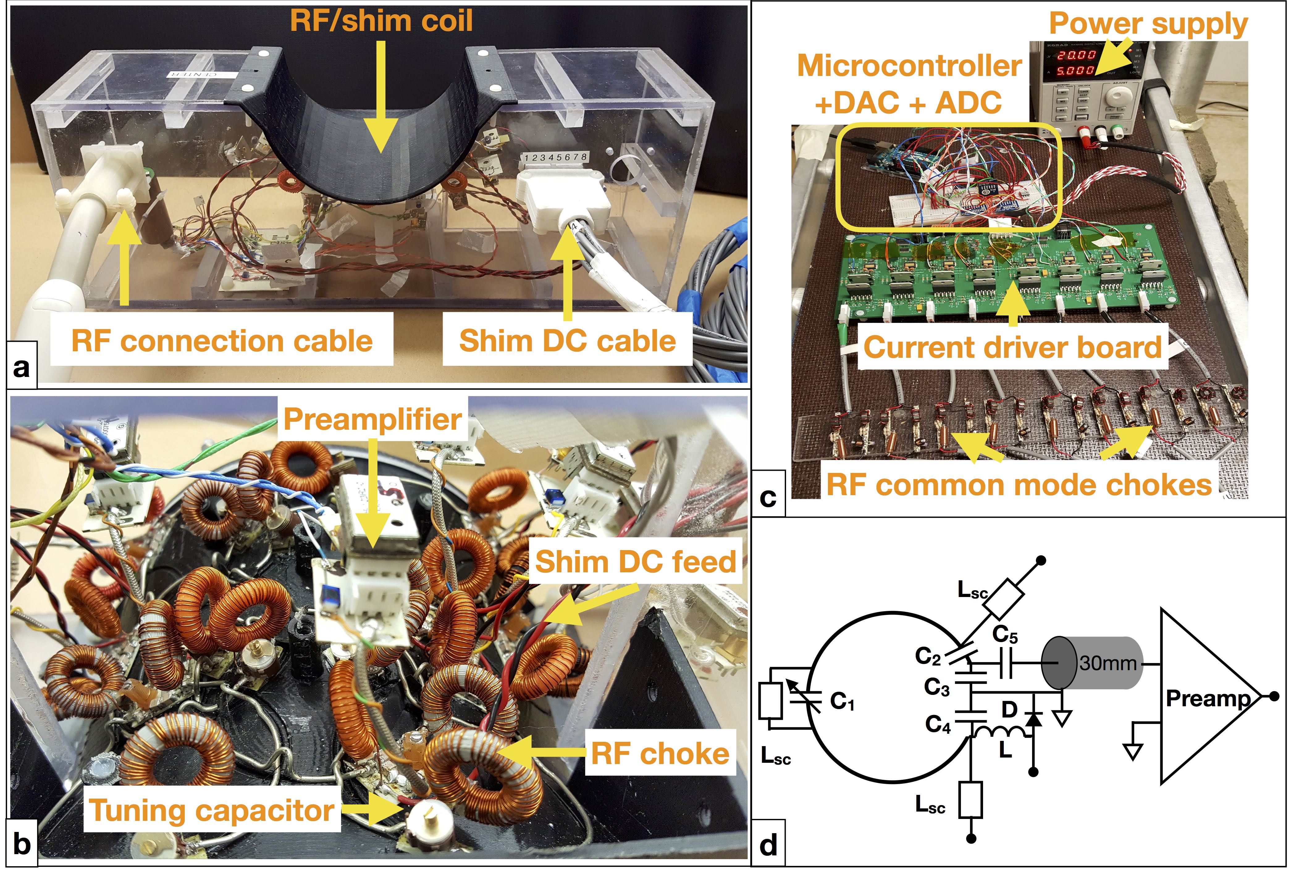
Design: An integrated ΔB0/Rx coil array was constructed featuring 8 x 55 mm diameter loops of 16-AWG copper mounted on a close-fitted holder adapted to the neck region (Figure 1). The elements were disposed in a 3-3-2 configuration being geometrically decoupled to reduce interactions between neighboring elements. In addition, low input impedance preamps (MPB-127R73-90, Hi-Q.A. Inc., ON, Canada) were closely mounted to the loops to improve the decoupling between non adjacent elements. 1uH toroidal chokes are used to bridge the tuning capacitors on each RF loop in combination with 1000 pF capacitors to block the DC in the RF feed. Shim currents are controlled using a current driver board5 and a microcontroller. The desired values are updated from the microcontroller via SPI interface to an octal DAC (AD5628), which drives 8 push-pull amplifiers. A current sense resistor, included in the feedback loop of the amplifiers, was used to measure the actual DC current values circulating in the coils. The voltage drops were read with two 4-channel ADCs (ADS1015) connected on the I2C interface of the microcontroller. RF common mode currents during transmission phase are reduced in the DC feed cables by using high impedance RF chokes and parallel resonant LC circuits at the output of the current driver board.
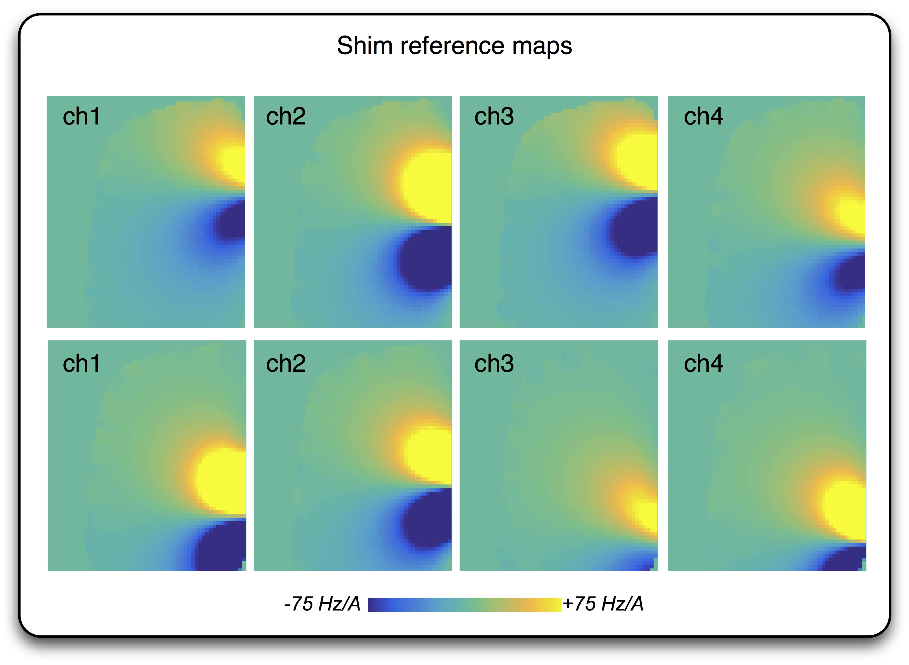
Calibration: Shim reference maps in Hz/A were acquired on a
small phantom (2L distilled water doped with 7.5 g NISO4 X 6H2O + 10 g NACL) in a manner similar to [6]: for each channel, field maps were
acquired with ± 400 mA circulating in the shim; the difference between the
two field maps was divided by the difference in current (800 mA), followed by
low-pass filtering to reduce the effect of noise (Figure 2).
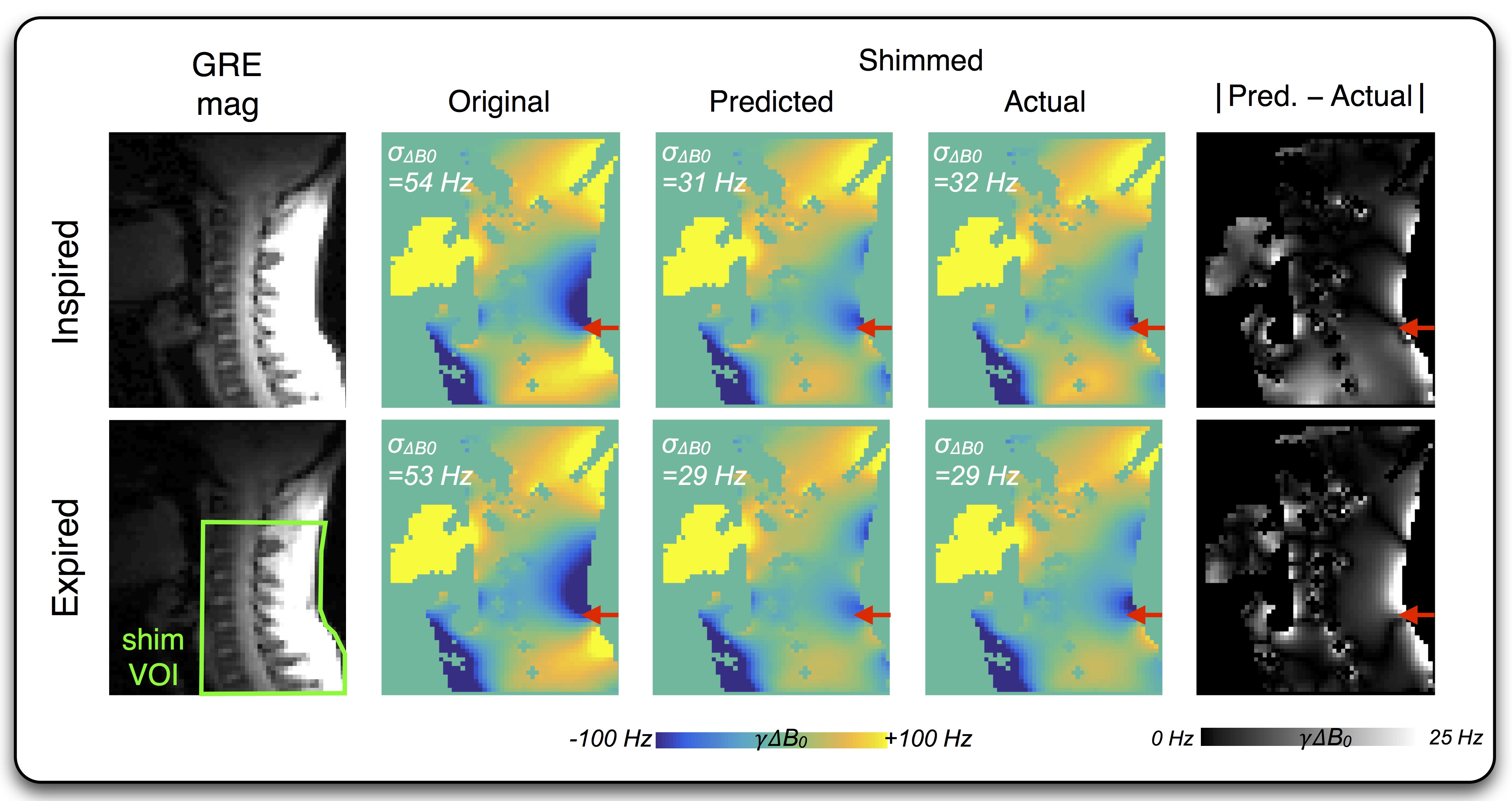
In vivo experiment: GRE field maps of a single subject were
acquired separately under inspired and expired breath-hold conditions with
acquisition and phase processing parameters similar to those described in a
previous work [6]. A shim volume of interest (VOI) was designated, encompassing
the inferior portion of the cervical spinal cord and the region posterior to
it. Using the reference maps, optimal shim currents were calculated
independently for the two breath-holds, limiting the max absolute current per
channel to 400 mA. Field maps were reacquired for each of the two breath-holds upon
setting the shims.
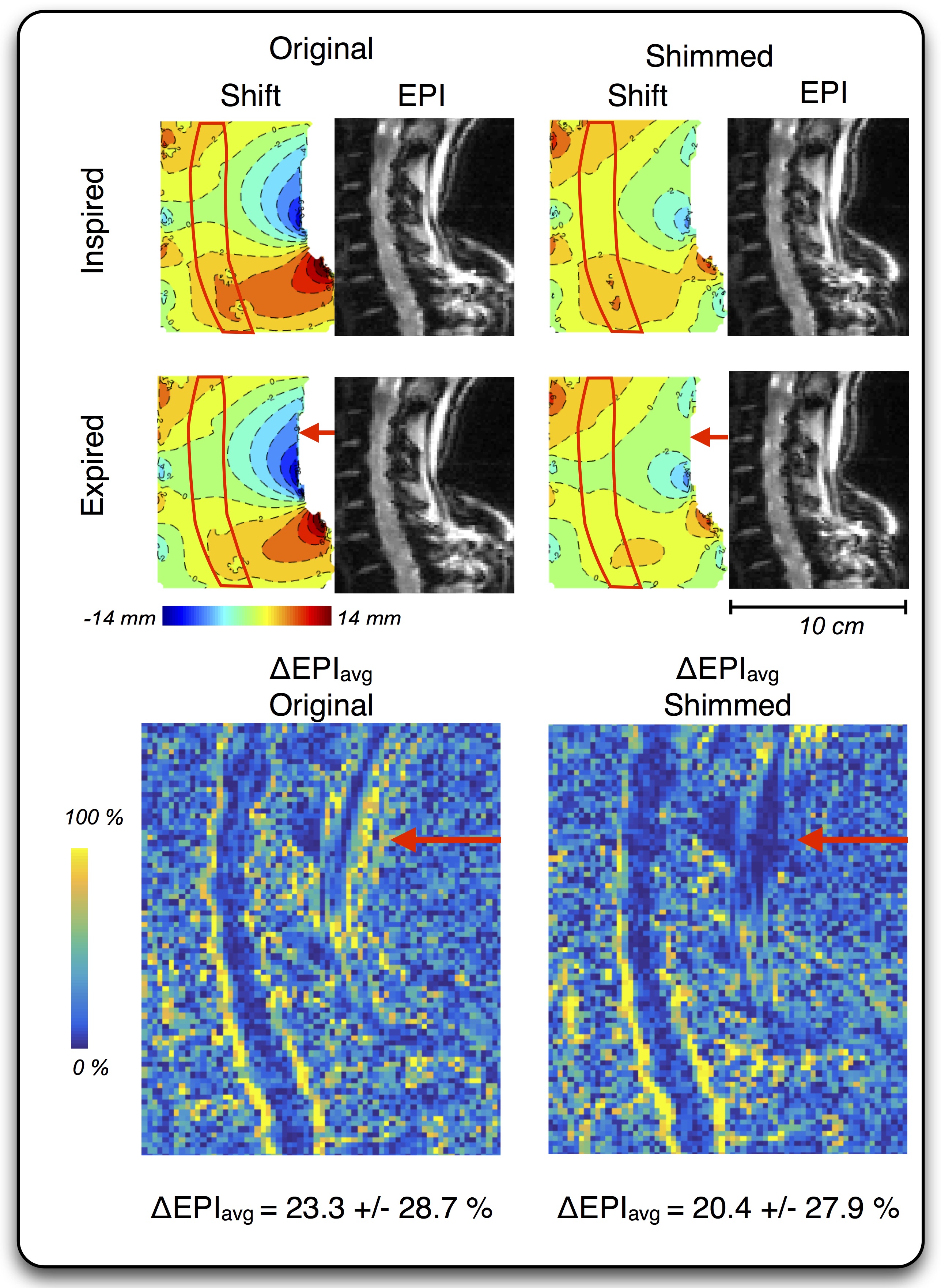
The effect of the optimized shims was further explored on a 2D single-shot GRE-EPI
using the following parameters: TE=16 ms, volume TR=500 ms; flip angle 50°; bandwidth=1630
Hz/pixel; R=2 acceleration factor using GRAPPA; partial Fourier=6/8; spatial resolution=1.5x1.5
mm2 in-plane, with 3 sagittal slices of thickness 3.0 mm centered on
the spinal cord, for an effective FOV=192x192x9 mm3. EPI were
acquired for both breath-holds, in both shim-off and shim-optimized states.
Results and Discussion
Over the shim VOI, application of the ΔB0/Rx array reduced the standard deviation of the field from its original values of 53.6 and 52.9 Hz (inspired and expired, respectively) down to 32.1 and 28.6 Hz, amounting to respective improvements in field homogeneity of 40% and 46% (Figure 3). Figure 4 shows that EPI voxel shift over the same region, as calculated from the scaled absolute field maps, was reduced from 2.71 ± 2.1 mm and 2.5 ± 2.1 mm (inspired and expired) down to 1.8 ± 1.2 and 1.4 ± 1.3 mm, with the maximum shift for the breath-holds reduced from an initial 13.1 mm to 8.0 mm. The mean-normalized percent-difference of the EPI (ΔEPIavg = 200% x | EPIin – EPIex|/ (EPIin + EPIex) ) was reduced 12.5% from an original 23.3 ± 28.7 % to 20.4 ± 27.9 % (median ± standard deviation).Though limited to a single subject, this preliminary proof-of-concept demonstrate the dual potential of the integrated ΔB0/Rx design for high-quality signal reception along with ΔB0 correction for both static and respiration-induced ΔB0 distortions. Forthcoming experiments will include the characterization of Rx SNR and, once a heat-sink is incorporated to allow for shim currents > 400 mA, real-time shimming using a set of respiratory bellows.
Acknowledgements
Study funded by the Canada Research Chair in Quantitative Magnetic Resonance Imaging (JCA), the Canadian Institute of Health Research [CIHR FDN-143263], the Canada Foundation for Innovation [32454, 34824], the Fonds de Recherche du Québec - Santé [28826], the Fonds de Recherche du Québec - Nature et Technologies [2015-PR-182754], the Natural Sciences and Engineering Research Council of Canada [435897-2013] and the Quebec BioImaging Network. The authors thank the Unité de Neuroimagerie Fonctionnelle (University of Montreal) for the support.References
1. Verma, T. & Cohen-Adad, J. Effect of respiration on the B0 field in the human spinal cord at 3T. Magn Reson Med 72, 1629–36 (2014).2. Stroman, P. W. et al. The current state-of-the-art of spinal cord imaging: methods. Neuroimage 84, 1070–81 (2014).
3. Stockmann, J. et al. An Integrated 32ch RF-Shim Array Coil for Improved B0 Shimming of the Brain at 7 Tesla. in Proceedings of the 25th Annual Meeting of ISMRM, Honolulu, 2017. Abstract 967.
4. Germain, G. et al. Optimization of geometry for combined RF/shim coil arrays for the spinal cord. in Proceedings of the 24th Annual Meeting of ISMRM, Singapore, 2016. Abstract 1154.
5. Arango, N., Stockmann, J. P., Witzel, T., Wald, L. & White, J. Open-source, low-cost, flexible, current feedback-controlled driver circuit for local B0 shim coils and other applications. in Proceedings of the 24th Annual Meeting of ISMRM, Singapore, 2016. Abstract 1157.
6. Topfer, R. et al. A 24-channel shim array for the human spinal cord: Design, evaluation, and application. Magn Reson Med 76, 1604–1611 (2016).
Figures