0788
MT-Based Detection of Semi-Solids without Background Water Signal.1AMRI, LFMI, NINDS, NIH, Bethesda, MD, United States
Synopsis
Brain tissue can be modeled as a system with a water pool and an invisible pool of semi-solids. The exchange between these pools can be measured by saturating the semi-solids and observing the decrease in water signal after exchange. This requires making a differential measurement, comparing data with and without saturation. Here we present a variation of the STEAM sequence, adding water suppression, which allows for observation of exchanging spins in a single acquisition without a water background signal. The method was demonstrated in both a phantom and human subject at 3 T.
Introduction
Magnetization transfer (MT), the
exchange of spins between tissue water and semi-solids, is an important
contrast mechanism in human brain imaging. It dominates gray-white matter T1
contrast and it is frequently used to study the pathological loss of myelin. MT
MRI generally relies on differentiating small MT-related effects from a large
background signal, making it sensitive to contamination by confounds. Here we present an MT MRI approach that
overcomes this by generating an MT effect in the absence of a background
signal.
Theory
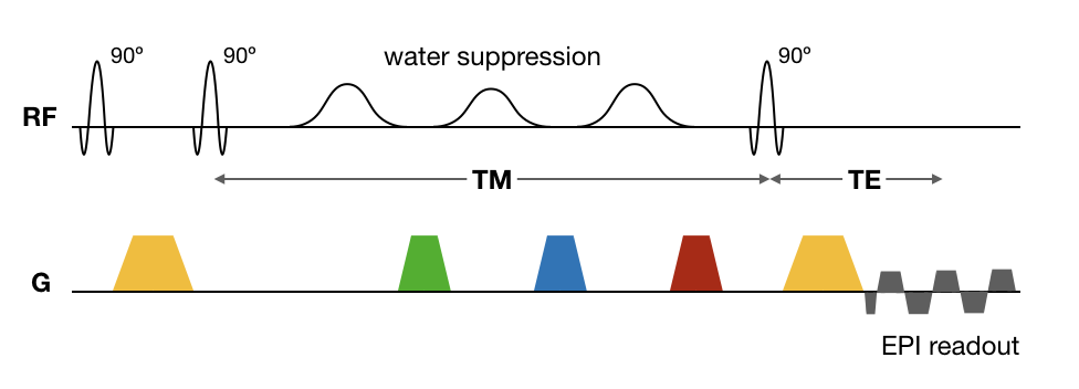
Our approach is based on a STEAM sequence, which generates a spatial modulation pattern in Mz of the water using two 90° pulses (Fig. 1). During the first part of the mixing time (TM), this pattern transfers to the semi-solids, after which any remaining pattern in the water Mz is eliminated by water suppression (WS) pulses at the center of TM. During the remainder of TM, MT effects return some of the semi-solid Mz pattern back to water. This pattern is then visualized by a third 90° pulse and subsequently imaged with an EPI readout.Methods
The method was tested with both a water and hair conditioner phantom and on a human subject. The data were acquired on a Siemens Prisma 3 T, using a 32-channel head receive coil. The acquisition was a single 3 mm slice EPI, with 64x48 resolution over a 240x180 mm2 FOV, echo time 22 ms, repetition time 5 s. Repeating the scan with a longer echo time (~1 ms) and combining the two images served to cancel the fat signals. The MT-STEAM sequence had a 5 ms separation between the first two pulses (center-to-center); the TM was varied from 40 to 3000 ms. All three pulses had a (nominal) 90° flip angle. The water suppression [1] used three Gaussian pulses of 8 ms (sigma 1.3 ms). The flip angles were optimized to 90, 80 and 100° by the minimizing the signal at TM 40 ms. The phase of the three STEAM pulses was cycled in 180° steps, yielding all combinations over 8 repetitions. An EPI with a single excitation served as amplitude reference.
For comparison, a MT saturation recovery experiment was also performed, with an on-resonance 6 ms MT saturation pulse [2] and a delay times varying between 8 and 600 ms.
Results
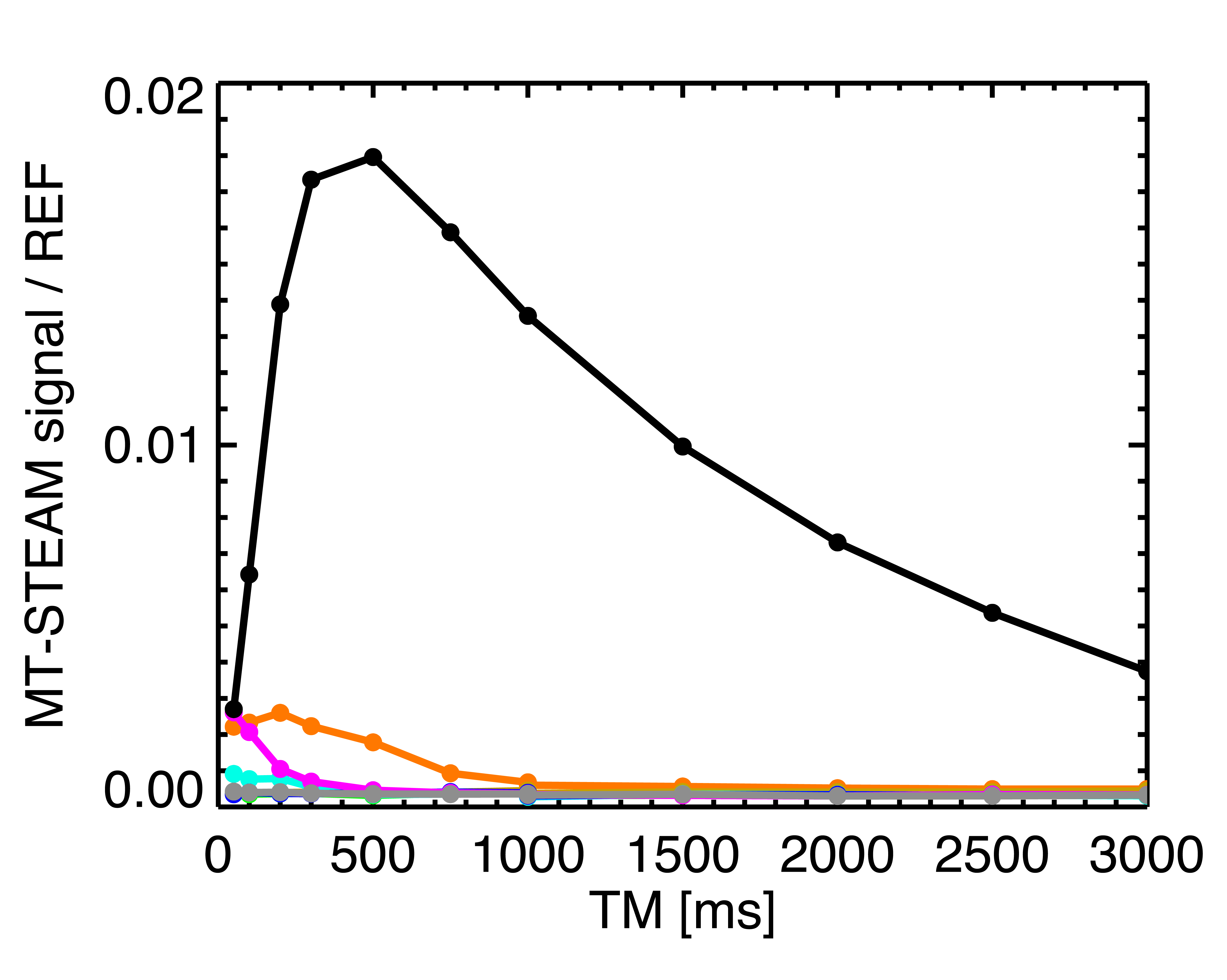
Both the phantom and human data showed the expected phase cycling pattern, proving that the observed signals are indeed generated by a stimulated echo. The WS performed well in the human subject and the hair conditioner phantom (Fig. 2,4), however residual signal was 3% in the water phantom. The TM dependence of the STEAM signal, shown in Fig. 3, corresponds to the expected shape. The MT contribution peaks around 400 ms, about twice the time it takes to reach a maximum in a MT saturation experiment (data not shown).
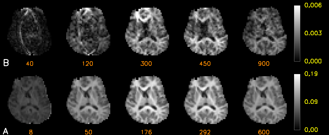
Fig. 4 shows the results of the human subject, demonstrating that the MT-STEAM signal has the expected TM dependence and image contrast. It also shows that the amplitude of the effect is limited, because of the reliance on a sequential, bi-directional exchange involved and losses inherent to STEAM. For comparison, the MT saturation is also shown in Fig. 4, scaled to the same reference EPI data.
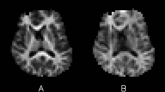
In Fig. 5 the square of the MT saturation is compared to the MT-STEAM results. The MT-STEAM image was scaled 5 times higher than the MT saturation. STEAM images without water suppression showed a factor 0.25 relative intensity, two times lower than what would be theoretically expected for a STEAM echo.
Discussion
The MT-STEAM approach can be used to selectively image the signals from exchanging spins, without a non-exchanging background. This reduces confounding effects, including those from background signal instabilities due to e.g. motion. The phase-cycling shows that the detected signals do originate from the intended coherence pathway and the TM dependence conforms to the expected shape of a bi-directional exchange process, consistent with the MT saturation measurements. In principle, this reliance on bi-directional exchange may allow study of the diffusion properties of magnetization in the semi-solids, whose signal is otherwise invisible to their short T2. Nevertheless, the losses due to bi-directional exchange and those inherent to the STEAM sequence limit the available signal to about 1% of the (water) baseline signal. At higher field strength this fraction is expected to increase substantially, as the available MT signal increases due to the longer T1 of the semi-solids at higher B0[3].Acknowledgements
No acknowledgement found.References
1. R.J. Ogg, P.B. Kingsley, J.S. Taylor. WET, a T1- and B1-Insensitive Water-Suppression Method for in Vivo Localized 1H NMR Spectroscopy. J. Magn. Reson. B 104, 1994, 1-10.
2. P. van Gelderen, X. Jiang, J.H. Duyn. Rapid measurement of brain macromolecular proton fraction with transient saturation transfer MRI. Magn Reson Med. 77, 2017, 2174-2185.
3. P. van Gelderen, X. Jiang, J.H. Duyn. Effects of magnetization transfer on T1 contrast in human brain white matter. Neuroimage 128, 2016, 85-95.
Figures