0758
Double DANTE: an improved method for high-resolution intracranial vessel wall imaging1Department of Biomedical Engineering & Physics, Academic Medical Center, Amsterdam, Netherlands, 2Department of Radiation Oncology, The Netherlands Cancer Institute, Amsterdam, Netherlands, 3Department of Radiology, Academic Medical Center, Amsterdam, Netherlands, 4A.A. Martinos Center for Biomedical Imaging, MGH, Harvard Medical School, Boston, MA, United States, 5MR Clinical Science, Philips Healthcare, Markham, ON, Canada, 6Department of Radiology, Utrecht Medical Center, Utrecht, Netherlands, 7Spinoza Centre for Neuroimaging, Amsterdam, Netherlands
Synopsis
We present the use of Double DANTE as an improved method for DANTE prepared 3D TSE imaging of intracranial arteries. DANTE preparation inherently leads to banding artifacts, which become increasingly visible at high-resolution imaging. We show that Double DANTE strongly reduces banding separation without changes in the DANTE pulse train duration, and more importantly, without compromising flow suppression of blood or CSF. Double DANTE therefore enables high-quality intracranial vessel wall MRI at 0.6mm isotropic resolution.
Introduction
The use of a DANTE prepared 3D TSE sequences for intracranial vessel wall imaging (VWI) has received increased interest, demonstrating effective suppression of both blood and slow flowing CSF1-5. An unfortunate property of the DANTE pulse train is the occurrence of a banding phenomenon across the image, which becomes increasingly visible at higher resolution. While the banding separation Δr is related to the pulse interval τ and gradient strength G, given by $${\Delta}r=\frac{2\pi}{\tau{\gamma}G}$$ these parameters also affect flow-suppression efficiency1, effectively making application of DANTE challenging at high isotropic spatial resolutions.
In this work, we propose an alternative DANTE preparation, dubbed ‘Double DANTE’ or ‘D-DANTE, first suggested for use in NMR spectroscopy6. We show that compared to regular DANTE, D-DANTE can reduce banding separation without changes in any of the DANTE parameters, except for the phase cycling scheme. Furthermore, we demonstrate that D-DANTE enables high-resolution intracranial VWI without sacrificing flow suppression performance.
Methods
Experiments were performed on a 3T Philips Ingenia MRI scanner. DANTE prepulses were implemented as illustrated in Figure 1A, with user-defined input of the number of RF pulses N, flip angle α, gradient strength (G), gradient interval τ and phase increment ΔΦ between RF pulses. A D-DANTE sequence is created by interleaving two pulse trains, in which odd pulses have ΔΦ=0°, while even pulses have ΔΦ>0°. Figure 1B shows phantom data where D-DANTE decreases banding separation by a factor 2 or 4 by choosing either ΔΦ=22.5°or 180°. For this particular purpose, the gradient strength was chosen too be very low (2mT/m) to exaggerate the banding effect.
To compare DANTE and D-DANTE in terms of CSF suppression, Bloch-simulations of the DANTE preparation were performed in MATLAB similar to Viessmann et al.3 using an ensemble of 1000 individual spins and the following parameters: N=300, τ=1ms, G=22.5mT/m, α=8°. DANTE was performed with ΔΦ=180°, while for D-DANTE, ΔΦ was either 22.5° or 180°. For CSF, a linear velocity profile within the simulated voxel was assumed with an average velocity of 2cm/s.
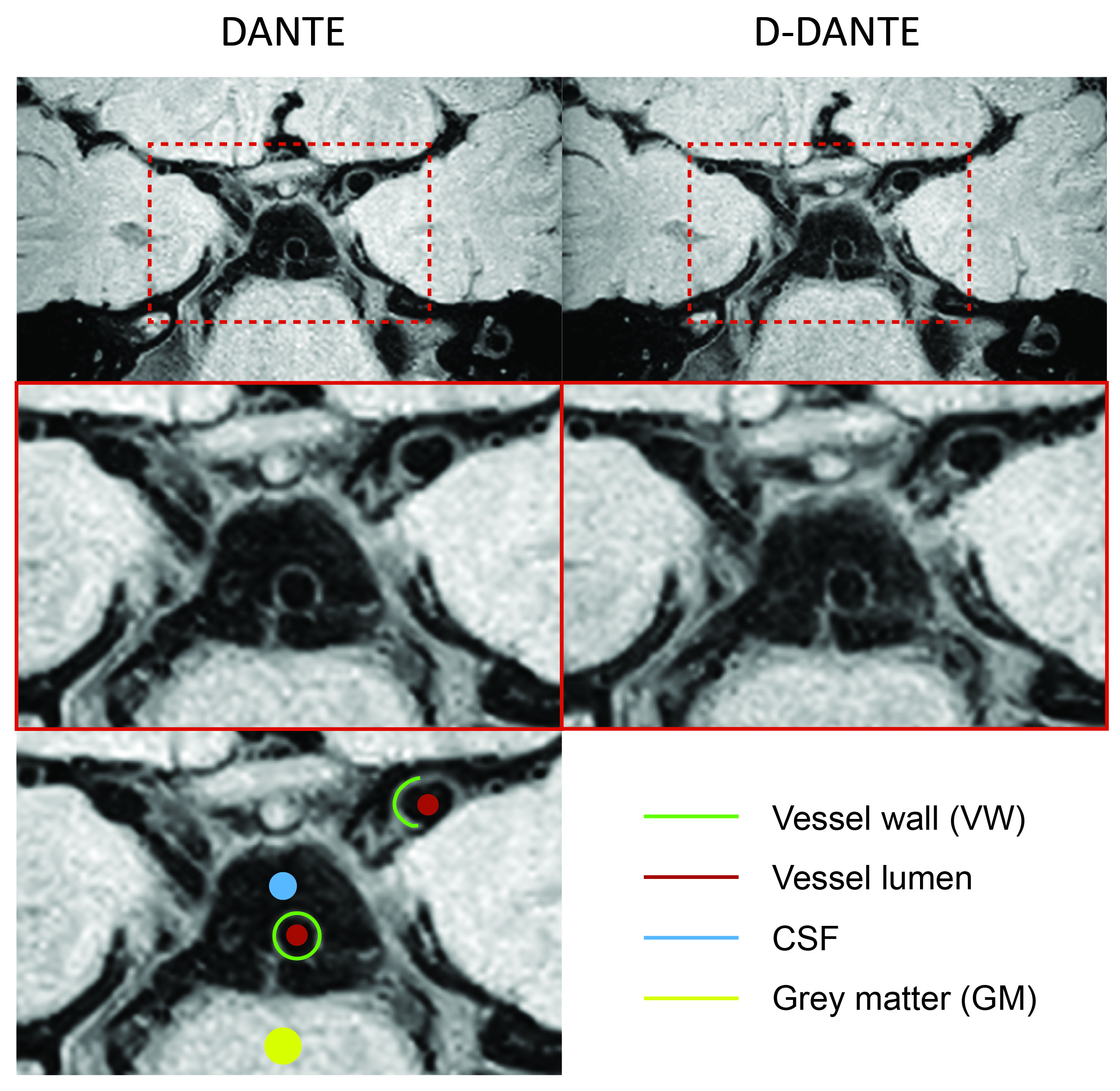
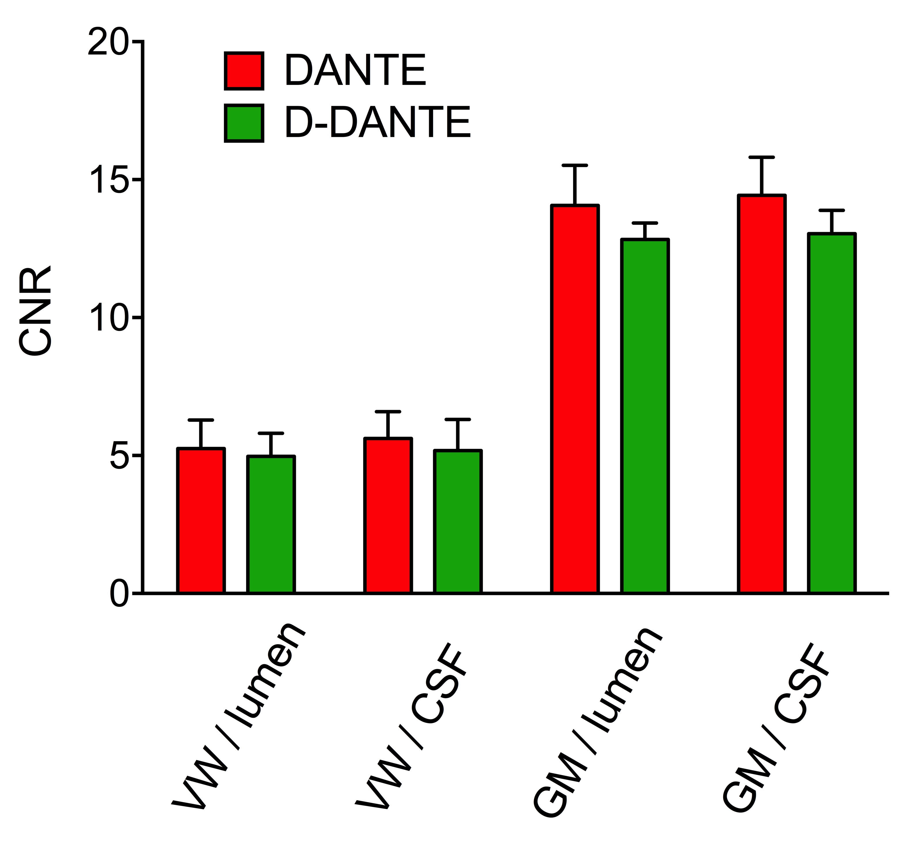
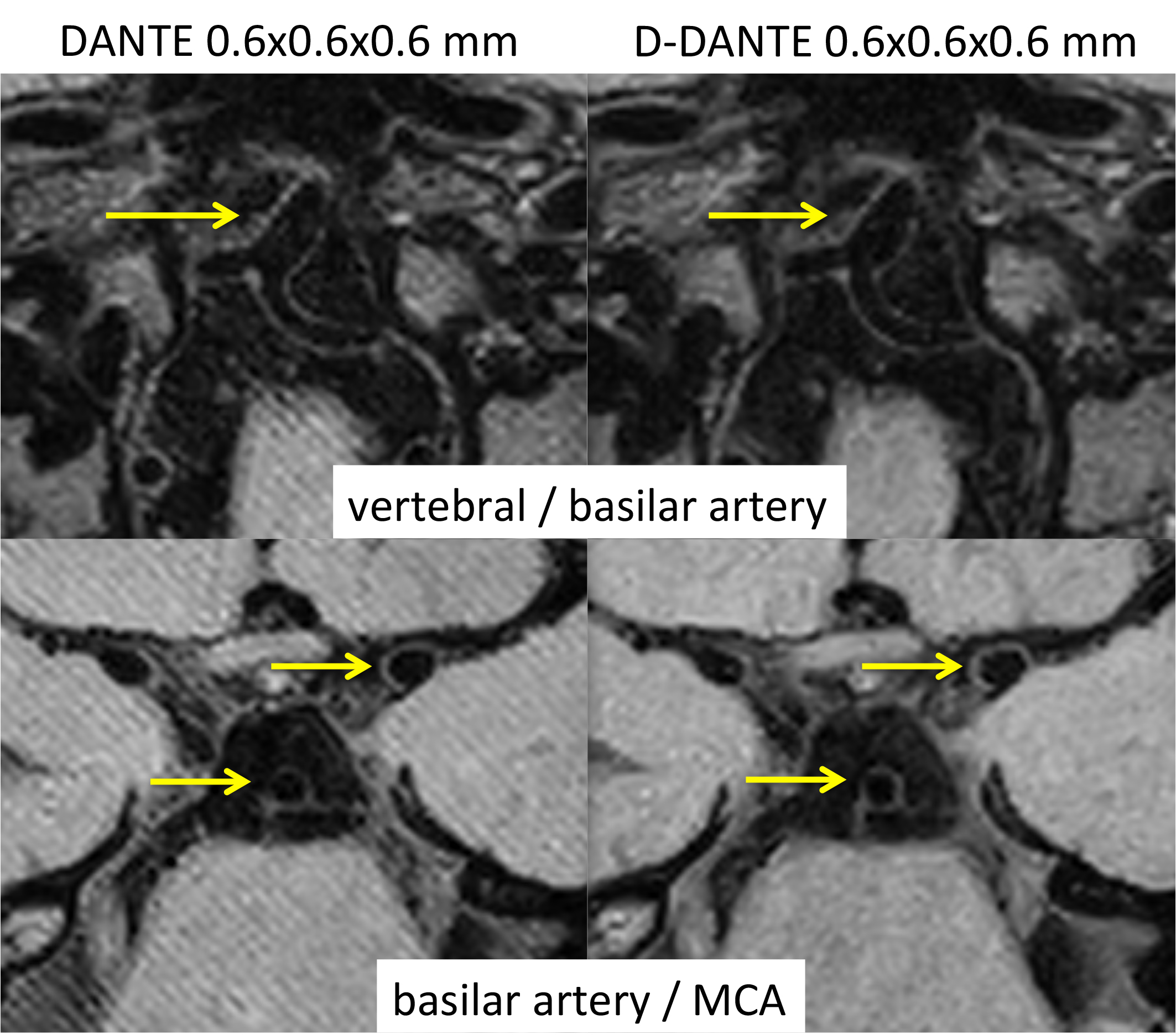
To assess flow suppression efficiency of D-DANTE in vivo, 5 healthy volunteers were scanned using a DANTE prepared VFA TSE sequence with AntiDrive7. DANTE parameters were equal to those in simulations, except only ΔΦ=22.5° was used for D-DANTE. TSE readout parameters were as follows: TR/TE=1500/46ms, TSEfactor=56, startup echoes=6, FOV=200×180×60mm3, resolution=0.5×0.5×1mm3, acquisition time = 4min30s. The anisotropic resolution was chosen to prevent banding artifacts for regular DANTE when comparing results to D-DANTE. Each scan was followed by a noise scan with equal imaging and reconstruction parameters, but with RF and gradients turned off. From the resulting images, CNR was calculated between VW/lumen, VW/CSF, grey matter (GM)/lumen, and GM/CSF. For VW, the average was taken from the basilar and middle cerebral artery (MCA), see Figure 3. Differences in CNR between DANTE and D-DANTE were assessed using multivariate repeated measures ANOVA with p<0.05 as level of statistical significance. Finally, to illustrate the performance of DANTE/D-DANTE at high isotropic resolution, the previously described scans were repeated using a voxel size of 0.6×0.6×0.6mm3 at an acquisition time of 6min30s.
Results and Discussion
Simulation results in Figure 2 show changes in longitudinal magnetization (Mz) during the 300ms DANTE preparation. As expected, VW magnetization is mostly preserved during DANTE preparation, while flowing CSF tissue is effectively saturated. More importantly, vessel wall/CSF contrast is only modestly compromised using D-DANTE, with a slightly better performance at low ΔΦ (22.5°) as compared to ΔΦ=180°. In vivo imaging showed very similar results (Figure 3 & 4), in which D-DANTE seemed to cause a small decrease in CNR values, although this was not statistically significant. When applying VWI at 0.6mm isotropic resolution (Figure 5), clear banding artifacts were noticed for DANTE, possibly making the images unsuitable for diagnosis. In contrast, D-DANTE showed no banding and clear delineation of all major intracranial arteries. Note that the increase in resolution only affected total acquisition time, but not the D-DANTE preparation itself. In fact, the application of D-DANTE in general, also for other vascular beds, will allow more flexibility in choosing the gradient strength and pulse interval τ. This could in turn facilitate preventing the occurrence of banding if gradient strength is limited or the incidence of high acoustic noise levels commonly associated with DANTE at specific values for τ.Conclusion
Double DANTE preparation is an attractive alternative to regular DANTE prepared TSE for intracranial VWI applications. Double DANTE can provide high-resolution (0.6 mm isotropic), high-quality CSF/blood suppressed vessel wall imaging without the banding artifacts banding artifacts that limit the spatial resolution for regular DANTE.Acknowledgements
No acknowledgement found.References
1 Li L et al. DANTE-prepared pulse trains: A novel approach to motion-sensitized and motion-suppressed quantitative magnetic resonance imaging. Magn Reson Med 2012; 68: 1423-1438.
2 Li L et al. Black-blood multi contrast imaging of carotid arteries with DANTE-prepared 2D and 3D MR imaging. Radiology 2014; 273: 560-569.
3 Viessmann et al. T2-Weighted Intracranial Vessel Wall Imaging at 7 Tesla Using a DANTE-Prepared Variable Flip Angle Turbo Spin Echo Readout (DANTE-SPACE). Magn Reson Med 2017; 77: 655-663
4 Wang J et al. Joint blood and cerebrospinal fluid suppression for intracranial vessel wall MRI. Magn Reson Med 2016; 75: 831-838.
5 Xie et al. Improved Black-Blood Imaging Using DANTE-SPACE for Simultaneous Carotid and Intracranial Vessel Wall Evaluation. Magn Reson Med 2016; 75:2286–2294
6 Geen H et al. Selective excitation at two arbitrary frequencies. The Double-DANTE sequence. J Magn Reson 1989; 81: 646-652.
7 Harteveld et al. High-resolution intracranial vessel wall MRI in an elderly asymptomatic population: comparison of 3T and 7T. Eur Radiol. 2017; 27(4): 1585-1595.
Figures
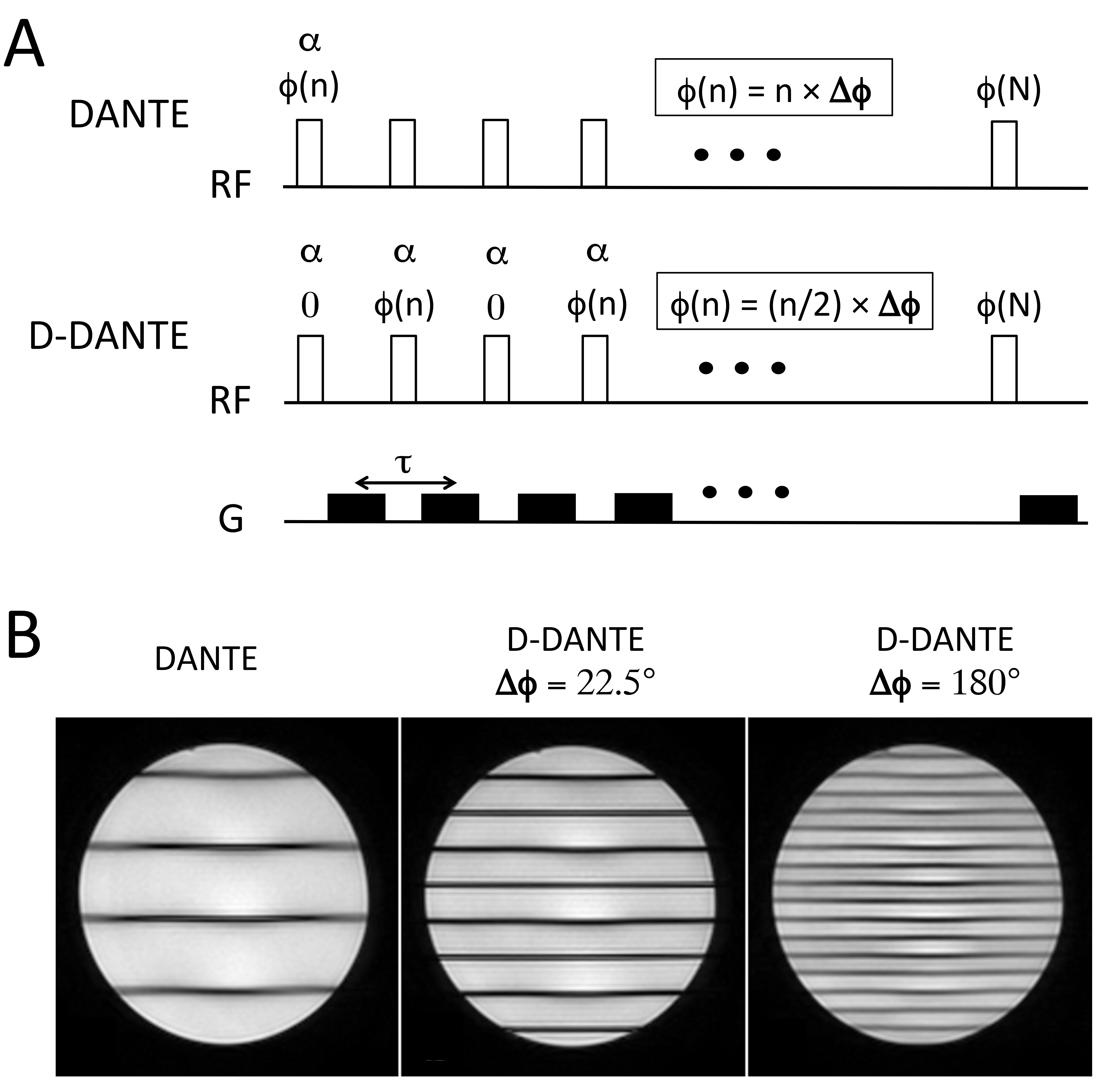
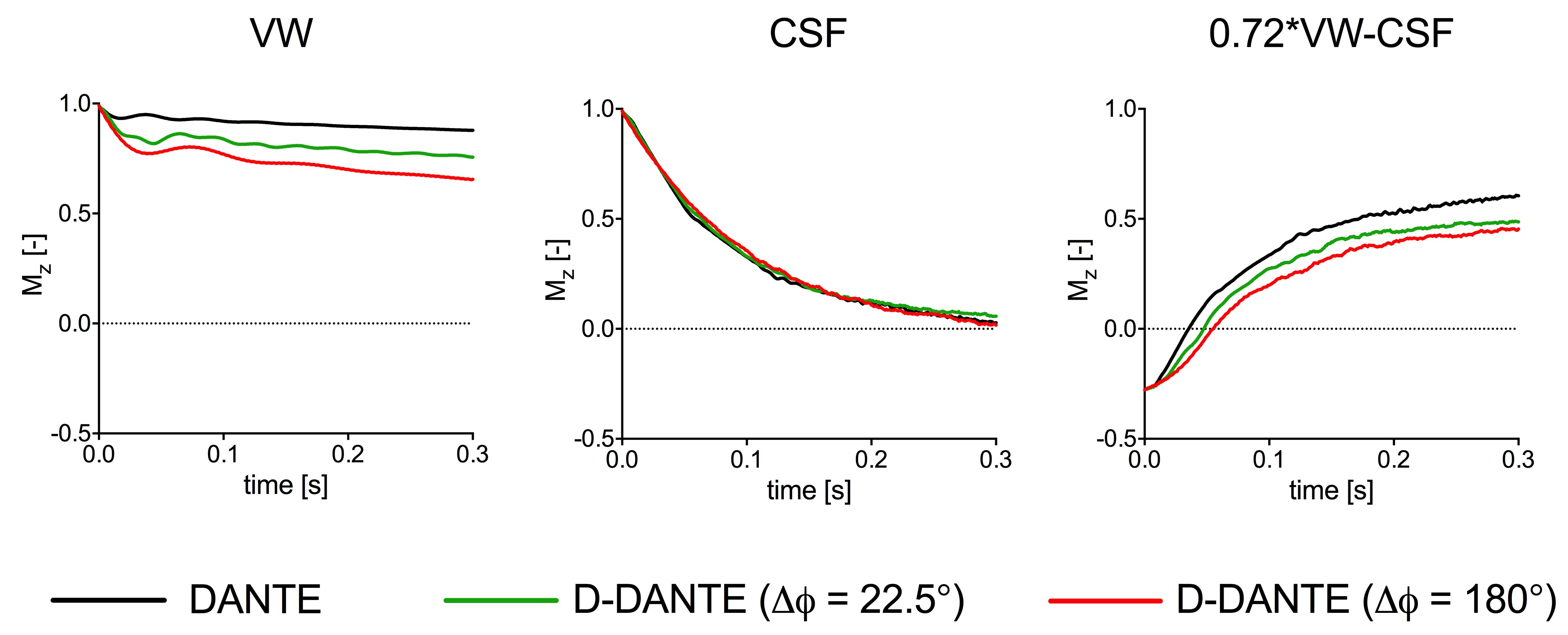
Figure 2: Results from Bloch-simulations indicating the change in longitudinal magnetization (Mz) during DANTE/D-DANTE preparation for different tissues (VW = vessel wall, CSF = cerebral spinal fluid). The right panel indicates VW/CSF contrast, in which differences in proton density were taken into account (contrast = 0.72VW-CSF). For all simulations the following parameters were used: N=300, τ=1ms, G=22.5mT/m, α=8°. Relaxation effects were also included (T1VW = 1000ms, T1CSF = 3500, T2VW = 55ms, T2CSF = 160ms)