0746
An accurate and efficient infarction segmentation method for diffusion weighted images using a deep convolutional neural network1Academy for Advanced Interdisciplinary Studies, Peking University, Beijing, China, 2College of Engineering, Peking University, Beijing, China, 3Department of Neurology, Peking Union Medical College Hospital, Beijing, China, 4Department of Neurology, Peking University First Hospital, Beijing, China
Synopsis
Accurate identification of infarcted regions of the brain is critical in management of stroke patients. An efficient and accurate method based on a deep convolutional neural network for segmentation of infarcts in the diffusion-weighted images is proposed.
Introduction
Accurate and automatic segmentation of acute ischemic infarct from magnetic resonance imaging is of great importance in clinical trials. Manual delineation is labor intensive, exhibits great variability due to unclear infarct boundaries, and most importantly, is not practical due to urgent time requirement for prompt therapy. Currently, modern deep learning techniques have the potential to provide a more reliable, fully-automated solution. In this study, a deep convolutional neural network was adopted for efficient lesion segmentation in diffusion weighted images with high accuracy.Methods
522 patients with acute ischemic stroke were enrolled in this study. All the participants were scanned at 3T GE Discovery MR 750 scanner with a diffusion weighted imaging sequence, b value is 1000. Other parameters are: TR = 3000 ms, TE= 65.4 ms, slice thickness = 6mm, matrix size =256 x 256. A total of 2040 DWI images with acute ischemic stroke lesions were collected in this study. The manual segmentation of infarct lesion serves as a ground truth for the automatic technique. We used 1240 DWI images for training and the rest 800 images for testing.
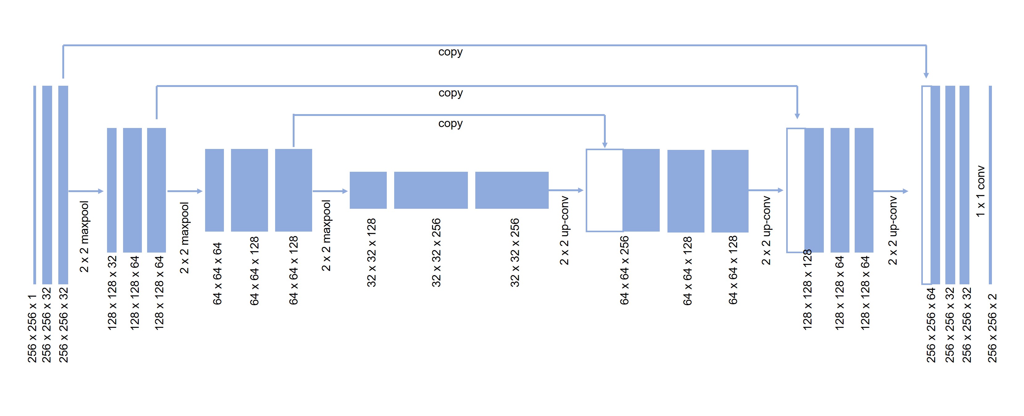
The u-net[1] was adapted for lesion segmentation by reducing the number of down sampling layers in the original model from four to three, since our images were roughly half the size as those considered by the u-net authors. We also zero pad our convolutions to keep the images the same size. The model was implemented in Keras. To prevent the network from memorizing just the training examples, elastic deformations was implemented for data augmentation. As is also common, there is a large class imbalance since most of the pixels are background. Normalizing the pixel intensities to lie between 0 and 1. The input of the model is the MRI image the output is a segmentation mask, a pixel-by-pixel mask that indicates whether each pixel is part of the lesion or the background (Figure 1).
To quantitatively evaluate the segmentation performance, the Dice ratio was used to measure the overlap between automated and manual segmentation results.
Results
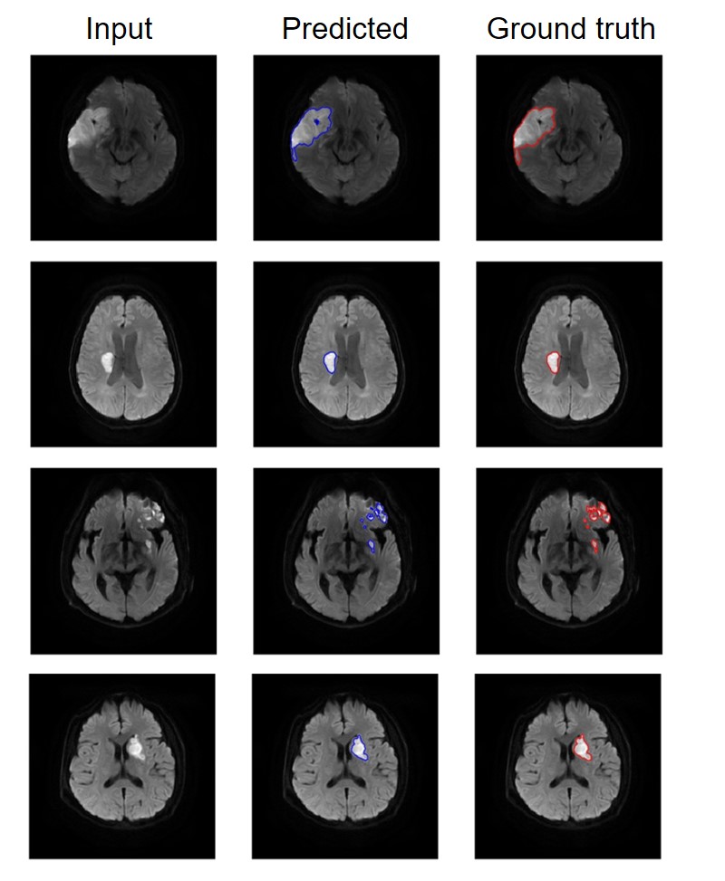
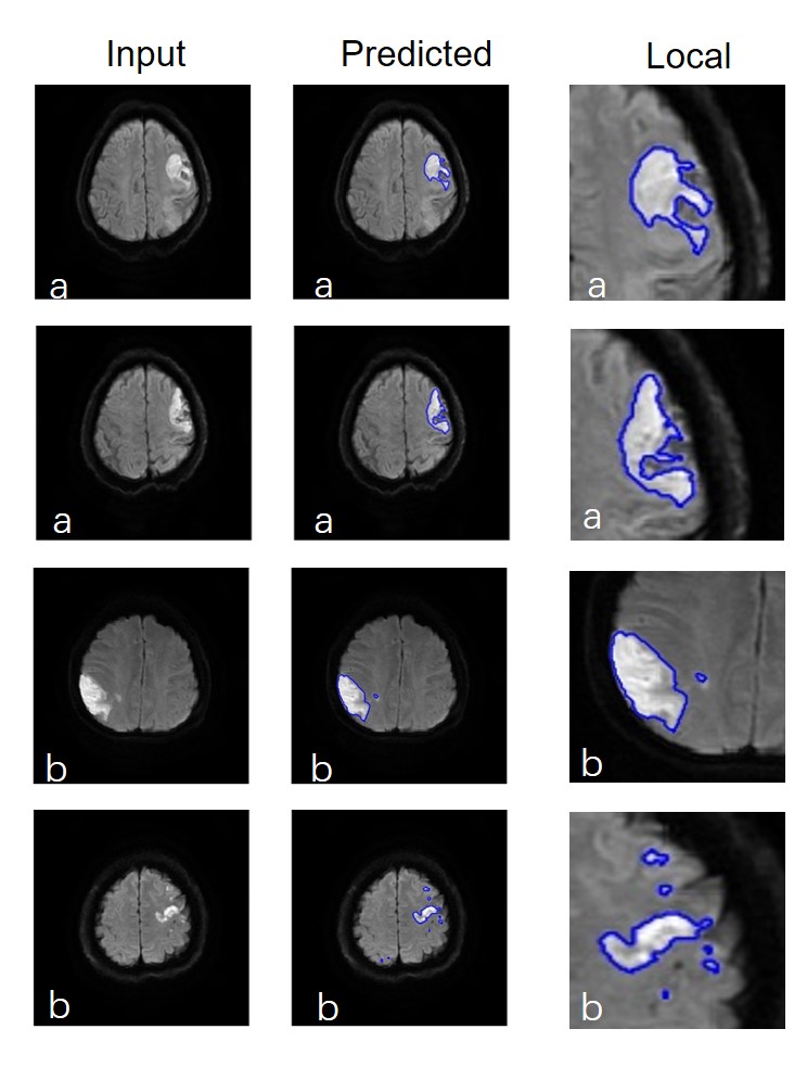
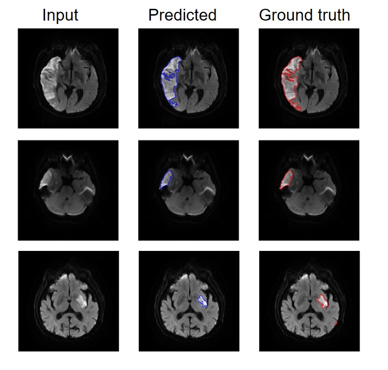
The algorithm performed very well with few exceptions. Dice coefficient between the ground truth and the automatic segmentation is 0.87. Example segmentations on patients with infarct lesions is given in Figure 2. Moreover, this method was able to achieve both complex edges (Figure 3b) and small lesion (Figure 3b) detection. As is shown in figure 4, although the intensity and first order statistical features are almost identical for both infarct and artifact regions[2], artifacts and infarcts were effectively distinguished by this algorithm.Discussion
Segmentation is an important step in many medical image analyses. However, manual segmentation is tedious, time consuming, and subjective. Speed and accuracy of are critical in clinical acute stroke management. In this study, we adopt the u-net for acute infarcts automatic segmentation, which could speed up the assessment of stroke region for stroke management applications. The u-net based method takes about 1 seconds to perform segmentation of one image. Even though artifacts and infarcts have similar imaging characteristics, the algorithm can efficiently discriminate between them. Besides, this method attained comparable dice coefficient to manual segmentation and strong capability to identify small lesions and complex lesion edges.Conclusion
Automatic segmentation of the acute ischemic infarcts lesions using deep learning with a convolutional neural network is accurate and efficient. This method could be a potential tool for clinicians to quantify the acute ischemic stroke and assist the decision making especially for thrombolytic therapy.Acknowledgements
No acknowledgement found.References
1. Ronneberger, O., P. Fischer, and T. Brox, U-Net: Convolutional Networks for Biomedical Image Segmentation, in Medical Image Computing and Computer-Assisted Intervention – MICCAI 2015: 18th International Conference, Munich, Germany, October 5-9, 2015, Proceedings, Part III, N. Navab, et al., Editors. 2015, Springer International Publishing: Cham. p. 234-241.
2. Prakash, K.B., et al., Identification, Segmentation, and Image Property Study of Acute Infarcts in Diffusion-Weighted Images by Using a Probabilistic Neural Network and Adaptive Gaussian Mixture Model. Academic Radiology, 2006. 13(12): p. 1474-1484.
Figures