0725
Increased BBB leakage to water but not gadolinium in a rat model of Alzheimer’s disease1Division of Neuroscience and Experimental Psychology, The University of Manchester, Manchester, United Kingdom, 2Bioxydyn Ltd, The University of Manchester, Manchester, United Kingdom, 3Bioxydyn Ltd & Division of Informatics, Imaging, and Data Sciences, The University of Manchester, Manchester, United Kingdom
Synopsis
The presence of blood-brain barrier (BBB) dysfunction in patients with Alzheimer’s disease (AD) is unclear. This study uses a novel MRI approach to study AD and age-related alterations in BBB leakage to water (PSw) and gadolinium (Ktrans) in a transgenic rat model of AD. We show PSw is increased in transgenic animals relative to wild-types but that Ktrans is independent of genotype. This study demonstrates the benefit of probing the BBB with molecules of different sizes, and suggests measurements of BBB permeability to water are more sensitive to AD-related BBB alterations than estimates of gadolinium leakage.
Introduction
The presence of increased blood brain barrier (BBB) permeability due to Alzheimer's disease (AD) is uncertain, but may be a critical early event in disease onset and progression by altering clearance of amyloid-β. Two recent MRI studies have identified increased BBB leakage to gadolinium contrast agents (Ktrans) in patients with mild cognitive impairment, providing initial evidence for early regional BBB damage1-2. However, a number of previous studies have also failed to detect AD-related alterations3-6. It is possible these inconsistent results reflect an insensitivity of gadolinium-based contrast agents to detect the degree and type of BBB damage present in AD. We hypothesized that MRI measurements of trans-endothelial water exchange would be more sensitive to subtle BBB damage than Ktrans, and thus be able to more easily detect early disease effects. A novel multi-flip angle multi-echo (MFAME) acquisition was used to simultaneously measure BBB flow-extraction product to gadolinium-DOTA (Ktrans) and water (PSw) within a single acquisition. In a transgenic rat model of familial AD (TgF344-AD), we show increased PSw in transgenic animals relative to wild-types, whereas Ktrans is altered with age only.Methods
Two cohorts of TgF344-AD rats were scanned on a 7T Bruker BioSpec system (Bruker Corporation, Billerica, USA): 5 transgenic (TG) and 5 wildtype (WT) aged 13 months, and 8TG/5WT aged 18 months. The young cohort were scanned twice (Δt = 1.5 weeks) to estimate scan-rescan reproducibility of MRI measures. All experiments were carried out in accordance with the Animal Scientific Procedures act 1986 and approved by the University of Manchester Local Ethical Review Committee.
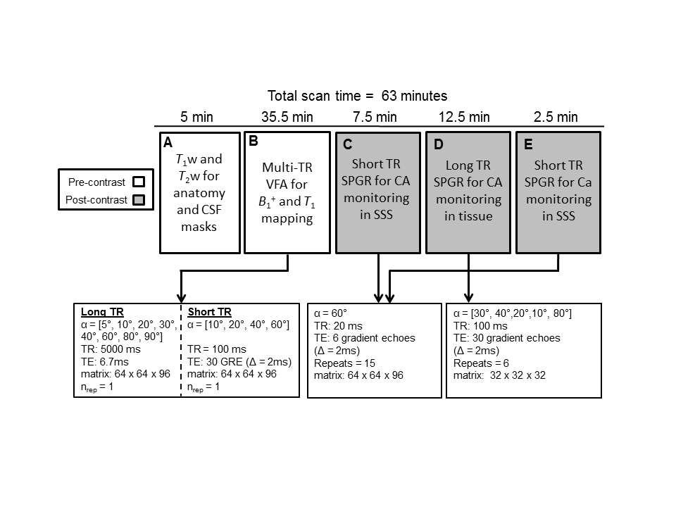
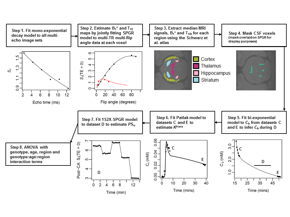
The MFAME-MRI protocol is shown in Figure 1. Dataset A was collected for brain region delineation, CSF masking, and lesion detection. Dataset B enabled combined measurement of B1+ and native T1. Datasets C and E were collected for estimation of Ktrans and were designed to have short TR and high spatial resolution, to minimise both sensitivity to transvascular water exchange in tissue and partial volume effects in the superior sagittal sinus (SSS), respectively. Dataset D was collected to estimate PSw, and designed with long TR, low spatial resolution, and multiple flip angles, each optimised to maximize sensitivity to transvascular water exchange. All rats were anesthetized with 4% isoflurane and maintained with 2.5% isoflurane in 100% O2. Gd-DOTA (0.5 mmol.kg-1) was injected intravenously with a pump at 1 mL.min-1 on the 6th volume of dataset C. The data analysis pipeline is shown in Figure 2. Ktrans was estimated by fitting the Patlak model to datasets C and E. PSw was estimated by fitting a 2-site one-exchange model7 to dataset D. The effect of genotype, age, and brain region (plus interaction terms) on PSw and Ktrans were estimated using three-way ANOVA. Post-hoc tests on TG/WT differences, corrected for multiple comparisons, were undertaken using Tukey’s Honest Significant Differences (HSD) method. All model fitting and statistical analyses were performed in R (Version 3.1, Austria).
Results
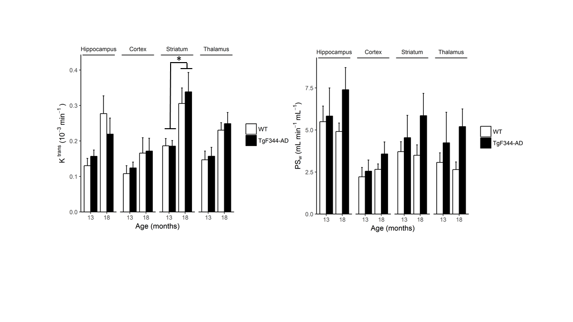
Scan-rescan reproducibility (CoV) values averaging over all regions and rats were 20% and 32% for Ktrans and PSw respectively, but were not significantly different (P = 0.080). In a three-way ANOVA, the effect of age on Ktrans was significant (P = 0.0036) but the effect of genotype was not (P = 0.096). The opposite effect was observed for PSw, with a significant effect of genotype (P = 0.0044) but not age (P = 0.080). Interaction terms were not significant (all P > 0.05). Region was a statistically significant factor for Ktrans but not PSw. Figure 3 shows TG and WT mean values and s.e.m for Ktrans and PSw in the hippocampus, cortex, striatum and thalamus. Post-hoc testing revealed no significant TG/WT differences in Ktrans or PSw at the regional level. A significant age-dependent effect was observed for Ktrans in the striatum (P = 0.0068).Discussion
The observed age-related increase in Ktrans agrees with previous work in human studies1. Our inability to detect genotype-dependent alterations in Ktrans agrees with most previous MRI studies but disagrees with recent work by Montagne et al.1 and Van de Haar et al.2. However, our sample sizes were small and possibly underpowered for this purpose. Despite this, we demonstrated a significant genotype effect on PSw suggesting BBB water permeability provides increased sensitivity and/or specificity for detection of AD-related BBB damage than Ktrans. It is currently unclear whether the observed changes in PSw are a result of increased sensitivity to structural changes in endothelial cell structures and functions (e.g. loss of tight-junctions) or sensitivity to altered water-channel expression9 (e.g. AQP4).Conclusion
TgF344-AD rats display increased BBB permeability to water compared to wild-type littermates.Acknowledgements
The purchase of the TgF344-AD rat breeding pairs was jointly supported by the European Union's Seventh Framework Programme (FP7/2007-2013) under grant agreement n°HEALTH-F2-2011-278850 (INMiND) and Alzheimer Research UK network funds. The maintenance and breeding of the TgF344-AD strain was funded by the European Union's Seventh Framework Programme (FP7/2007-2013) under grant agreement n°HEALTH-F2-2011-278850 (INMiND) and EPSRC grant (EP/M005909/1). The MRI facility is supported through an equipment grant from BBSRC UK (BB/F011350). Scanning of animals was funded through EPSRC grant (EP/M005909/1).References
1. Montagne, A., Barnes, S. R., Sweeney, M. D., et al. Blood-brain barrier breakdown in the aging human hippocampus. Neuron, 2015; 85(2), 296-302.
2. Van de Haar, H., Burgmans, S., Jansen, J. F. et al. Blood-brain barrier leakage in patients with early Alzheimer disease. Radiology, 2016; 281(2), 527-535.
3. Schlageter, N. L., Carson, R. E., & Rapoport, S. I. Examination of Blood—Brain Barrier Permeability in Dementia of the Alzheimer Type with [68Ga] EDTA and Positron Emission Tomography. Journal of Cerebral Blood Flow & Metabolism; 1987; 7(1), 1-8.
4. Caserta, M. T., Caccioppo, D., Lapin, G. D., Ragin, A., & Groothuis, D. R. Blood–brain barrier integrity in Alzheimer's disease patients and elderly control subjects. The Journal of neuropsychiatry and clinical neurosciences, 1998; 10(1), 78-84
5. Starr, J. M., Farrall, A. J., Armitage, P., McGurn, B., & Wardlaw, J. Blood–brain barrier permeability in Alzheimer's disease: a case–control MRI study. Psychiatry Research: Neuroimaging, 2009; 171(3), 232-241
6. Van de Haar, H. J., Burgmans, S., Hofman, P. A., Verhey, F. R., Jansen, J. F., & Backes, W. H. Blood–brain barrier impairment in dementia: Current and future in vivo assessments. Neuroscience & Biobehavioral Reviews, 2015; 49, 71-81.
7. Schwarzbauer, C., Morrissey, S. P., Deichmann, R., et al. A. Quantitative magnetic resonance imaging of capillary water permeability and regional blood volume with an intravascular MR contrast agent. Magnetic resonance in medicine, 1997; 37(5), 769-777.
8. Schwarz, A. J., Danckaert, A., Reese, T., et al., A. A stereotaxic MRI template set for the rat brain with tissue class distribution maps and co-registered anatomical atlas: application to pharmacological MRI. Neuroimage, 2006; 32(2), 538-550.
9. Iliff, J. J., Wang, M., Liao, Y., et al. A paravascular pathway facilitates CSF flow through the brain parenchyma and the clearance of interstitial solutes, including amyloid β. Science translational medicine, 2012; 4(147),
Figures