0724
Imaging disruption of blood-brain-barrier (BBB) in Mild Cognitive Impairment without using contrast agent1Department of Radiology, Johns Hopkins University, Baltimore, MD, United States, 2Department of Neurology, Johns Hopkins University, Baltimore, MD, United States, 3Department of Psychiatry and Behavioral Sciences, Johns Hopkins University, Baltimore, MD, United States
Synopsis
Disruption of BBB in AD has received increasing attention due to its potential role in amyloid accumulation and clearance. However, measurement of BBB leakage in AD has been proven challenging, especially when using non-contrast techniques. In this study, we measured BBB permeability to water in MCI patients using a novel technique that does not require any contrast agent. It was found that MCI patients have a higher permeability-surface-area-product (PS), i.e. leaky BBB, compared to elderly controls. Individuals with higher PS values had poorer cognitive performance and more severe vascular inflammation. These findings support the role of BBB disruption in AD.
Purpose
Extensive literature suggests that damage to the blood-brain-barrier (BBB) is an early event in Alzheimer’s disease (AD), and precedes brain atrophy1. A recent hypothesis of AD pathogenesis suggests that BBB leakage allows pathogens and neurotoxins to gain entry into the brain, which then triggers the immune response of the brain and results in excessive accumulation of amyloid deposits, causing neurodegeneration2,3. Current methods using gadolinium contrast-agent are primarily sensitive to major breakdown of BBB, but its ability to detect small disruption of BBB at early stage of AD is still controversial4,5. Here, we measured BBB permeability to water in Mild Cognitive Impairment (MCI) patients using a novel technique that does not require any exogenous contrast agent: water-extraction-with-phase-contrast-arterial-spin-tagging (WEPCAST) MRI6. We demonstrated a disruption of BBB in this early stage of AD by revealing that BBB in MCI patients has a greater permeability to water compared to elderly controls. The relationships between BBB permeability and vascular inflammation and cognitive performance were also examined.Materials and Methods
Participants
13 elderly subjects were recruited, including 5 MCI patients (M/F:3/2, Age:71.6±13.4yrs) and 8 cognitively normal controls (M/F:4/4, Age:66.4±5.2yrs).
MRI Experiments
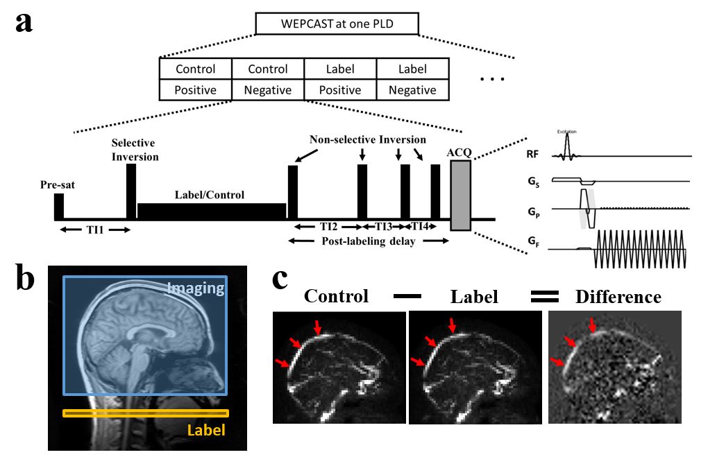
BBB permeability to water was quantified by a novel technique, WEPCAST MRI (Figure 1a), which selectively measures ASL signal in main draining veins of the brain, e.g. superior sagittal sinus (SSS), using a phase-contrast-encoded acquisition6. WEPCAST measures the extraction fraction (E) of water (in its first-pass through capillary) and, when combined with CBF (f) measurement, can provide an estimation of BBB permeability in terms of permeability-surface-area product (PS): $$$PS=-ln(1-E)·f$$$7.
All subjects were studied on a 3T Philips System. For WEPCAST MRI, a mid-sagittal imaging slice, a labeling duration of 2000ms, and a post-labeling delay (PLD) of 4000ms were used. Five background suppression pulses were employed. The encoding velocity of WEPCAST was set to be 15 cm/s. Global CBF was quantified using a conventional phase-contrast MRI.
Blood Test and Cognitive Assessment
Other tests were performed as part of the study protocol. Hematocrit and homocysteine, a plasma inflammatory marker, were measured with standard blood test procedures. The cognitive assessment used a battery of tests, including Hopkins Verbal Learning Test, Montreal Cognitive Assessment, Trail Making Test A & B, Digit Span Test, Wechsler Memory Scale Logical Memory Test, Digit Symbol Substitution Test, Stroop Color Word Test, Category Fluency Test and National Adult Reading Test.
Statistical Analysis
Linear regression analysis was performed to compare PS values between MCI and elderly control group, using age as a covariate. A multivariate linear regression model was used to examine the relationship between PS and blood test/cognitive assessment results, after correcting for age.
Results and Discussion
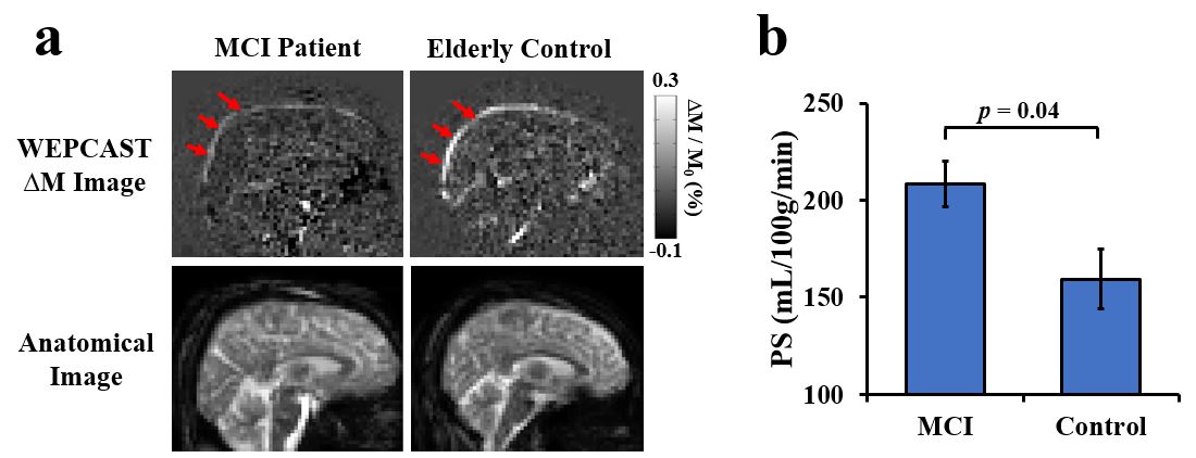
Figures 1b and c show typical slice positioning of WEPCAST MRI and representative phase-contrast-encoded control, label, and difference images. Note the complete suppression of static and tissue perfusion signals, leaving only vessel signals. Figure 2a shows representative WEPCAST difference images (ΔM) in a MCI patient and a control subject. Compared to the normal control, the patient has a lower ΔM in the SSS (arrows), suggesting a higher BBB permeability. Figure 2b shows bar plots summarizing PS values from all participants. The MCI group revealed a higher PS value (i.e. leaky BBB) compared to elderly control group (MCI: 208.4±15.4 mL/100g/min, Control: 159.4±11.2 mL/100g/min, p=0.040).
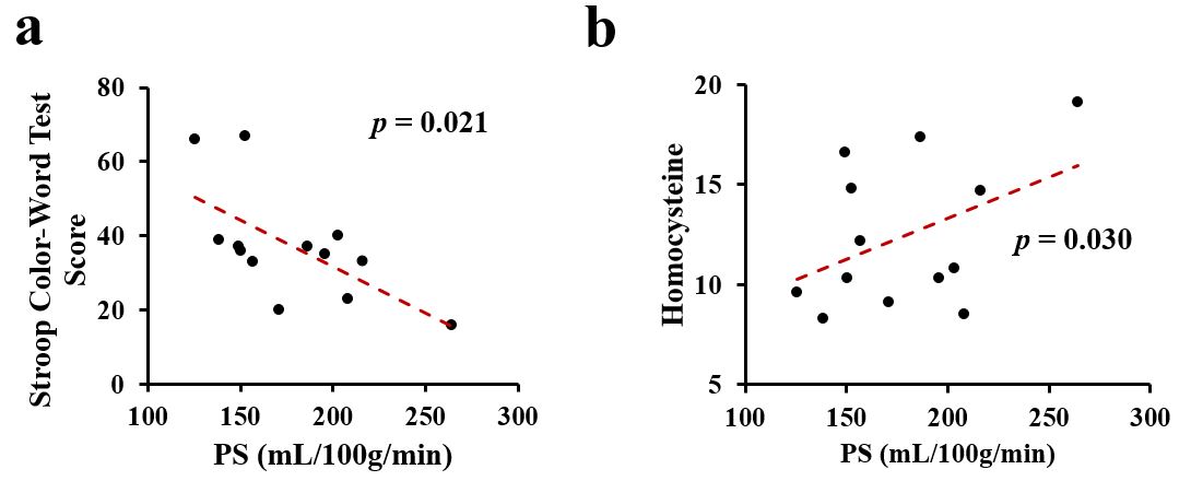
Next, we investigated the relationship between PS and cognitive performance. Regression analysis suggested an inverse association (p=0.021) (Figure 3a) between PS and Stroop test score (a test of executive function). Individuals with a higher PS (i.e. leaky BBB) tend to have a poorer executive function. Additionally, a positive relationship between PS and the time to complete a Trail Making Task A was found (p=0.031), i.e. individuals with a leaky BBB tend to take longer to complete the task. Moreover, in the Adult Reading Test, individuals with a higher PS were found to have more errors (p=0.035), lower Verbal IQ (p=0.037) and lower Full Scale IQ (p=0.037).
Finally, we examined the potential association between PS and vascular inflammation. We found that plasma homocysteine, a marker of endothelial injury, was positively associated with PS (p=0.030) (Figure 3b). These findings suggest that BBB leakage in MCI is related to microvascular inflammation.
Conclusion
To our knowledge, the present work is the first study to examine BBB leakage in MCI patients without using contrast agent or any other invasive procedures. Compared to elderly controls, MCI patients revealed a higher PS, suggesting a BBB disruption. Altered permeability was also shown to be related to poorer cognitive performance and more severe vascular inflammation. These findings support the role of BBB disruption in AD pathogenesis.Acknowledgements
No acknowledgement found.References
1. Erickson MA, Banks WA. Blood-brain barrier dysfunction as a cause and consequence of Alzheimer's disease. J Cereb Blood Flow Metab 2013; 33: 1500-1513.
2. Zlokovic BV. Neurovascular mechanisms of Alzheimer's neurodegeneration. Trends Neurosci 2005; 28: 202-208.
3. Kumar DK, Choi SH, Washicosky KJ, Eimer WA, Tucker S, Ghofrani J, Lefkowitz A, McColl G, Goldstein LE, Tanzi RE, Moir RD. Amyloid-beta peptide protects against microbial infection in mouse and worm models of Alzheimer's disease. Sci Transl Med 2016; 8: 340ra372.
4. Montagne A, Barnes SR, Sweeney MD, Halliday MR, Sagare AP, Zhao Z, Toga AW, Jacobs RE, Liu CY, Amezcua L, Harrington MG, Chui HC, Law M, Zlokovic BV. Blood-brain barrier breakdown in the aging human hippocampus. Neuron 2015; 85: 296-302.
5. Starr JM, Farrall AJ, Armitage P, McGurn B, Wardlaw J. Blood-brain barrier permeability in Alzheimer's disease: a case-control MRI study. Psychiatry Res 2009; 171: 232-241.
6. Lin Z, Li Y, Su P, Mao D, Wei Z, Pillai JJ, Moghekar A, Van Osch M, Ge Y, Lu H. Non-contrast assessment of blood-brain-barrier permeability with MRI. Magn Reson Med 2017; (in revision).
7. Crone, C, The permeability of capillaries in various organs as determined by use of the ‘indicator diffusion’ method. Acta Physiologica Scandinavica. 1963; 58: 292–305.
Figures