0706
High resolution single-vessel fMRI with the radial encoding method1Max Planck Institute for Biological Cybernetics, Tübingen, Germany, 2Graduate Training Centre of Neuroscience, Tübingen, Germany
Synopsis
A golden angle radial encoding (GARE) method was implemented to map BOLD signal from individual venules penetrating the rat somatosensory cortex with 50 μm spatial resolution. This real-time acquisition method makes it possible to detect the hemodynamic signal from individual vessel with much finer spatial scale than previously reported methods. It also provides high flexibility to define the field of view to only focus on the activated brain regions and increase the sampling rate for fMRI imaging. This GARE method improves the existing single vessel fMRI method with higher spatiotemporal resolution.
Introduction
Previously, we have applied a line-scanning fMRI method to reveal the contribution of vascular components to blood oxygenation level-dependent signal (BOLD) and the cerebral-blood-volume (CBV) signal with a spatial resolution of 100 µm and a sampling rate of 100 ms. However, the line-scanning method reshuffled the k-space acquisition so that each image was reconstructed from data acquired along the entire experimental time series with a fast sampling rate, but not in real time1,2. Here, we applied the golden angle radial encoding (GARE) method to map BOLD signal from individual venules penetrating the rat somatosensory cortex with 50 μm spatial resolution. This continuous-golden-angle acquisition scheme compares favorably with conventional cartesian schemes, as it provides spatial/temporal freedom by grouping any arbitrary number of projections in the azimuthal direction, therefore enabling imaging acquisition acceleration and improving the spatial resolution with a small field of view. The repeated sampling in the k-space center ensures an overall uniform contrast in the center of field of view (FOV) with radial fMRI so as to focus on the small region-of-interest (ROI) for activated cortex. This work further improves the high spatiotemporal resolution single-vessel fMRI studies on neurovascular coupling.Methods
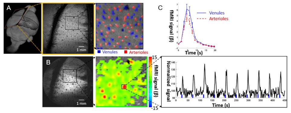
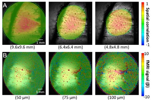
All images were acquired with a 14.1 T/26cm horizontal bore magnet (Magnex), interfaced to an AVANCE III console (Bruker) and equipped with a 12 cm gradient set, capable of providing 100 G/cm with a rise time of 150 μs (Resonance Research). A transreceiver surface coil with 6mm diameter was used to acquire fMRI images. The radial projections spaced with a constant azimuthal increment of golden-angle (111.25o). The FOV along the radial encoding direction was kept to only cover the cortical regions of interest. Radial encoding fMRI: A 2D radial encoding sequence was applied to map the fMRI signal with the following parameters: TE: 8 ms; TR: 19.738 ms; projections: 76; matrix: 96x96; slice thickness: 500 μm; in plane resolution: 100x100 µm. In order to map the sensory-evoked single vessel fMRI, electrodes were placed on the forepaw to deliver trains of 300 μs, 2 mA pulses at 3Hz during 4s in each fMRI epoch. The anatomical arteriole-venule (A-V) map was detected by 2D multiple gradient echo (MGE) imaging with the following parameters: TE: 2.5, 5, 7.5, 10, 12.5, 15 ms; TR: 50 ms; slice thickness: 500 μm; flip angle: 55°; matrix: 192x192; in-plane resolution: 50x50 μm. The single vessel map is acquired by averaging the MGE images acquired from the second echo to the forth echo, where the venule voxels showed as dark dots due to fast T2* decay, but arteriole voxels remain bright dots due to the in-flow effect. To demonstrate the spatial/temporal freedom of radial encoding fMRI, imaging acceleration was achieved with 38 radial projections (TE 8: ms; TR: 13.159 ms) for a reduced FOV (4.8x4.8 mm) to cover the responsive cortical region. High spatial resolution (50, 75, and 100 μm in-plane) real-time single-vessel fMRI images were recorded with 150, 100, and 75 projections, respectively.Results
Fig 1 shows sensory-evoked BOLD signal with GARE-fMRI in the forepaw S1 cortex upon electrical sitmulation of the forepaw. It demonstrates the peak BOLD primarily overlapped with venule volxels (blue marker, Fig 1A) with the time course of the positive BOLD signal from a selected venule (Fig 1B) and averaged velules (Fig 1C). Fig 2 demonstrates that GARE images with arbitrary FOV (9.6x9.6 mm, 6.4x6.4 mm, and 4.8x4.8 mm), while maintaining the contrast and spatial correlation in the cortical ROI (Fig 2A). The venule-specific BOLD signal was shown in the GARE-fMRI iamges with in-plane resolution from 50 to 100 μm (Fig 2B). Thus, the golden-angle radial encoding fMRI method allows us to detect the single vessel fMRI signal in a much finer spatial scale than previously reported methods.Acknowledgements
This work was supported by the Max-Planck-Society and the Graduate Training Centre of Neuroscience of Tübingen.References
1 Yu, X. et al. Sensory and optogenetically driven single-vessel fMRI. Nat Methods 13, 337-340, doi:10.1038/nmeth.3765 (2016).
2 Silva, A. C. & Koretsky, A. P. Laminar specificity of functional MRI onset times during somatosensory stimulation in rat. Proc Natl Acad Sci U S A 99, 15182-15187, doi:10.1073/pnas.222561899 (2002).
Figures