0620
Prospective motion and B0 shim correction for MRS at 7 Tesla1University of Minnesota, Minneapolis, MN, United States
Synopsis
The goal of this study was to demonstrate the feasibility of prospective motion and shim correction for MRS at 7 Tesla. We combined an optical tracking system for motion correction and a shim navigator for first-order B0 shim correction into a semi-LASER sequence. The new sequence was validated in the prefrontal cortex, a region sensitive to motion due to susceptibility effects induced by the proximity of the nasal cavity. Results show excellent performance, with similar spectral quality (signal-to-noise ratio and linewidth) before, during, and after motion when both motion and shim navigators are used.
Purpose
Prospective motion and shim corrections have been successfully demonstrated for human brain 1H MRS at 3 T with EPI volumetric1 or spiral2 navigators. Another approach used an optical tracking system3,4, but without prospective shim correction. The aim of the present study was to demonstrate the feasibility of prospective motion and shim correction for human brain MRS at 7 T, using an optical camera for motion correction and a FASTMAP-like shim navigator for linear shim correction, and assess performance in the prefrontal cortex (PFC), a region sensitive to motion due to susceptibility effects induced by the proximity of the nasal cavity.Methods
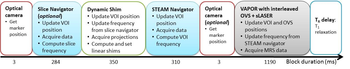
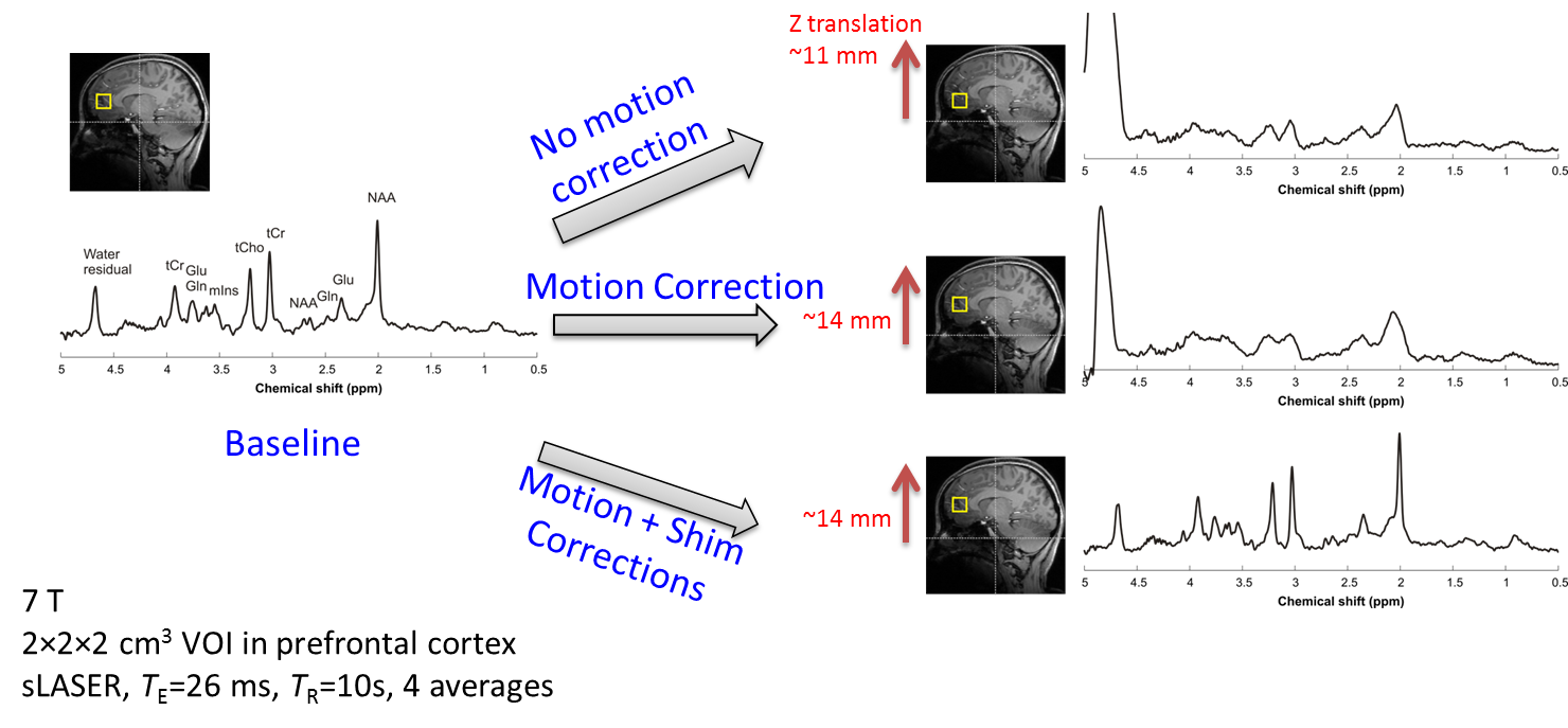
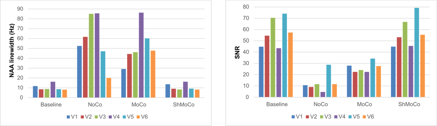
Healthy subjects (n = 6) were scanned on a 7 T Siemens scanner with a Nova 32-channel receive head coil. A VOI of 20×20×20 mm3 was positioned in the PFC using T1-weighted MPRAGE images. B0 shimming was achieved using FAST(EST)MAP which resulted in water linewidth of 13±1 Hz. All MRS data were acquired using a semi-LASER sequence (sLASER5). The sequence was modified to incorporate real-time motion, frequency and shim correction during every repetition time (TR). Motion was tracked using a single optical camera6. Dynamic B0 shimming was achieved by acquiring three 1D projections along X, Y, Z using a 2D-RF pulse2,7. Two frequency navigators were also implemented: one slice navigator placed before the dynamic shim module and one STEAM navigator immediately before the water suppression module (Figure 1). To validate the prospective motion and shim corrected sequence, sLASER spectra (16 transients) were obtained under three different conditions: 1) motion and shim corrections off (denoted as NoCo); 2) motion correction on and shim correction off (denoted as MoCo); and 3) shim and motion corrections on (denoted as ShMoCo). To mimic motion, the patient table was moved during the sLASER acquisition. The first four transients were acquired without any movement, then the table was manually moved by a few mm every other transient for 8 transients, resulting in a cumulated Z-translation between 10 to 20 mm. The final four transients were acquired without further motion. All navigators (frequency and shim) had small flip angles to minimize disturbance of steady-state magnetization.Results and Discussion
Spectra acquired from one subject with the three different conditions (NoCo, MoCo and ShMoco) are shown in Figure 2. At baseline, i.e. without motion, high-quality spectra were obtained with narrow linewidth and high SNR (mean tNAA linewidth = 10 ± 3 Hz, mean tNAA SNR = 57 ± 3, n = 6) (Figure 3). After motion in NoCo condition, the uncorrected VOI position was closer to the nasal cavity resulting in greatly degraded linewidth, SNR and water residual (linewidth 20-84 Hz, SNR 4-29). In MoCo condition, although the VOI position was corrected during motion, the linewidth and SNR still much worse than baseline (linewidth 29-34 Hz, SNR 22-34). With ShMoCo, the spectral quality was identical to that obtained without motion (linewidth 11 ± 3 Hz, SNR 58 ± 13, n=6).
We chose to move the patient table in Z direction with a VOI in the PFC and a large cumulated displacement of up to 20 mm to mimic a worst case scenario. In practice, subject motion is often smaller and VOIs are located in regions less sensitive motion. We obtained similar performance when asking the subject to perform random head movements (data not shown). The first frequency navigator (slice navigator before the shim module) is useful only in the case of large motion. In such a case, the slice navigator prevents the shim module from being significantly off-resonance, which would result in suboptimal shim correction. Otherwise, good shim performance is obtained without the slice navigator. To our knowledge, this is the first report of real-time motion and shim correction at 7 T and also the first implementation of prospective and shim correction combining optical camera with shim navigator. One advantage of the optical camera over motion navigators is that the VOI position can easily be updated several times during the TR. For example, our sequence updates the voxel position not only before the shim navigator, but also just before the 90° excitation pulse in sLASER (Figure 1), ensuring that the voxel is always at the intended location.
Conclusion
Combining optical camera for prospective motion correction with a shim navigator for prospective B0 shim correction is feasible at 7 T. The quality of spectra acquired in the presence of motion is identical to that of spectra acquired without motion. Prospective motion and shim correction has the potential to greatly facilitate MRS studies, especially in patients and/or children populations.Acknowledgements
This work was supported by NIH grants: P41 EB015894, P30 NS076408. The authors would like to thank Drs Michael Herbst and Maxim Zaitsev for their help with XPACE, Drs Thomas Ernst and Xiaoping Wu for helpful discussions and Dr Andrea Grant for maintenance of the optical tracking system.References
1. Hess et al. Real-time motion and B0 corrected single voxel spectroscopy using volumetric navigators. Magn Reson Med. 2011 Aug;66(2):314-23.
2. Keating and Ernst. Real-time dynamic frequency and shim correction for single-voxel magnetic resonance spectroscopy. Magn Reson Med. 2012 Nov;68(5):1339-45.
3. Zaitsev et al. Single-voxel MRS with prospective motion correction and retrospective frequency correction. NMR Biomed. 2010 Apr;23(3):325-32.
4. Lange et al. Spectroscopic imaging with prospective motion correction and retrospective phase correction. Magn Reson Med. 2012 Jun;67(6):1506-14.
5. Öz and Tkáč. Short-echo, single-shot, full-intensity proton magnetic resonance spectroscopy for neurochemical profiling at 4 T: validation in the cerebellum and brainstem. Magn Reson Med. 2011 Apr;65(4):901-10.
6. Zaitsev et al. Magnetic
resonance imaging of freely moving objects: prospective real-time
motion correction using an external optical motion tracking system. Neuroimage. 2006 Jul 1;31(3):1038-50.
7. Reynaud et al. Fast low-specific absorption rate B0 -mapping along projections at high field using two-dimensional radiofrequency pulses. Magn Reson Med. 2015 Mar;73(3):901-8.
Figures