0617
Improving Variable-Density Single-Shot Fast Spin Echo with Deep-Learning Reconstruction Using Variational Networks1Electrical Engineering, Stanford University, Stanford, CA, United States, 2Global MR Applications and Workflow, GE Healthcare, Menlo Park, CA, United States, 3GE Global Research Centre, GE Healthcare, Herzliya, Israel, 4Radiology, Stanford University, Stanford, CA, United States, 5GE Global Research Centre, GE Healthcare, Niskayuna, NY, United States
Synopsis
In this work, a deep-learning-based reconstruction approach using a variational network (VN) was developed to accelerate the variable density single-shot fast spin echo (VD SSFSE) reconstruction. The image quality of this approach was clinically evaluated compared to standard parallel imaging and compressed sensing (PICS). The VN approach achieves improved image quality with higher perceived signal-to-noise ratio and sharpness. It also allows real-time image reconstruction of VD SSFSE sequences for practical clinical deployment.
Introduction
Single-shot fast spin echo (SSFSE) has been widely used for T2-weighted imaging of the abdomen and pelvis. Half-Fourier acquisitions in clinical practice1 or more recently variable density (VD) sampling are usually necessary to shorten the echo train length2. Recently, VD sampling and variable refocusing flip angles have been shown to reduce scan time, enable compressed sensing reconstruction, and allow full k-space coverage with clinically relevant echo times (TE)2. However, reconstructing VD-sampled k-space usually requires a combination of parallel imaging and compressed sensing (PICS), which may take from 30s to 1min per slice due to the iterative optimization. Given fast acquisition times of ~500ms/slice, conventional PICS techniques have reconstruction lags and queues that are unacceptable in a clinical setting. In addition, conventional PICS reconstruction often requires empirical tuning of several parameters, which can result in suboptimal image quality in a significant fraction of cases, particularly in pediatric applications that have a wide range of patient sizes.
Recently, a variational network (VN)3 has been proposed to reconstruct images of the knee by unfolding a PICS reconstruction and learning the regularization. This accelerates conventional PICS, and enables reconstruction times of under 0.2s/slice. The purpose of this work was to accelerate the VD SSFSE reconstruction with a VN, and to clinically evaluate the image quality of this approach compared to standard PICS.
Methods
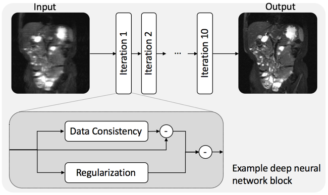
A VN3 with 10 iterations of data-processing blocks was implemented. Each data-processing block contains a data consistency and a regularization bypass3. Coil sensitivity maps, which were precomputed with ESPIRiT4, the acquired VD-sampled k-space, and a zero-filling reconstruction were used as inputs to the network.
With IRB approval and informed consent, abdominal SSFSE acquisitions in 600 pediatric subjects were collected and reconstructed with L1-CS-SENSE4, which was then taken as ground truth during training. An additional 150 samples were used for validation. Each sample consisted of a coronal slice acquired with a 32-channel torso coil (NeoCoil, Pewaukee, WI) and a VD SSFSE pulse sequence with an acceleration factor of 3.25x. Other imaging parameters were: TE 96ms, repetition time (TR) 470ms, bandwidth +/-83.3kHz, acquisition matrix 320 × 202, field of view (FOV) 300-480mm × 210-384mm, initial flip angle 130°, minimum flip angle 80°, center flip angle 100°, and last flip angle 45°. Training was performed on a single NVIDIA GeForce GTX 1080Ti GPU for 784 epochs (~1 day).
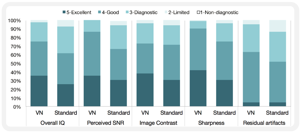
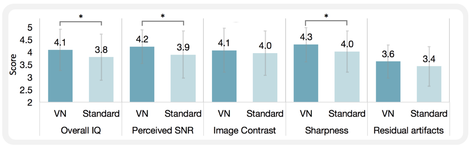
To test the performance of the trained model, datasets of 27 pediatric abdominal cases were collected with the same parameters as the training datasets. For each case, the FOV was prescribed based on the size of the patient. Standard PICS (L1-CS-SENSE) reconstruction was performed for comparison with a constant regularization factor of 0.002 and 30 iterations. Both these parameters were chosen based on the results of previous studies aimed at optimizing PICS2. Reconstructed images were evaluated in independent blinded fashion by three radiologists in terms of overall image quality (IQ), perceived signal-to-noise ratio (SNR), image contrast, sharpness, and residual artifacts with scores ranging from 1 (non-diagnostic) to 5 (excellent). Wilcoxon tests were performed to test the hypothesis that there was no significant difference between the VN approach and the standard PICS reconstruction.
Results
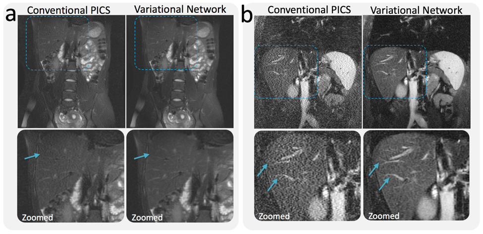
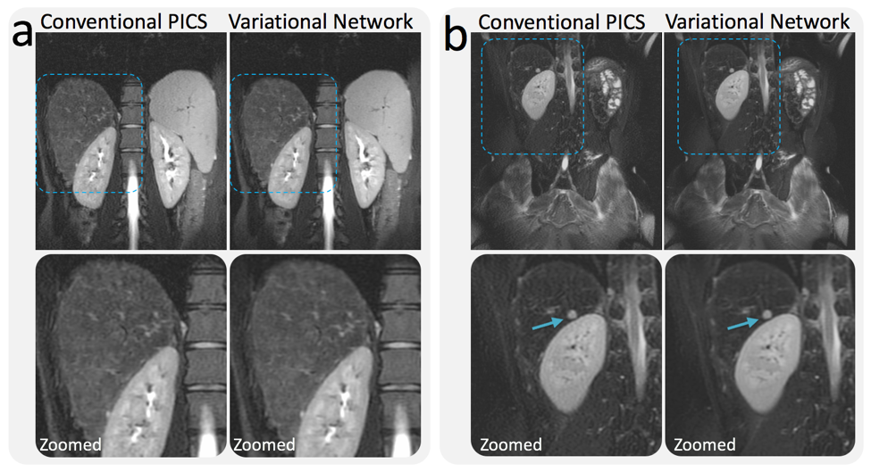
Compared with conventional PICS reconstruction, the VN approach achieved improved perceived SNR (P=0.01) and improved sharpness (P<0.001), with no significant difference in image contrast (P=0.24) and residual artifacts (P=0.07). In terms of overall image quality, VN performed significantly better than PICS (P=0.02) (Fig. 2 and 3). 4 out of 27 PICS reconstructions had residual artifacts attributable to spike noise, which were considerably reduced with the VN approach (Fig. 4). Examples of VN and PICS reconstructions in two patients are shown in Fig. 5.Discussion
As demonstrated in a previous study3, the VN is able to learn the regularization in each iteration of a PICS reconstruction. Compared with standard PICS with constant regularization and number of iterations, this provides flexibility in applying adaptive regularization levels to different cases. Although trained with the PICS reconstruction as ground truth, the VN was able to learn the average regularization among all training samples. Therefore, it functions as a denoising network in cases with low SNR to improve the overall quality of the PICS reconstruction.Conclusion
This work implemented and evaluated a VN approach for accelerated reconstruction of VD SSFSE acquisitions. Compared to the conventional PICS reconstruction, the VN approach achieves improved image quality with higher perceived SNR and sharpness. Further, the VN approach allows real-time image reconstruction of VD SSFSE sequences for practical clinical deployment.Acknowledgements
GE Healthcare, NIH R01 EB009690.References
1. Semelka RC, Kelekis NL, Thomasson D, Brown MA, Laub GA. HASTE MR imaging: description of technique and preliminary results in the abdomen. J Magn Reson Imaging 1996;6:698–699.
2. Taviani V, Litwiller DV, Tamir JI, Loening AM, Hargreaves BA, and Vasanawala SS. Variable Density Compressed Sensing Single Shot Fast Spin Echo. Proc. Intl. Soc. Mag. Reson. Med. 2016; 24: 618.
3. Hammernik K, Klatzer T, Kobler E, Recht MP, Sodickson DK, Pock T, Knoll F. Learning a Variational Network for Reconstruction of Accelerated MRI Data. arXiv preprint arXiv:1704.00447. 2017 Apr 3.
4. Uecker M, Lai P, Murphy MJ, Virtue P, Elad M, Pauly J, Vasanawala SS, Lustig M. ESPIRiT - An Eigenvalue Approach to Autocalibrating Parallel MRI: Where SENSE meets GRAPPA. Magn Reson Med 2014; 71:990-1001.
Figures