0609
Dynamics of adipose tissue and liver fat: Effects of acute exercise1German Diabetes Center Düsseldorf, Düsseldorf, Germany
Synopsis
Impaired adipose tissue fat uptake may lead to liver fat accumulation. Acute exercise releases adipose tissue fat, which also increases liver fat. Here we examine how a single bout of HIIT exercise affects the dynamics of adipose tissue and liver fat by MRS and an insulin clamp. Subjecting participants to a single bout of HIIT resulted in an improved response to insulin in both adipose tissue and liver. These results suggest that adipose tissue sequesters harmful saturated fats that otherwise would accumulate in the liver and that exercise improves the uptake of these fats thereby leading to long-term health effects.
Introduction
Impaired uptake of fat in adipose tissue can lead to accumulation of fat in the liver 1. There is still, however, uncertainty as to what extent the adipose depots regulate liver fat accumulation. Insulin is the major regulator fat uptake in adipose tissue, driving fatty acids (FA) into adipose tissue through activating lipoprotein lipase. Exercise on the other hand releases FAs from adipose tissue, which acutely increases liver fat 2. In contrast, continuous physical activity is protective of liver fat accumulation, indicating delayed beneficial effects of acute exercise. What these delayed effects are and on what timescale they are manifested is unknown 2. Recently we observed that a 4-hour insulin clamp increases the saturated fat content in adipose tissue, reflecting insulin stimulated uptake of saturated fats 3. In accordance with this, we observed a decrease in liver fat content during the insulin clamp, thus reflecting redistribution of saturated fat from the liver to adipose tissue 3. The insulin clamp, however, only decreased liver fat in healthy volunteers but failed to do so in type 2 diabetic (T2D) patients 3. Thus, the redistribution of fat from liver to adipose tissue seems to be impaired in T2D. In this study, we examine how a single bout of high intensity interval training (HIIT) affects the dynamics of adipose tissue and liver fat by MRS using the hyperinsulinemic-euglycemic clamp.Methods
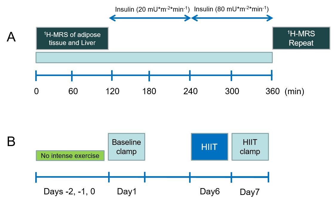
Ten male T2D (age 58.1±5.0y, BMI 31.3±2.2 kg/m2) and 10 age and BMI matched male controls (CON) (age 54.3±5.2y, BMI 30.7±2.2 kg/m2) were included in the study. The participants were examined on two separate days 1 week apart. On both days, the participants underwent a hyperinsulinemic-euglycemic clamp, with identical MRS measurements before and after the clamp. The first day served as a baseline measurement and was preceded by 3 days of restraint from intense physical activity. The day before the second clamp, the participants underwent a 1 hour supervised session of HIIT. On the day after the HIIT session, the participants again underwent a second clamp. The study timeline is illustrated for the clamp days in Figure 1A and for the whole week in Figure 1B. All MRS measurements were conducted on a 3T MR scanner (Philips Achieva, Best, The Netherlands). Proton spectra were acquired from abdominal deep subcutaneous adipose tissue (DSAT) for determining FA composition indices, and from liver for determining liver fat content (HCL), as previously described 4. DSAT spectra were analysed for unsaturation (=CH/CH2) and saturated chain length (CH2/CH3), while liver spectra were analysed for HCL. Spectra were faithfully localized between before and after measurements.Results
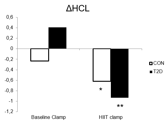
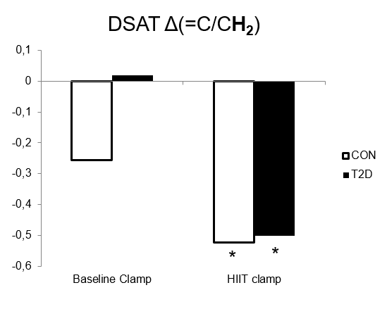
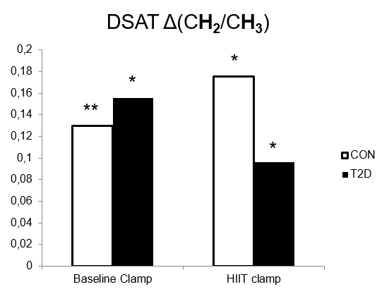
The baseline clamp increased CH2/CH3 in DSAT for both CON (5.63±0.15 to 5.76±0.19, P=0.002) and T2D (5.56±0.19 to 5.69±0.18, P=0.022), while unsaturation showed no change in either group. HCL tended to decrease during the baseline clamp in CON from 2.78% to 2.55% (P=0.25), while it tended to increase in T2D (13.96% vs 14.37%, P=0.92). In the HIIT clamp CH2/CH3 again increased in DSAT for both CON (5.54±0.17 to 5.71±0.18, P=0.038) and for T2D (5.59±0.19 to 5.74±0.25, P=0.013). In contrast to baseline, the HIIT clamp decreased unsaturation in DSAT for both CON (14.46±1.32 to 13.94±1.28, P=0.017) and for T2D (13.68±1.27 to 13.18±1.49, P=0.045). Also in contrast to baseline, HCL decrease during the HIIT clamp in both CON (3.81% to 3.19%, P=0.029) and in T2D (13.95% vs 13.01%, P=0.007). The change in HCL and adipose tissue FA composition is shown in Figures 2, 3 and 4.Discussion
Here we show that adipose tissue and liver respond to insulin by redistributing saturated fats from liver to adipose tissue. This redistribution seems to be impaired in T2D. Subjecting participants to a single bout of HIIT one day prior to the clamp, resulted in an improved response to the insulin clamp in both adipose tissue and liver for CON and T2D. These results suggest that adipose tissue sequesters harmful saturated fats that otherwise would accumulate in the liver.Conclusions
A single bout of HIIT exercise improves the response of adipose tissue and liver to insulin, facilitating redistribution of harmful saturated fats from the liver to adipose tissue.Acknowledgements
We thank the participants for their invaluable contributions. The German Diabetes Center is funded by the German Federal Ministry of Health (Berlin, Germany) and the Ministry of Innovation, Science and Research of the State of North Rhine Westphalia (Düsseldorf, Germany). This study was supported in part by grants from the German Federal Ministry of Education and Research (BMBF) to the German Center for Diabetes Research (DZD e.V.), from the Helmholtz Alliance Imaging and Curing Environmental Metabolic Diseases (ICEMED) and the Schmutzler-Stiftung.References
1. McQuaid SE, Hodson L, Neville MJ, et al. Downregulation of adipose tissue fatty acid trafficking in obesity: a driver for ectopic fat deposition? Diabetes. 2011 Jan;60(1):47-55.
2. Loher H, Kreis R, Boesch C, Christ E. The Flexibility of Ectopic Lipids. Int J Mol Sci. 2016 Sep; 17(9): 1554.
3. Magn Reson Mater Phy (2017) 30(Suppl 1): page S250, abstract 263.
4. Lundbom J, Hakkarainen A, Söderlund S, et al. Long-TE 1H MRS suggests that liver fat is more saturated than subcutaneous and visceral fat. NMR Biomed. 2011 Apr;24(3):238-45.
Figures