0608
Dietary intervention can induce changes in hepatic fat content during the day detectable by MR spectroscopy1MR-Unit, Dept. Diagnostic and Interventional Radiology, Institute for Clinical and Experimental Medicine, Prague, Czech Republic
Synopsis
We analyzed in vivo hepatic fat content changes during the day under different dietary interventions (fasting, fat alone, fat + fructose, fat + glucose, and glucose alone). Single voxel 1H MR spectroscopy of the liver in ten healthy volunteers was used at 3T MR system. We demonstrated that it is possible to induce the hepatic fat content changes by appropriately chosen dietary interventions and that such changes can be detected noninvasively by 1H MR spectroscopy.
Introduction
Non-alcoholic liver steatosis is frequently diagnosed
in many developed countries and can progress into the liver steatohepatitis,
fibrosis, cirrhosis, and even to hepatocellular carcinoma. There is only limited
knowledge about the hepatic fat content (HFC) changes during the day and it
remains to be determined whether such changes can be detected using MR methods.
It can be assumed that the lipid accumulation in the liver is affected by nutrient
turnover in the circulation, especially by turnover of triglycerides,
non-esterified fatty acids (NEFA), and glucose. Therefore, we analyzed whether changes
of HFC induced by different dietary interventions during the day can be
detected using
1H MR
spectroscopy (MRS) of the liver.Subjects & Methods
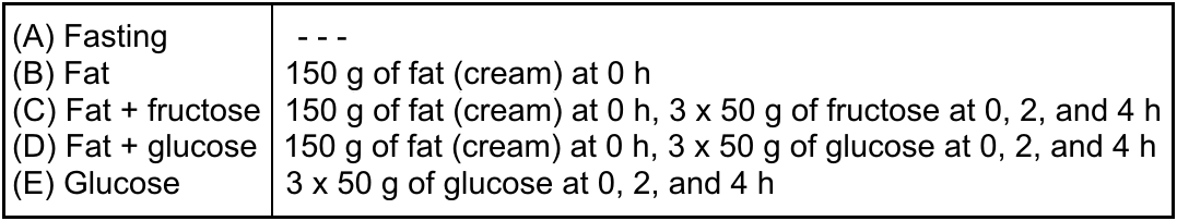
Ten healthy male volunteers without liver steatosis participated in the study (age 36±10 years, BMI 27±3 kg/m2, HOMA-IR 1.4±0.7 and the HFC measured by MRS 1.8±0.8%). They underwent five experiments with different dietary interventions, see Table. Single voxel spectroscopy at 3T MR system was used for the HFC estimation and spectra were measured in the morning after overnight fast, and three and six hours later.
PRESS sequence with TR/TE=4500/30 ms and voxel size VOI=40x30x25 mm in the 6th liver segment was applied in breath hold (1 acquisition, three repetitions). The localization was carefully checked during all subsequent measurements. Data were evaluated using LCModel according to the procedure described previously.1
The blood sampling for determination of triglycerides, NEFA, level of glucose and insulin were performed in the defined time intervals during all examinations. Standard ANOVA tests for repeated measurements were applied for statistical analysis. The p<0.05 was considered as statistically significant. The study was performed with the consent of the local ethical committee.
Results
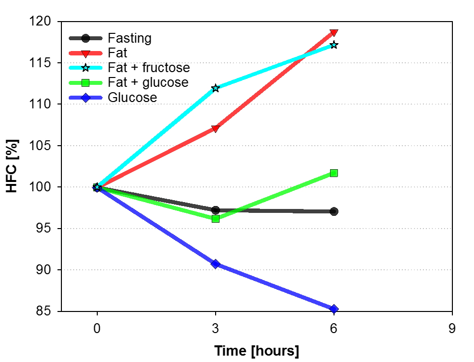
We found that fasting (A) did not change the HFC significantly from the morning session to the afternoon examination six hours later.
When 150 g of fat (cream) was provided to subjects (B), six hours later the HFC rose to 119±19% (p=0.012) of the morning value. The similar increase in HFC (117±18%, p=0.020) was observed when the experimental meal consisted of fat together with fructose (C). On the other hand, combination of fat with the same amount of glucose (D) did not induce significant HFC changes during experiment. Moreover, when glucose alone was administered to subjects (E), HFC dropped in six hours to only 85±13% (p=0.006) of the initial value (E), see Figure. The same results can be seen when absolute values of HFC are used.
Our results suggest that dietary glucose eliminates the impact of high-fat load on HFC and that the favorable effect of glucose on the HFC disappears when glucose is replaced by fructose.
Conclusion
We demonstrated that appropriately chosen diet induces
the hepatic fat content changes that can be detected by noninvasive 1H
MR spectroscopy. The presented experimental design can be a suitable model for
investigation of mechanisms of lipid accumulation in the liver and the pathogenesis
of non-alcoholic liver steatosis.Acknowledgements
The study was supported by Ministry of Health of the Czech Republic: AZV 16-28427A and Institutional support 00023001 IKEM.References
[1] Hajek M, Dezortova M, Wagnerova D, et al. Magn Reson Mater Phy 2011;24(5):297-304.Figures