0594
A connectome-wide investigation of the longitudinal effect of real-time fMRI amygdala neurofeedback emotional training on resting-state connectivity in combat veterans with PTSD1Laureate Institute for Brain Research, Tulsa, OK, United States, 2Laureate Psychiatric Clinic and Hospital, Tulsa, OK, United States, 3George Mason University, Fairfax, VA, United States, 4University of Arkansas, Fayetteville, AR, United States, 5University of Oklahoma, Norman, OK, United States
Synopsis
We introduced a longitudinal Multivariate Distance Matrix Regression (LMDMR) analysis for a connectome-wide study of the longitudinal effect on resting-state fMRI functional connectivity (rsfMRI-fc) without a priori seed definition. We applied this analysis to investigate the effect of real-time fMRI amygdala neurofeedback training in combat veterans with PTSD. The analysis revealed a significant interaction between a decrease in hyperarousal symptom and an increase in rsfMRI-fc between the precuneus and the left superior frontal region. This result suggests that enhanced regulation of emotional memory retrieval helped reduce PTSD symptoms.
Introduction
Resting-state fMRI functional connectivity (rsfMRI-fc) has been widely used to investigate abnormalities and on treatment effects on spontaneous brain activity in psychiatric disorders1-3. rsfMRI-fc is usually evaluated by defining a seed region a priori and examining correlations between the signals in the seed area and other brain regions. This approach, however, could limit the discovery in pre-assumed connectivities. Recently, a connectome-wide approach for rsfMRI-fc that utilizes Multivariate Distance Matrix Regression (MDMR) analysis has been proposed4. MDMR does not require a priori seed definition and can examine whole brain voxel-by-voxel connectivity. In the current study, we introduced an extension of MDMR - longitudinal MDMR (LMDMR) analysis - for analyzing connectome-wide effects in the longitudinal studies. We employed LMDMR to investigate the effect of real-time fMRI neurofeedback (rtfMRI-nf) training on resting-state functional connectivity in combat veterans with posttraumatic stress disorder (PTSD).Methods
Twenty-two combat veterans with PTSD completed three sessions of rtfMRI-nf emotional training and the pre- and post-training resting-state scan sessions. Participants were asked to upregulate their neurofeedback signal from the left amygdala (LA, experimental group [PTSD-exp), N=16) or parietal region not involved in emotion processing (control group [PTSD-ctrl], N=6) while recalling a positive autobiographical memory. Resting-state fMRI scans were performed in separate days (about a week before and after the rtfMRI-nf training sessions). AFNI (http://afni.nimh.nih.gov/afni/) and afni_proc.py script were used for analyses.
The distance matrix of the resting-state connectivity maps before and after the training was subject to longitudinal MDMR analysis. The design matrix included session, group, session by group interaction, age, and head motion regressors as well as subject-wise factor variables. The subject-wise regressors had 1 at a pair of a same subject’s samples and 0 for the others to regress out subject-wise average effect so that the longitudinal analysis could find the session effect on within-subject connectivity difference5. This design matrix (X) was rank-deficient due to collinearity between the subject-wise regressors and age and motion regressors. To address this rank deficiency we orthogonalized X using singular value decomposition (SVD)6, and the design matrix X was decomposed to X=USVT. MDMR analysis can be described as G=Xβ=USVTβ, where G is a centered negative distance matrix4. Pseudo-F value can be evaluated by F=[tr(HG)/(m-1)]/[tr[(I-H)G]/(n-m)], where H=UUT. The pseudo-F value’s significance was evaluated by a permutation test and thresholded by p<0.005 voxel-wise and by cluster-extent p<0.05.
The regions with a significant main effect of interest in the MDMR were used as seed regions for post-hoc connectivity analysis. A seed-based post-hoc analysis for the significant regions with the MDMR was done in the original resolution images. The statistical test of the post-hoc analysis was done with a linear mixed-effect (LME) model analysis for longitudinal design. We also performed a longitudinal MDMR analysis with additional regressors of PTSD symptoms (the Clinician-Administered PTSD Scale (CAPS) for DSM-IV7) and their interactions with session and group to examine an association between connectivity and symptom change.
Results
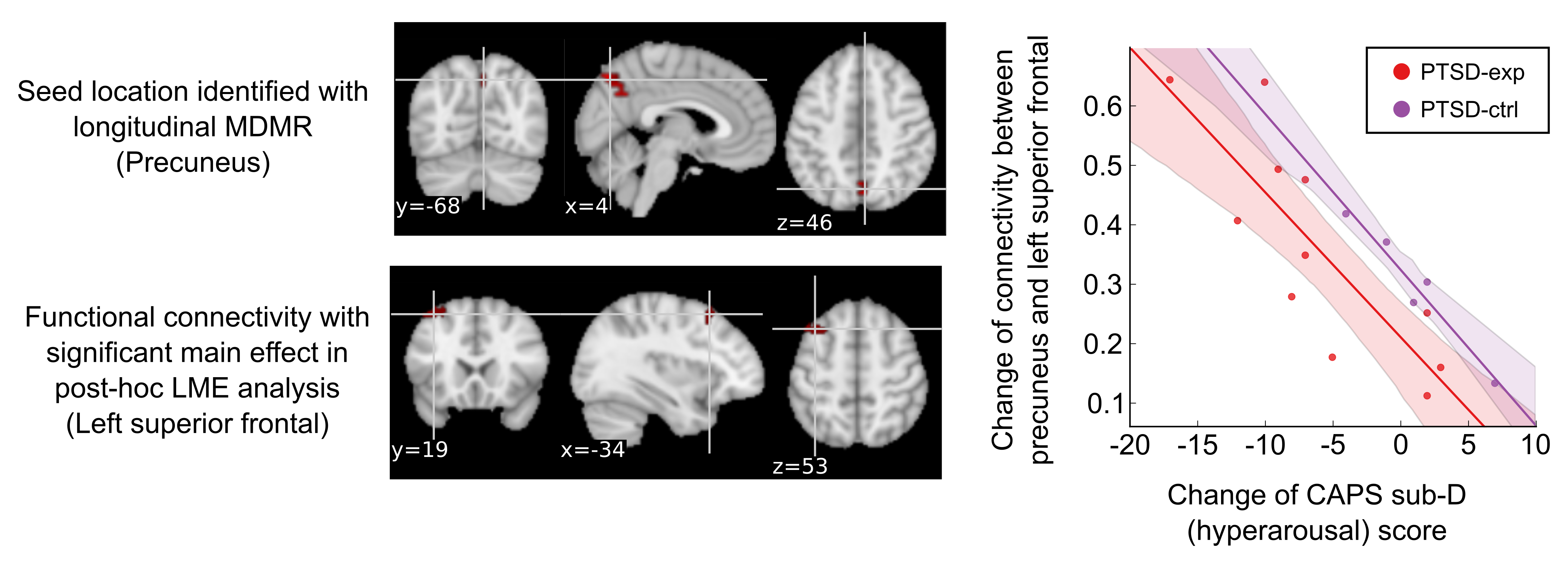
PTSD symptoms significantly decreased after the training (CAPS total scores, p=0.004; CAPS subscale C, p=0.020; CAPS subscale D, p=0.004). In the LMDMR analysis, no significant effect of session or interaction between session and group was found when no symptom regressor was included in the analysis. When the change in CAPS sub-D (hyperarousal) score was included in the model, longitudinal MDMR found a significant cluster in the right precuneus region (Fig. 1). The post-hoc LME revealed a significant interaction between session and CAPS sub-D score change in the connectivity between the precuneus and the left superior frontal region. The increase in this connectivity was significantly correlated with a decrease in CAPS sub-D (hyperarousal) for the PTSD-exp group (p=0.002). The PTSD-ctrl group also showed a similar trend but was not statistically significant (p=0.072).Discussion
The precuneus and the left superior frontal region have been implicated in memory retrieval8 and emotion regulation9, respectively. The increase in functional connectivity between these regions might be due to repetitive positive memory retrieval during the neurofeedback training. Interestingly, the increase in this connectivity was associated with a decrease in PTSD symptoms, which suggests regulation of emotional memory retrieval helped reduce PTSD symptoms.Conclusion
A connectome-wide approach using longitudinal MDMR enables exploratory analysis of the longitudinal effect on whole brain connectivity without a priori hypothesis and allows for the investigation of treatment effects on brain functional connectivity. LMDRM revealed a significant interaction between a decrease in hyperarousal symptoms and an increase in rsfMRI-fc between the precuneus and the left superior frontal region in veterans with PTSD. The rtfMRI-nf LA training enhanced regulation of emotional memory retrieval and reduced PTSD symptoms.Acknowledgements
This research was supported by W81XWH-12-1-0697 award from the U.S. Department of Defense, the Laureate Institute for Brain Research (LIBR), and the William K. Warren Foundation.References
1. Scheinost, D., T. Stoica, J. Saksa, et al., Orbitofrontal cortex neurofeedback produces lasting changes in contamination anxiety and resting-state connectivity. Transl Psychiatry, 2013;3:e250.
2. Yuan, H., K.D. Young, R. Phillips, et al., Resting-state functional connectivity modulation and sustained changes after real-time functional magnetic resonance imaging neurofeedback training in depression. Brain Connect, 2014;4(9):690-701.
3. Fonzo, G.A., M.S. Goodkind, D.J. Oathes, et al., Selective Effects of Psychotherapy on Frontopolar Cortical Function in PTSD. Am J Psychiatry, 2017:appiajp201716091073.
4. Shehzad, Z., C. Kelly, P.T. Reiss, et al., A multivariate distance-based analytic framework for connectome-wide association studies. Neuroimage, 2014;93 Pt 1:74-94.
5. Winkler, A.M., G.R. Ridgway, M.A. Webster, et al., Permutation inference for the general linear model. Neuroimage, 2014;92:381-97.
6. Mandel, J., Use of the Singular Value Decomposition in Regression Analysis. The American Statistician, 1982;36(1):15-24.
7. Blake, D., F. Weathers, L. Nagy, et al., Clinician-Administered PTSD Scale for DSM-IV (CAPS-DX). National Center for Posttraumatic Stress Disorder, Behavioral Science Division, Boston VA Medical Center, Boston, MA, 1995.
8. Brewin, C.R., J.D. Gregory, M. Lipton, et al., Intrusive images in psychological disorders: characteristics, neural mechanisms, and treatment implications. Psychol Rev, 2010;117(1):210-32.
9. Ochsner, K.N., J.A. Silvers, and J.T. Buhle, Functional imaging studies of emotion regulation: a synthetic review and evolving model of the cognitive control of emotion. Ann N Y Acad Sci, 2012;1251:E1-24.
Figures