0589
“Multi-Layer Connectome” for Robust Multi-Subject Brain Network Analysis and its Application to Baby Connectome Development Study1University of North Carolina at Chapel Hill, Chapel Hill, NC, United States, 2Brain and Cognitive Engineering, Korea University, Seoul, Republic of Korea
Synopsis
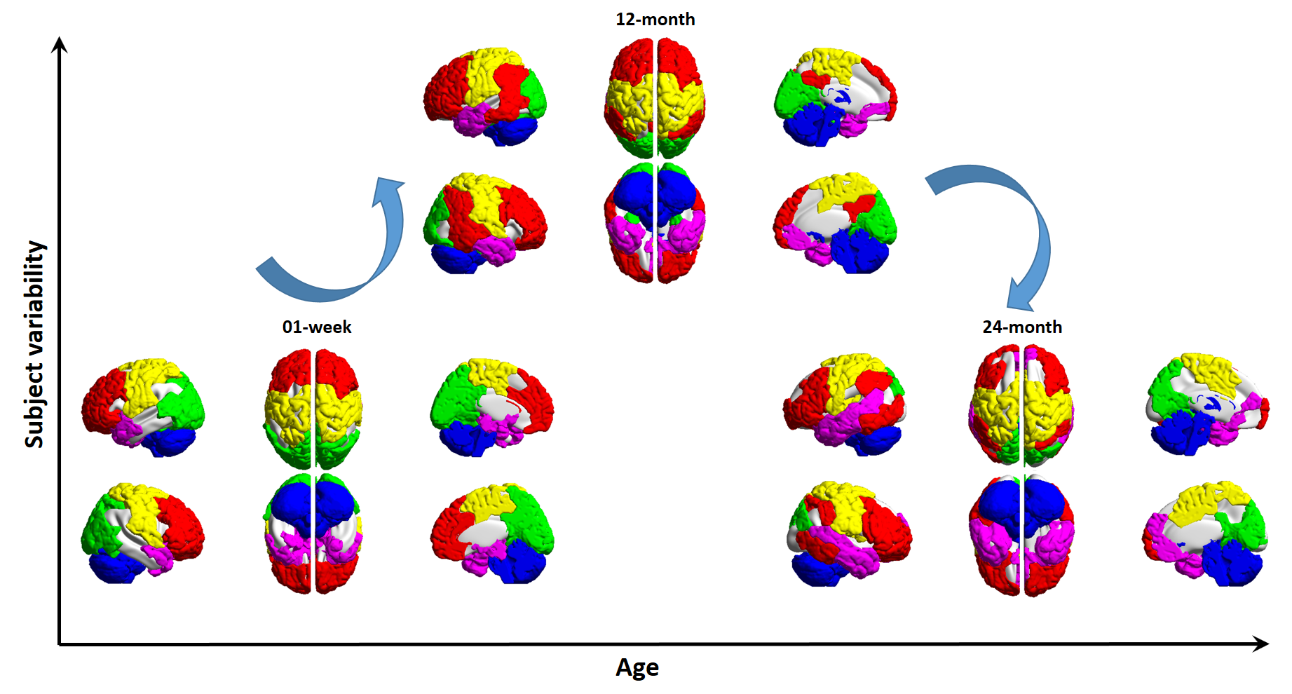
We propose a multi-layer connectome analysis method that extends the existing majority of single-layer brain network studies. In this method, multiple subjects’ connectome constitutes a multi-layer hyper-network with hyper-edges across layers. Result from applying this method to delineating neonatal brain development indicates that our method can capture robust group-level modules while keeping meaningful individual variability. The “increasing functional segregation/integration” model is further refined by us with a “consistent large-scale functional segregation/integration” with “rewiring-induced module refinement”, as well as an invert-U-shaped subject variability in modular structure in the first two years of life.
Introduction
Early brain development from neonate is a fascinating yet to date largely unknown. In such pivotal maturing processes, characterizing brain functional connectome evolution is one of the jewels. The “increasing functional segregation/integration” hypothesis seems to be a favorable model of developmental connectome [1-4]. However, such an overall summary of the developmental connectome lacks accurate spatiotemporal details. How and where does functional segregation/integration take place in the early developing brain is still unclear. Many graph-theoretical-analysis-based studies suggest increasing local and global efficiency for the early brain connectome. If this is the case, the brain should grow more and more efficient with great improvement of the small-worldness. However, increasing evidence suggests that, while developing in a dramatic way, our brain seems quite stable in general, with consistent connectivity infrastructures and relative robustness to the massive rewiring. This leads to an interesting hypothesis, that from the systems neuroscience point of view, the large-scale brain networks (i.e., modules) are relatively stable in their quantity, with rewiring-induced variations in network patterns for each of the functional networks. This study is to test this hypothesis with a newly proposed method.Methods
Community detection based on inter-regional FC is an ideal method to tackle the “segregation/integration” problem. Via maximizing intra-modular while minimizing inter-modular FC, community detection algorithms could detect relatively isolated sets of brain regions as a few functional networks, well balancing segregation/integration. However, traditional algorithms are mostly optimization algorithms involving free parameters and suffering stochastic errors. More importantly, these inherent issues make the community detection severely influenced by the resting-state fMRI data noise, making the result highly variable across subjects. Traditional methods often generate a group-level (with averaging) FC network and then apply community detection from an extensively group-averaged connectome, which could cancel noise but, more dangerously, eliminate individual differences, a particularly interesting problem in developmental neuroscience [5].
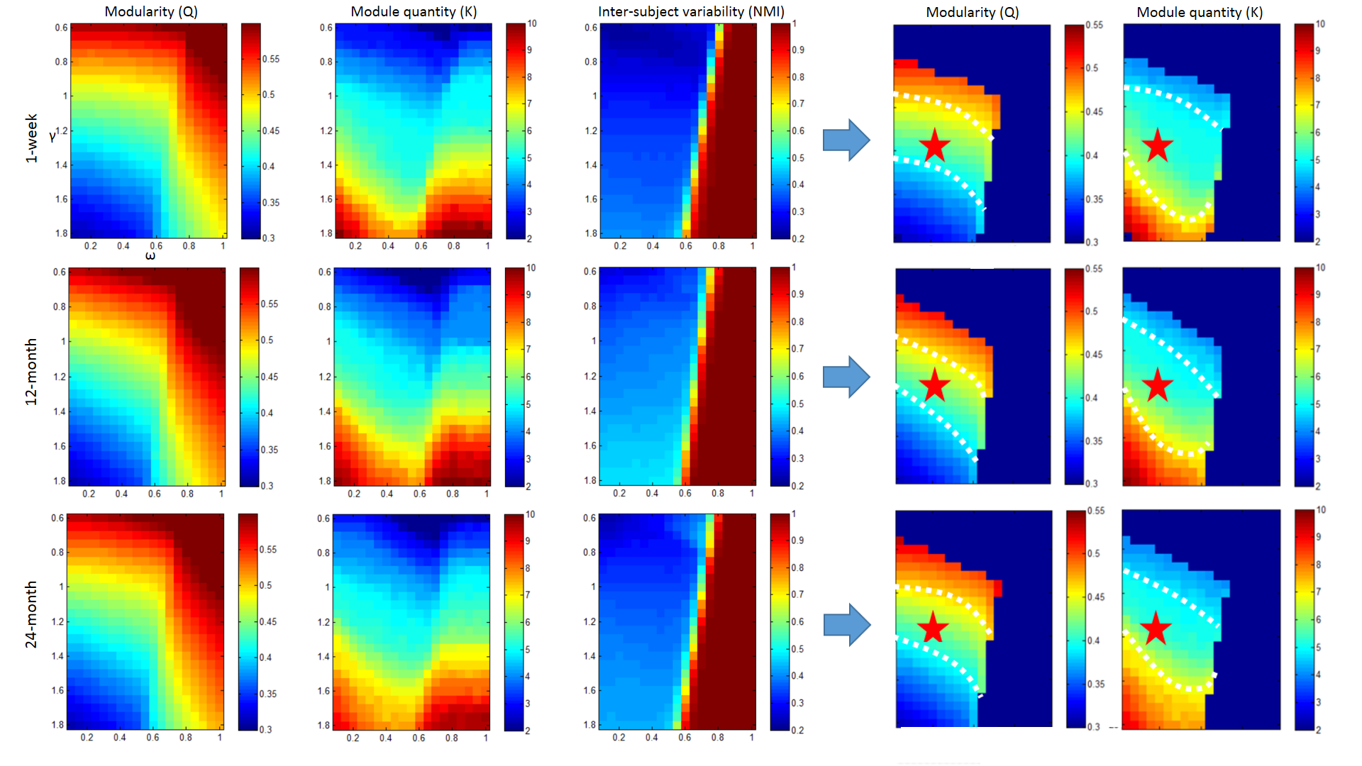
We implement a new method, characterizing multi-layer network properties, to solve these issues. In our method, each subject’s FC network constitutes one layer of a multi-layer “hyper-network” of all subjects, with hyper-edges connecting corresponding nodes (brain regions) between the different subjects. With the construction of a larger adjacency matrix for this hyper-network, a generalized Louvain algorithm [6] can be applied to detect functional modules for each subject in each layer, after a “group constraint” imposed by the cross-layer hyper-edges. Two parameters, one controlling the resolution (size) of the modules, one controlling the strength of the hyperedges, are optimized based on heuristic measures with consideration of stochastic variability. Our method has following merits: 1) detecting consistent modules for a group of infants; 2) maintaining individual differences; and 3) parameters are optimized, not arbitrarily predefined.
We apply this method to a longitudinal neonate&infant resting-state fMRI dataset with repetitive scans on ages of 1-week, 12-months, and 24-months (for data details see [7]). We aim to investigate the modularity development in the first two years of life, and test for our hypothesis on the consistent segregation/integration of the developing brain.
Results
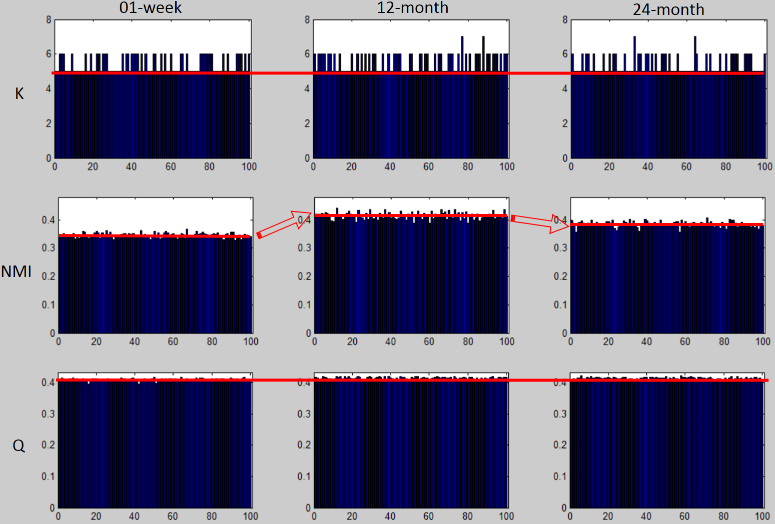
Parameter optimization was achieved based on the consideration of modularity (Q), module quantity and inter-subject variability (Fig. 1). Final multi-layer module detection result was chosen from 100 runs (Fig. 2) of the method with the maximum Q value.
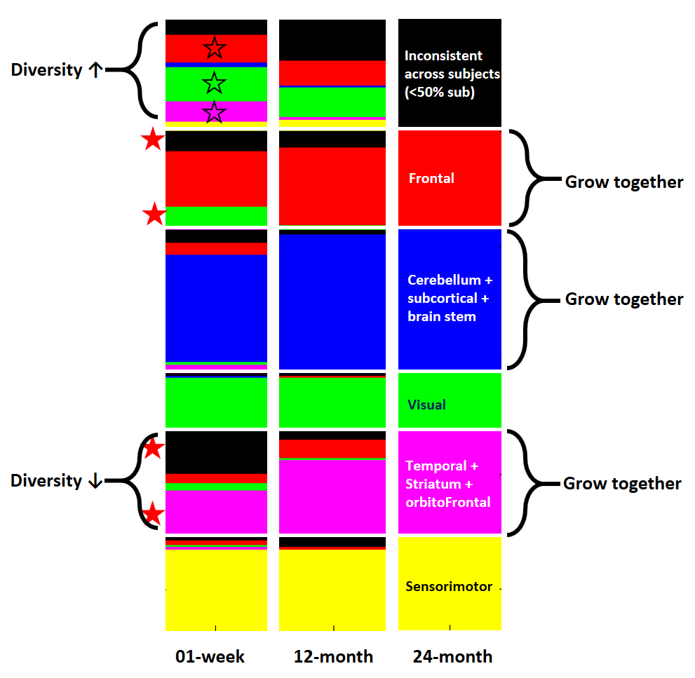
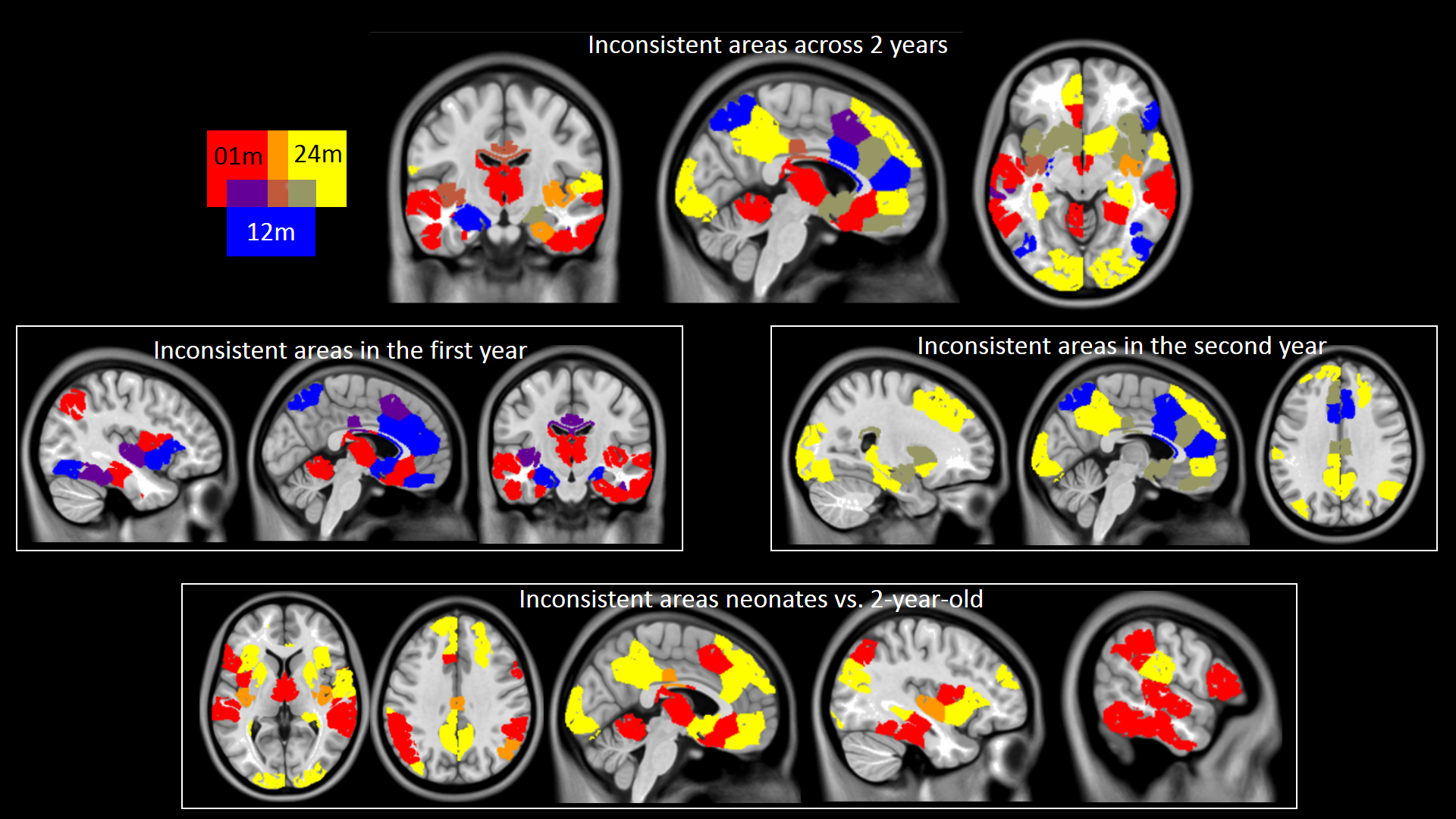
Based on both 100 realizations of module detection (Fig. 2) and the best module detection among all runs, we found a first increasing then decreasing subject variability in modular structure, but relatively stable modularity and module quantity. Multi-layer group-wise modularity was 0.417, 0.420 and 0.421 for neonates, 1-year-olds, and 2-year-olds. Subject variability in modular structure was 0.359, 0.431 and 0.396, respectively. We found both frontal, visual and temporal areas growing diverse with increasing subject variability. More regions are growing together into the frontal module, and regions in temporal, striatum and orbitofrontal cortices with formerly high individual variability grow together with decreased diversity (Fig. 3). Interestingly, the inconsistent regions grow spatially from medial subcortical to lateral cortical regions (Fig.4).
Conclusion
We conclude with our hypothetic model (Fig. 5), i.e., the large-scale brain modular structures are quite stable in their quantity, with rewiring-induced variations in several higher cognition-related modules, and an invert-U-shape subject variability.Acknowledgements
We acknowledge the Baby Connectome Project supported by NIH.References
[1] Cao, M., Huang, H., Peng, Y., Dong, Q., He, Y., 2016. Toward Developmental Connectomics of the Human Brain. Front Neuroanat 10, 25.
[2] Gao, W., Lin, W., Grewen, K., Gilmore, J.H., 2016. Functional Connectivity of the Infant Human Brain: Plastic and Modifiable. Neuroscientist.
[3] Zuo, X.N., He, Y., Betzel, R.F., Colcombe, S., Sporns, O., Milham, M.P., 2017. Human Connectomics across the Life Span. Trends Cogn Sci 21, 32-45.
[4] Di Martino, A., Fair, D.A., Kelly, C., Satterthwaite, T.D., Castellanos, F.X., Thomason, M.E., Craddock, R.C., Luna, B., Leventhal, B.L., Zuo, X.N., Milham, M.P., 2014. Unraveling the miswired connectome: a developmental perspective. Neuron 83, 1335-1353.
[5] Gao, W., Elton, A., Zhu, H., Alcauter, S., Smith, J.K., Gilmore, J.H., Lin, W., 2014. Intersubject variability of and genetic effects on the brain's functional connectivity during infancy. J Neurosci 34, 11288-11296.
[6] Mucha, P.J., Richardson, T., Macon, K., Porter, M.A., Onnela, J.P., 2010. Community structure in time-dependent, multiscale, and multiplex networks. Science 328, 876-878.
[7] Zhang, H., Yin, W., Lin, W., Shen, D., 2017. Early Brain Functional Segregation and Integration Predict Later Cognitive Performance. In: Wu, G., Laurienti, P., Bonilha, L., Munsell, B. (Eds.), Connectomics in NeuroImaging (CNI2017). Springer, Quebec, Canada.
Figures