0586
Parallel transmission kT-points and CAIPIRIHNA accelerated MP2RAGE for ultra-high field anatomical imaging1Cognitive Neuroscience, Maastricht University, Maastricht, Netherlands, 2MBIC - Maastricht Brain Imaging Center, Maastricht University, Maastricht, Netherlands, 3Center for Magnetic Resonance Research, University of Minnesota, Minneapolis, MN, United States, 4Advanced Clinical Imaging Technology, Siemens Healthcare AG, Lausanne, Switzerland, 5Department of Radiology, University Hospital of Lausanne, Lausanne, Switzerland, 6LTS5, École Polytechnique Fédérale de Lausanne (EPFL), Lausanne, Switzerland
Synopsis
The MP2RAGE imaging sequence is widely used for T1 weighted anatomical imaging, especially at 7T, where B1 inhomogeneity may degrade the image quality. This study demonstrates an improved MP2RAGE sequence featuring kT-points parallel transmission excitation and CAIPIRINHA 2D acceleration for improved T1 weighted anatomical images of the living human brain at 7 T and 9.4 T. High resolution (0.6 mm and 0.45 mm isotropic) data with high contrast-to-noise ratio allow for improved cortical segmentation, e.g. for cortical laminar investigations. We further demonstrate a 6-fold accelerated whole brain CAIPIRINHA acquisition at 0.6 mm resolution in only 4:10 min.
Introduction
The
MP2RAGE imaging sequence has proven highly useful for T1 weighted anatomical
imaging of the brain at 3T and 7T1. Here we demonstrate an extension of the pulse
sequence featuring kT-points parallel transmission2,3 for excitation and a CAIPIRINHA acceleration
scheme4 allowing for high contrast-to-noise images at
high resolution.Methods
Scans were performed on a 7T research system (Siemens Healthcare, Erlangen, Germany) with a Nova Medical 8TX/32RX head coil, as well as a 9.4T research system (Siemens Healthcare) for human imaging with a 16TX/31RX dedicated head coil5. SAR monitoring was performed using “protected mode” settings derived from previous numerical coil simulations. A prototype MP2RAGE imaging pulse sequence was modified to accommodate kT-points excitation, acceleration along the partition (kz) direction, in addition to the phase-encoding (ky) direction, and optional use of CAIPIRIHNA4 sampling. In this modified sequence, GRAPPA autocalibration lines are acquired in a separate GRE kernel rather than being integrated into the echo-trains. Standard B1 shimmed inversion pulses were used for all acquisitions.
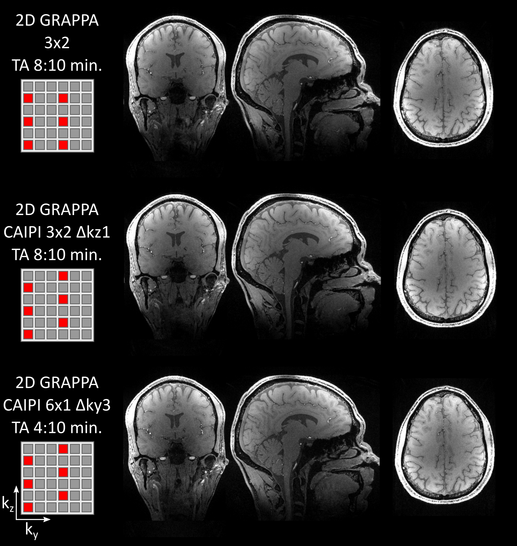
The following imaging parameters were used unless specified otherwise: TE/TR/TI1/TI2 = 2/5000/700/2500 ms, matrix size 384x384x256, 0.6 mm isotropic nominal resolution, acceleration factor (AF) 3×2 (phase-encoding lines x partition), nominal flip angles 7°/5°. kT-points protocols used 8 kT points with an RF subpulse spacing of 0.10-0.14 ms (matching the maximum RF amplitude of the B1 shim reference excitation). Two CAIPIRINHA-accelerated protocols were acquired either with the same acquisition time as the 2D GRAPPA protocol (AF = 3×2 Δky3), or a shorter scan duration (6×1 Δky3). Both CAIPIRINHA patterns lead to the same aliasing pattern.
For segmentation, the combined ("uniform") MP2RAGE images were intensity masked using the second inversion contrast. The brain was extracted using FSL5.0 BET and segmented using FSL5.0 FAST with default settings. No further (manual or automated) adjustments were made for best comparability.
Results
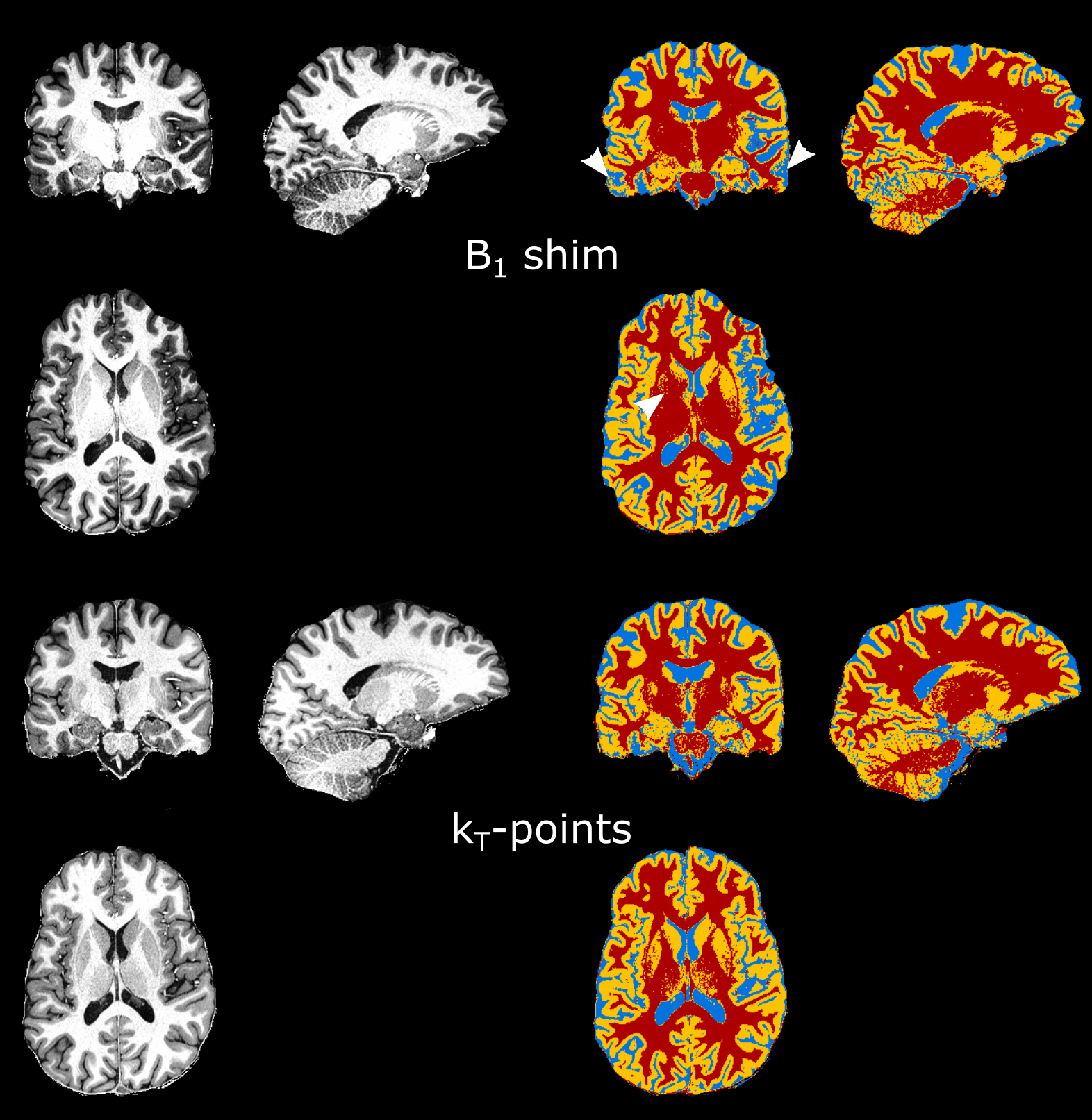
Figure 1 shows an example kT-points excitation k-space trajectory and the concomitant RF amplitudes at the 8 points in 8 TX channels. On the right, the predicted flip angle distribution is shown for 7 T and 9.4 T for a B1-shim (top) and the kT-points excitation (bottom). Figure 2 displays cuts of the combined images, and the segmentation results, for the B1 shim and the kT-points excitation at 7 T. Marked segmentation improvements are highlighted by white arrow heads.
Figure 3 shows a comparison of different CAIPIRINHA pattern schemes. No marked difference between the non-shifted 2D GRAPPA acceleration and either of the CAIPIRINHA accelerations was observed, but it should be noted that the 6×1 Δky3 acquisition reduces the acquisition time almost by a factor of two.
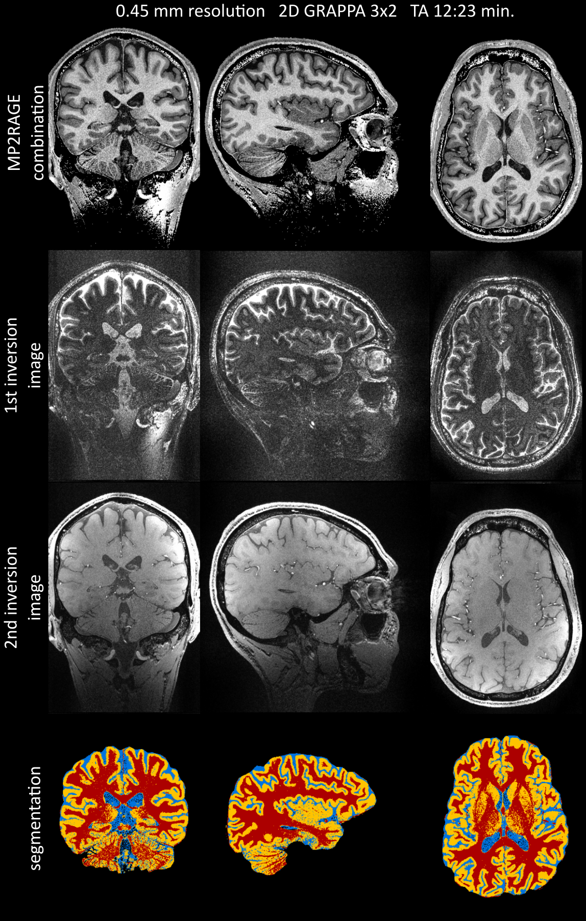
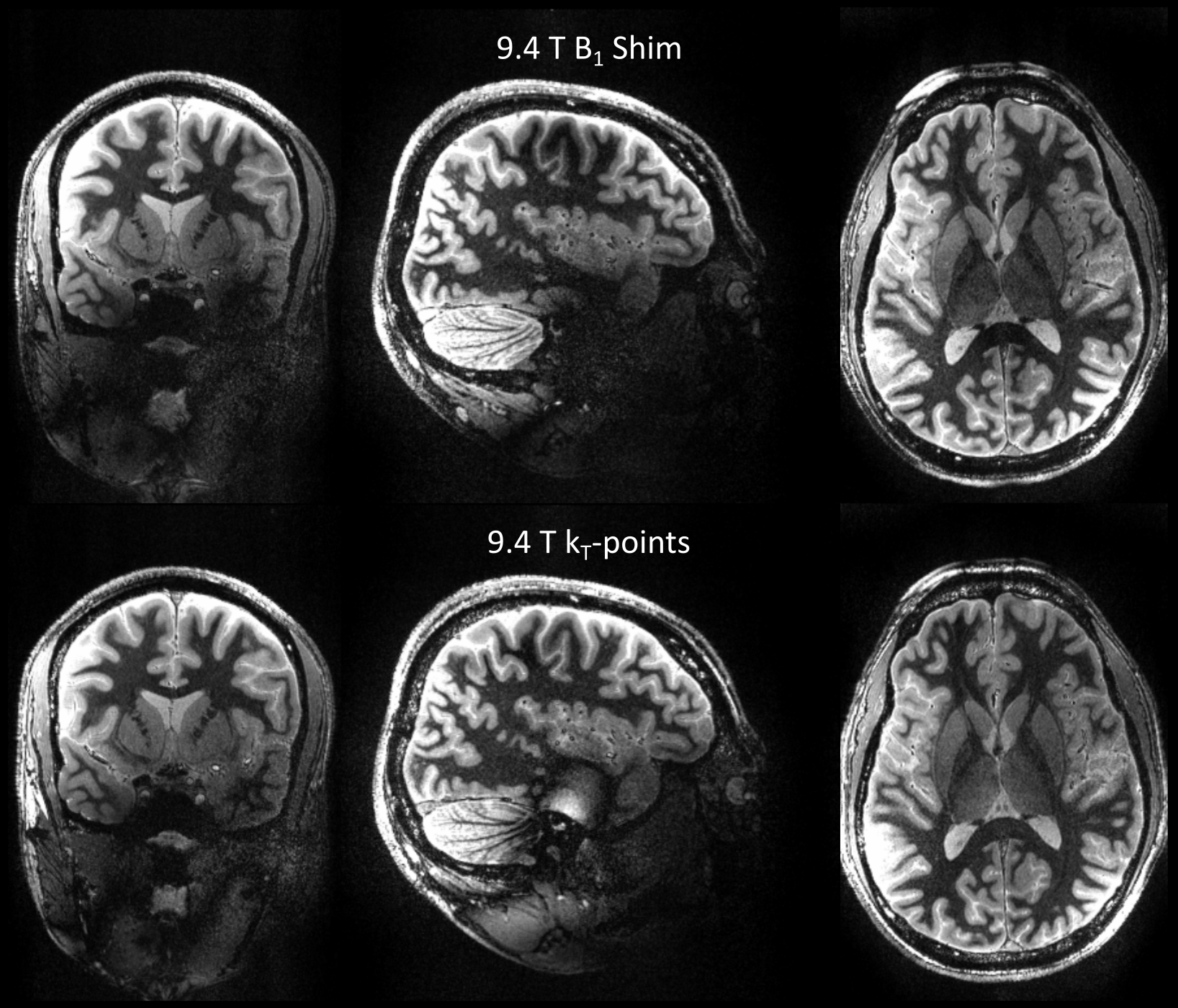
Figure 4 displays high resolution results of the 0.45 mm isotropic resolution acquisition. Figure 5 shows a comparison of a B1 shim and kT-points excitation at 9.4 T. kT-points.
Discussion and Conclusion
We demonstrate improved flip angle homogeneity using kT-points parallel transmission, and increased flexibility with CAIPIRINHA regarding acquisition time tradeoffs in MP2RAGE anatomical brain images at both 7T and 9.4T. Compared to single-channel transmit or B1 shimming, kT-points reduces flip angle inhomogeneity across the whole brain leading to improved segmentation results. Inversion imperfections still hamper the image quality, but may also be addressed using pTX inversion pulses6. Superior B1 homogeneity results in more homogeneous image signal and contrast, which is essential especially for automated image processing such as segmentation. The advantage over more B1 sensitive non-pTX acquisitions is demonstrated. 2D acceleration allows for either high resolution protocols without prohibitively long echo trains or compromised contrast and point-spread function due to changing T1 contrast7, or more flexible investment of most or all of the acceleration capability along the slow encoding dimensions, in order to achieve considerable reductions in acquisition time as demonstrated in the CAIPIRINHA 6×1 Δky3 protocol (see also Brenner et al., 20148). It should be noted that the proposed improvements to the sequence are readily compatible with other extensions such as multi-echo or additional inversion contrasts.Acknowledgements
The authors would like to thank Dr. Desmond Tse for support on pTX imaging and Dr. Stephan Kannengiesser at Siemens Healthineers, Erlangen, for support on the prototype CAIPIRINHA ‘IcePAT’ implementation. This work was supported by the Netherlands Organization for Scientific Research (NWO; VIDI grant 864-13-012 to F.D.M., VICI grant 453-12-002 to E.F, and VIDI grant 016-178-052 to B.A.P.).References
1. Marques JP, Kober T, Krueger G, van der Zwaag W, Van de Moortele PF, Gruetter R. MP2RAGE, a self bias-field corrected sequence for improved segmentation and T1-mapping at high field. Neuroimage. 2010;49(2):1271-1281. doi:10.1016/j.neuroimage.2009.10.002.
2. Cloos MA, Boulant N, Luong M, et al. kT -points: short three-dimensional tailored RF pulses for flip-angle homogenization over an extended volume. Magn Reson Med. 2012;67(1):72-80. doi:10.1002/mrm.22978.
3. Tse DHY, Wiggins CJ, Ivanov D, et al. Volumetric imaging with homogenised excitation and static field at 9.4 T. Magn Reson Mater Physics, Biol Med. 2016;29(3):333-345. doi:10.1007/s10334-016-0543-6.
4. Breuer FA, Blaimer M, Mueller MF, et al. Controlled aliasing in volumetric parallel imaging (2D CAIPIRINHA). Magn Reson Med. 2006;55(3):549-556. doi:10.1002/mrm.20787.
5. Shajan G, Kozlov M, Hoffmann J, Turner R, Scheffler K, Pohmann R. A 16‐channel dual‐row transmit array in combination with a 31‐element receive array for human brain imaging at 9.4 T. Magn Reson Med. 2014;71(2):870-879.
6. Cloos MA, Boulant N, Luong M, et al. Parallel-transmission-enabled magnetization-prepared rapid gradient-echo T1-weighted imaging of the human brain at 7T. Neuroimage. 2012;62(3):2140-2150.
7. van der Kouwe AJ, Benner T, Salat DH, Fischl B. Brain morphometry with multiecho MPRAGE. Neuroimage. 2008;40(2):559-569. doi:10.1016/j.neuroimage.2007.12.025.
8. Brenner D, Stirnberg R, Pracht ED, Stöcker T. Two-dimensional accelerated MP-RAGE imaging with flexible linear reordering. Magn Reson Mater Physics, Biol Med. 2014;27(5):455-462. doi:10.1007/s10334-014-0430-y.
Figures