0538
“In vivo” Field-Cycling relaxometry of tumours. Evidence for the role of the intracellular water lifetime as tumour biomarker.1University of Torino, Torino, Italy, 2University of torino, Torino, Italy, 3Stelar srl, Mede (PV), Italy
Synopsis
This work aims at developing an innovative diagnostic strategy, based on the "in vivo" measurements of longitudinal relaxation times at low and ultra-low magnetic fields with Fast Field Cycling FFC-NMR to obtain quantitative information on tumour metastatic potential, due to different water content and mobility, that is invisible to standard MRI. Preliminary results show that the endogenous contrast between normal and diseased tissue, due to differences in T1, is much greater at low field and the shape of the relaxation dispersion profiles may be used as a reporter of the molecular dynamical processes, biomarkers of the disease grade.
Introduction
Magnetic resonance imaging (MRI) has had a key role in the field of oncology over the last several decades. The MRI diagnostic power arises basically from the differences in the longitudinal and transverse proton relaxation times between healthy and pathological tissues. However, at the high field strength of the currently available scanners (1-7T), changes in T1 do not appear sensitive enough to report on peculiar aspects of the tumour stage. However, there is a diffuse opinion that, at low magnetic field strength, the marked increase of R1 observed in biological tissues might be beneficial to improve the MRI diagnostic potential in tumour phenotyping. (1-3) Herein it is shown that the in vivo acquisition of 1/T1 Nuclear Magnetic Resonance Dispersion (NMRD) profiles (from 0.01 to 10 MHz, proton Larmor frequency) fully supports this expectation as the observed R1s at low magnetic fields (<0.2 T) “in vivo”, on preclinical animal models, allow a clear discrimination between tumours endowed with different metastatic potential.Methods
1/T1 NMRD profiles are
acquired on Fast Field Cycling relaxometers able to switch the magnetic field
between different field strengths, during the measurement procedure. A field
cycle overcomes the problem of the low sensitivity at low magnetic fields and
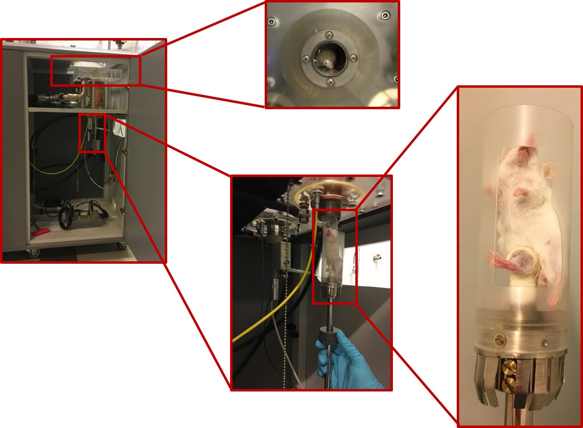
allows rapid acquisition. The currently
available relaxometers are designed for liquid samples measurements. Therefore,
in order to host a mouse, the commercially available relaxometer had to be
modified with the implementation of a 40 mm 0.5T Field Cycling magnet and a
dedicated 11mm solenoid detection coil placed around the mouse’s leg (Fig. 1)
where is located the tumour xenograft prepared with mouse mammary adenocarcinoma cells, namely TSA,
4T1, 168Farn, injected in the leg muscle. These cell display different
characteristics in terms of aggressiveness and metastatic potential (i.e.
168Farn<TSA<4T1).Results
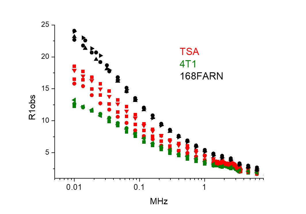
A simple inspection on the obtained NMRD profiles, allow us to clearly distinguish healthy from tumour tissues as the tumour tissues invariantly show lower R1 values in particular at low magnetic field strengths. The T1 elongation was different for the three tumour models (Fig.2) essentially reflecting their different aggressiveness. To better understand this behavior one needs to recall that each R1 in the profile represents an average of the R1 values of water molecules in different tissue microenvironments: the extracellular space (R1ex) and the intracellular compartment characterized by a more restricted water molecules mobility (R1in), being Vex and Vin the respective volume fractions. Water molecules can cross the barriers between the two compartments thus contributing to mixing, at some extent, the relaxation rates of the intracellular and extracellular compartments. Therefore, kin (water exchange rate from the intra- to the extra-cellular space) have to be introduced in the model used for the fitting of the Mz decay. According to this bicompartimental model, the time evolution of MZ is dependent on the relationship between the absolute values of the “relaxation” term,|R1in -R1ex|, and an “exchange” term |kin + kex|, also defined as the NMR “shutter-speed”. (4) The most striking result from the fitting procedure is the large variation of kin to indicate that the water exchange rate across the plasmalemma membrane is a distinctive hallmark that differentiates muscle (representative of healthy cells) and tumour cells. This finding clearly reports on the peculiar characteristics of the given tumour cell type. In fact, the intracellular water lifetime tin (tin =1/kin) values obtained for three breast cancer cell lines are inversely proportional to their metastatic potential (4T1>TSA>168Farn).Discussion
The herein reported results open new horizons for the non-invasive evaluation of tumour metabolic phenotypes, by providing useful information related to the tumour metastatic propensity. The simultaneous fitting of Mz over an extended range of magnetic field strengths allows attaining a good estimation of Kin that is crucial to cell function. Cell water content and volume are related to the concentration of intracellular osmotic active compounds as well as to the extracellular tonicity. Ion pumps or active transporters up/down regulation occurring in the presence of a pathological state, can be exploited as a specific reporter of the cellular state. kin reports on the activities of a number of transporters (GLUT-1, Na+/K+ ATP-ase and collectively it may represent an hallmark of tumor cells aggressiveness.Conclusion
We may conclude that the measurement of transmembrane permeability provides insights for more specific assessments of the pathophysiogical status of tumours as well as of other biological tissues. Despite the FFC-NMR instrumentation is not endowed with spatial resolution, fundamental knowledge obtained in this study can open the route to new diagnostic horizons in oncology until now uncharted.Acknowledgements
This project has received funding from the European Union’s Horizon 2020 research and innovation programme under grant agreement No 668119 (Identify project)References
1. Field-cycling NMR relaxometry with spatial selection. Pine KJ, Davies GR, Lurie DJ. 2010, Magn Reson Med., Vol. 63, pp. 1698-702.
2. Magnetic field dependence of 1/T1 of protons in tissue. Koenig SH, Brown RD 3rd, Adams D, Emerson D, Harrison CG. s.l. : Invest Radiol., 1984, Vol. 19, pp. 76-81.
3. Feasibility of high-resolution one-dimensional relaxation imaging at low magnetic field using a single-sided NMR scanner applied to articular cartilage. Rössler E, Mattea C, Stapf S. s.l. : J Magn Reson., 2015, Vol. 251, pp. 43-51.
4. Intratumor mapping of intracellular water lifetime: metabolic images of breast cancer? al, Springer CJ et. s.l. : NMR Biomed., 2014, Vol. 27, pp. 760-73.
Figures