0531
Influence of Infant Feeding on Longitudinal Brain and Cognitive Development1Pediatrics, Brown University, Providence, RI, United States, 26Waisman Center, University of Wisconsin, Madison, WI, United States, 3Pediatrics, Memorial Hospital of Rhode Island, Providence, RI, United States
Synopsis
Infancy and early childhood are sensitive and rapid periods of brain growth that coincide with the emergence of nearly all cognitive, behavioral, and social-emotional functions. Brain growth, including myelination, is modulated modulated by neural activity and are responsive to environmental, genetic, hormonal, and other influences, including nutrition. He we use longitudinal neuroimaging to examine the influence of early infant nutrition, specifically breastfeeding, on brain and cognitive development.
BACKGROUND
Throughout early neurodevelopment, myelination helps provide the foundation for brain connectivity and supports the emergence of cognitive and behavioural functioning. Early life nutrition is an important and modifiable factor that can shape myelination and, consequently, cognitive outcomes. Differences in the nutritional composition between human breast and formula milk may help explain the functional and cognitive disparity often observed between exclusively breast versus formula-fed children. However, past cognitive and brain imaging studies comparing breast and formula feeding are often: cross-sectional; performed in older children and adolescents relying on parental recall of infant feeding; and generally treat formula-fed children as a single group despite the variability between formula compositions. Here we address some of these weakness by examining longitudinal trajectories of brain and neurocognitive growth in children exclusively breastfed versus formula-fed for at least 3 months as well as between children who received different formula compositions.METHODS
Magnetic resonance imaging data were acquired of 88 formula and 62 exclusively breast-fed (for at least 90days) infants matched for age (p=0.24), gestation (p=0.39), birth weight (p=0.52), gender (p=0.85), parent marital status (p=0.66) and parent education (p=0.9). 231 longitudinal datasets were obtained on the breastfed children, and 221 on the formula-fed children, at approximate 6month increments. The formula-fed group contained 21 (9 female) children who received formula #1; 28 (10 female) who received formula #2; and 39 (15 female) who received formula #3. To quantify myelination, mcDESPOT was used with age-optimised protocols for paediatric imaging [1]. The Mullen Scales of Early Learning [2] were used to assess overall cognitive, verbal, and non-verbal abilities. Nonlinear Mixed modelling using a Gompertz growth model [3] was used to investigate group differences in brain and cognitive development trends. Linear modelling was used for the cognitive data.
RESULTS
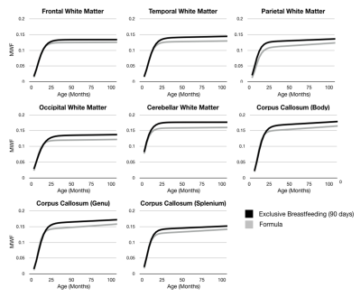
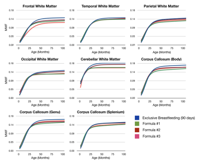
In all investigated brain regions, we find differential patterns of development (Fig. 1) with breastfed children qualitatively exhibiting a prolonged period of rapid development between 500 and 750 days of age, with an overall increase in myelin content by 2 years of age that persists throughout childhood. Statistically significant differences (p<0.000001) were found between all Gompertz growth model parameters in the frontal, temporal, and occipital white matter, and the body and genu of the corpus callosum. Examining differences between children who received different formula compositions (Fig. 2), we found significant differences in developmental patterns throughout the brain. In particular, we found children who received formula compositions with higher levels of DHA, ARA, choline, and sphingolipids (formulas #2 and #3) showed increased levels of myelin development.
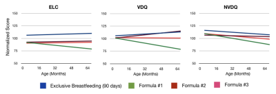
Differences in overall cognitive ability (ELC) and verbal (VDQ) and non-verbal (NVDQ) skills were also found between breast and formula-fed children (Fig. 3) and between children who received different formula compositions (Fig. 4).
DISCUSSION
Our results indicate that exclusive breastfeeding for at least 3 months is associated with improved myelination diffusely throughout the brain by 2 years of age, including early and late maturing brain regions and networks associated with a broad array of cognitive and behavioral skills. Supporting this structure-function link, we also show improved overall cognitive ability and rates of cognitive development, including verbal and non-verbal functioning, in breast-fed children versus those who received only infant formula. Investigating across infant formulas, we find an overall correspondence between brain development profiles and trajectories of cognitive maturation. Specifically, children who received Formula #1, which shows the slowest myelination profile across the majority of brain regions, also have the most pronounced decline in cognitive function across early childhood. Formula #2, which had the closest myelination trend to breastfeeding also exhibit cognitive trends that are most consistent with breastfeeding. These results add to the consensus that prolonged and exclusive breastfeeding plays an important role in early neurodevelopment and childhood cognitive outcomes.Acknowledgements
No acknowledgement found.References
[1] Deoni, S.C.L. et al., 2012. Investigating white matter development in infancy and early childhood using myelin water faction and relaxation time mapping. NeuroImage, 63(3), pp.1038–1053.
[2] Mullen, E.M., 1995. Mullen scales of early learning.
[3] Dean, D.C. et al., 2015. Characterizing longitudinal white matter development during early childhood. Brain structure & function, 220(4), pp.1921–1933.
Figures


