0511
Association Between Concussion-induced Dizziness and Damage of Thalamo-cortical Connectivity after mild Traumatic Brain Injury1Research Center of Translational Imaging, College of Medicine, Taipei Medical University, Taipei, Taiwan, 2Department of Anatomy and Cell Biology, School of Medicine, College of Medicine, Taipei Medical University, Taipei, Taiwan, 3Department of Biomedical Imaging and Radiological Sciences, National Yang-Ming University, Taipei, Taiwan, 4Department of Radiology, School of Medicine, College of Medicine, Taipei Medical University, Taipei, Taiwan, 5Department of Medical Imaging, Taipei Medical University Hospital, Taipei, Taiwan, 6Department of Medical Research, Taipei Medical University Hospital, Taipei, Taiwan
Synopsis
Dizziness is a frequent symptom of concussion, however neuroimaging evidence that supports clinical symptoms after mTBI was less explored. This study demonstrated the post-concussion damages of thalamo-cortical networks revealed by the axonal injuries of specific fiber tracts and the altered functional connectivity. The estimates of Dizziness Handicap Inventory significantly correlated to the functional connectivity of thalamic nuclei.
Background and Purpose
Dizziness is one of the prominent post-concussion symptoms that cannot be elucidated by the conventional imaging approaches1. The occurrence of dizziness caused by the mild traumatic brain injury (mTBI) is associated with the impairment of vestibular pathway which is composed of the vestibular nuclei, cerebellum, and thalamo-cortical circuits. We hypothesize that the mTBI-induced shear injury may structurally damage the thalamo-cortical tracts leading to the alterations of functional connectivity of thalamo-cortical networks and the subsequent post-concussion symptoms, e.g. the dizziness. We performed the tract-based analysis and functional connectome analysis to investigate whether the alterations of thalamo-cortical connections can support the occurrence of dizziness or other post-concussion symptoms.Materials and Methods
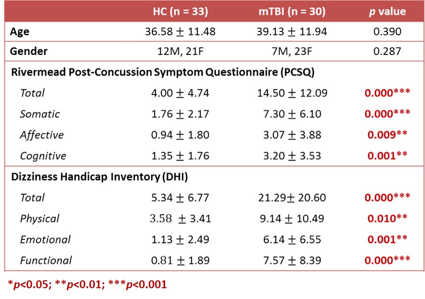
This study was approved by the local Institutional Review Board and the written informed consent was provided by each participant. Thirty patients with mTBI and 33 age- and gender-matched healthy controls (HC) were recruited (Table 1). Inclusion criteria for patients were witnessed closed-head trauma, no focal neurologic deficit, and initial Glasgow Coma Scale higher than 13. Clinical assessments were performed to evaluate post-concussion symptoms. MRI data, including a 3D T1-MPRAGE (TR/TE: 2300/3.26 ms; voxel size: 1.0x1.0x1.0 mm3), diffusion tensor imaging with 64 gradient directions and 10 sets of b0 (TR/TE: 7500/59 ms; voxel size: 0.86x0.86x3.0 mm3), and BOLD resting-state fMRI (TR/TE: 2000/20 ms; voxel size: 2.2x2.2x3.5 mm3, 190 volumes) were acquired on a 3T MR scanner (Siemens MAGNETOM Prisma). Patients received MR scans within 4 weeks after mTBI.
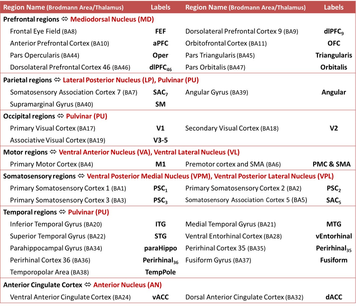
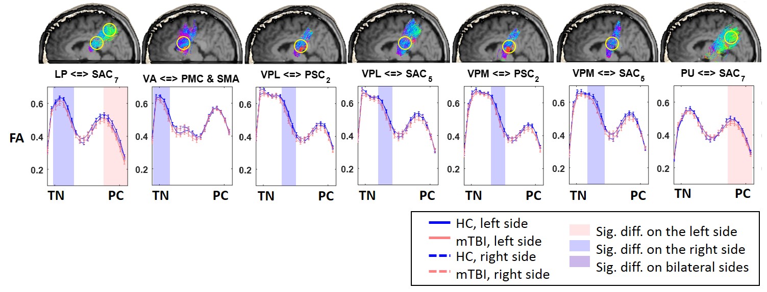
Seventy-six thalamo-cortical tracts between 8 thalamic nuclei (defined by the Thalairach atlas) and 31 cortical regions (defined by the Brodmann area) were automatically generated using the constrained spherical deconvolution and probabilistic tractography2,3(Table 2). A tract-based analysis (p < 0.05 with a constraint of cluster size) was applied to assess the difference of fractional anisotropy (FA) along each thalamo-cortical tract between mTBI and HC groups4.
The fMRI data were preprocessed using SPM8 with the standard procedures: corrected for slice timing, realigned, spatially normalized into the standard space, and spatially smoothed with a 6-mm FWHM Gaussian kernel5. The regional BOLD signal was then extracted by averaging voxel signals within the region, and regressing out the confounding effects of motion parameters and signals from the white matter and cerebrospinal fluid. The thalamo-cortical connectivity was estimated by calculating the Pearson’s correlation coefficient between regional BOLD signals (with bandpass-filtered between 0.01 and 0.10 Hz) followed by Fisher’s r-to-z transform. Two-sample t-test was used to determine the differences between groups (p < 0.05). Partial correlation coefficients with controlling age and gender effects were computed to reveal the relations between functional connectivity and post-concussion symptoms (p < 0.01).
Results and Discussion
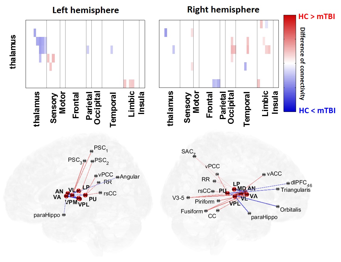
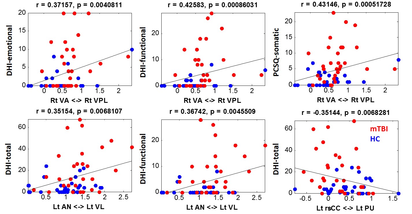
The mTBI patients had significant post-concussion symptoms (PCSQ) and elevated scores of Dizziness Handicap Inventory (DHI) compared to healthy controls (Table 1). Results of automatic tract-based analysis showed reduced FA values (reflecting impaired fiber integrities) of fiber tracts between thalamic nuclei and the connected cortical regions, including the premotor cortex, supplementary motor area, and several somatosensory cortices, in mTBI patients (Fig. 1). The identified locations of the injured thalamo-cortical tracts are consistent with the computational model of shear distribution within a simulated brain during a head impact6. The damage of thalamo-cortical tracts mainly involved the connections to the primary and associated somatosensory cortices, which can cause the dizziness and other somatic symptoms in mTBI patients (Table 1). The connectome analysis of thalamo-cortical connectivity also revealed abnormalities in the mTBI group. Compared to HC group, mTBI patients exhibited significant decreases of functional connectivity between thalamic nuclei and the somatosensory, visual, and posterior cingulate cortices (red lines/pixels in Fig. 2). Compensatory increments of functional connective between thalamic nuclei and several frontal regions were also noticed (blue lines/pixels in Fig. 2). Finally, the correlation analysis revealed that the abnormally elevated connectivity between thalamic nuclei, one of the characteristics of thalamo-cortical dysrhythmia7, were positively correlated to several DHI items and somatic symptoms measured by PCSQ (Fig.3). Disruption of thalamo-cortical connectivity between pulvinar nuclei and retrosplenial cingulate cortex also showed a significant correlation to the DHI scores (Fig. 3).Conclusions
This study reported the structural and functional alterations of thalamo-cortical connectivity in patients with mTBI and the corresponding relations with the dizziness and somatic post-concussion symptoms. A continuous study on a larger study population can be promising to identify reliable image biomarkers for assisting clinical diagnosis and therapeutic evaluation in mTBI patients.Acknowledgements
This study was funded in part by the Taipei Medical University (TMU103-AE1-B20) and the Ministry of Science and Technology (MOST 106-2314-B-038-024-MY2, MOST 104-2923-B-038 -003 -MY3), Taipei, Taiwan.References
1. Maskell F, Chiarelli P, Isles R. Dizziness after traumatic brain injury: overview and measurement in the clinical setting. Brain Inj. 2006;20:293-305.
2. Tournier JD, Calamante F, Connelly A. Robust determination of the fibre orientation distribution in diffusion MRI: non-negativity constrained super-resolved spherical deconvolution. NeuroImage 2007;35(4):1459-72.
3. Tournier J, Calamante F, Connelly A. MRtrix: diffusion tractography in crossing fiber regions. International Journal of Imaging Systems and Technology 2012;22(1):53-66.
4. Chen YJ, Lo YC, Hsu YC, et al. Automatic whole brain tract‐based analysis using predefined tracts in a diffusion spectrum imaging template and an accurate registration strategy. Human brain mapping 2015;36(9):3441-58.
5. Penny WD, Friston KJ, Ashburner JT, Kiebel SJ, Nichols TE, editors. Statistical parametric mapping: the analysis of functional brain images. Academic press; 2011 Apr 28.
6. Mendez CV, Hurley RA, Lassonde M, Zhang L, Taber KH. Mild traumatic brain injury: neuroimaging of sports-related concussion. The Journal of neuropsychiatry and clinical neurosciences. 2005 Aug;17(3):297-303.
7. Llinás R, Ribary U, Jeanmonod D, Cancro R, Kronberg E, Schulman J, Zonenshayn M, Magnin M, Morel A, Siegmund M. Thalamo-cortical dysrhythmia I. Functional and imaging aspects. Thalamus & Related Systems. 2001;1(03):237-44.
Figures