0490
Association between Vulnerability of Carotid Artery Atherosclerotic Plaques and White Matter Lesions in Asymptomatic Elderly Population: A MR Vessel Wall Imaging StudyYing Cai1, Yang Liu2, Qiang Zhang3, Weizhong Tian1, Chun Yuan3,4, Cheng Li5, Wei Wang2, and Xihai Zhao3
1Department of Radiology, Taizhou People's Hospital, Taizhou, China, 2Department of Radiology, Yangzhou First People’s Hospital, Yangzhou, China, 3Center for Biomedical Imaging Research, Department of Biomedical Engineering, Tsinghua University, Beijing, China, 4Department of Radiology, University of Washington, Seattle, WA, United States, 5Department of Radiology, Zhongda Hospital, Medical School of Southeast University, Nanjing, China
Synopsis
This study investigated the relationship between morphological and compositional characteristics of carotid artery atherosclerotic plaques and WML in asymptomatic elderly population using 3D multicontrast MR vessel wall imaging. We found that carotid artery plaque compositional characteristics, especially calcification, intraplaque hemorrhage, and high risk plaque, were significantly associated with the severity of WML, suggesting that vulnerable atherosclerotic plaque in carotid artery might be an independent indicator for cerebral ischemic lesions in asymptomatic elderly adults.
Introduction
Carotid artery atherosclerosis plays important role not only in acute ischemic stroke but also in white matter lesions (WML). It has been shown that carotid artery plaque burden was associated with WML [1,2]. However, the association between characteristics of carotid plaque compositions and WML is controversial [2-5]. This study sought to evaluate the relationship between morphological and compositional characteristics of carotid plaques and WML in asymptomatic elderly population using 3D multicontrast MR vessel wall imaging.Methods
Study sample: One hundred and twenty-five asymptomatic elderly subjects (mean age: 71.9±5.9 years, 57 males) were recruited from a pilot community study of Cardiovascular Risk of Older Population (CROP). MR imaging: Carotid arteries and brain were imaged on a 3.0T MR scanner with custom-designed 36-channel neurovascular coil [6]. The carotid multicontrast MR imaging protocol and parameters are as follows: 3D MERGE: fast field echo (FFE), TR/TE 9/4.2 ms, flip angle 6°; 3D SNAP: FFE, TR/TE 10/4.8 ms, flip angle 11°/5°; 3D TOF: FFE, TR/TE 20/4.9 ms, flip angle 20°. All three sequences were acquired with the same field of view of 16x16x4 cm3 and spatial resolution of 0.8x0.8x0.8 mm3. A routine T2-FLAIR sequence was acquired for brain imaging. Image interpretation: All MR images were reviewed by two experienced radiologists using custom-designed software with consensus. The morphology of carotid artery including lumen area (LA), wall area (WA), total vessel area (TVA), mean wall thickness (MWT), and normalized wall index (NWI=WA / [LA+WA] x 100%) was measured. Presence/absence of plaque compositions, such as calcification, lipid-rich necrotic core (LRNC), intraplaque hemorrhage (IPH), and high risk plaque (plaques with IPH and/or large LRNC (LRNC area/WA ≥40%) was determined. The WML was graded using published criteria [7]. Severe WML was defined as WML >3. Statistical analysis: Morphological and compositional characteristics of atherosclerotic plaques were compared among subjects with different WML grades using Kruskal-Wallis test. Logistic regression was performed to determine the odds ratio (OR) and corresponding 95% confidence interval (CI) of each plaque composition in discriminating severe WML. The study protocol was approved by institutional review board and the written informed consent of all subjects was obtained.Results
All 125 subjects were found to have cerebral WML with grades from 1 to 8. There were no significant differences in the morphological characteristics of carotid artery plaque among different WML grades (all p >0.05, Table 1). Increasing trend was found in the prevalence of calcification, IPH, and high risk plaque in carotid arteries with grades of WML increased (Table 1). Logistic regression analysis revealed that the OR of calcification, LRNC, IPH, and high risk plaque was 17.89 (95% CI 2.00-159.90, p=0.010), 2.76 (95% CI 0.49-15.66, p=0.252), 16.00 (95% CI 2.72-94.23, p=0.002), and 19.64 (95% CI 3.22-119.65, p=0.001) in discriminating severe WML. After adjusted for confounding factors, the associations of WML with calcification, IPH, and high risk plaque remained statistically significant (all p<0.05, Table 2). Figure 1 is an example of elderly adult who had severe WML and carotid artery vulnerable plaque.Discussion and Conclusion
In the present study, we found that carotid artery plaque compositional characteristics, especially calcification, IPH, and high risk plaque, were significantly associated with the severity of WML, suggesting that vulnerable atherosclerotic plaque in carotid artery might be an independent indicator for cerebral ischemic lesions in asymptomatic elderly adults.Acknowledgements
None.References
- Manolio TA, Burke GL, O'Leary DH, et al. Relationships of cerebral MRI findings to ultrasonographic carotid atherosclerosis in older adults: the Cardiovascular Health Study. CHS Collaborative Research Group. Arterioscler Thromb Vasc Biol. 1999;19:356-365.
- Kwee RM, Hofman PA, Gronenschild EH, et al. Association between carotid plaque characteristics and cerebral white matter lesions: one-year follow-up study by MRI. PLoS One. 2011;6:e17070.
- Pico F, Dufouil C, Lévy C, et al. Longitudinal study of carotid atherosclerosis and white matter hyperintensities: the EVA-MRI cohort. Cerebrovasc Dis. 2002;14:109-15.
- Altaf N, Daniels L, Morgan PS, et al. Cerebral White Matter Hyperintense Lesions are Associated with Unstable Carotid Plaques. Eur J Vasc Endovasc Surg. 2006;31:8-13.
- Altaf N, Morgan PS, Moody A, et al. Brain White Matter Hyperintensities Are Associated with Carotid Intraplaque Hemorrhage. Radiology. 2008;248:202-9.
- Zhou Z, Li R, Zhao X, et al. Evaluation of 3D multi-contrast joint intra- and extracranial vessel wall cardiovascular magnetic resonance. J Cardiovasc Magn Reson. 2015;17:41.
- Liao D, Cooper L, Cai J, et al. Presence and severity of cerebral white matter lesions and hypertension, its treatment, and its control. The ARIC Study. Atherosclerosis Risk in Communities Study. Stroke. 1996;27:2262-2270.
Figures
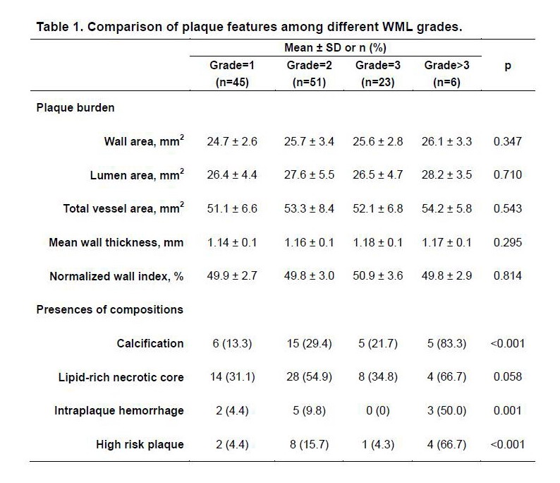
Table 1. Comparison of plaque features among different WML grades.
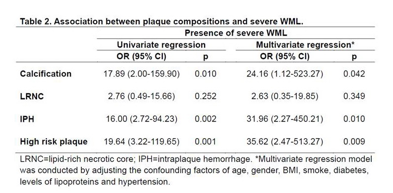
Table 2. Association between plaque compositions and severe WML.
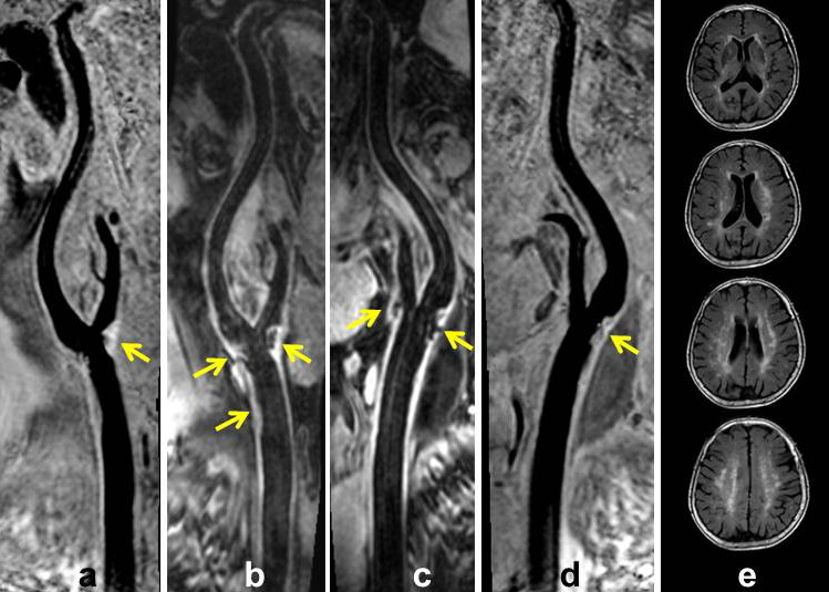
Figure 1. The MR images are from an 80 years old women. a and b represent SNAP and MERGE images for right carotid artery, and c and d represent MERGE and SNAP images for left carotid artery, respectively. Multiple atherosclerotic plaques (yellow arrows) were detected in bilateral carotid arteries. High risk plaque with IPH cahracterized by hyperintense on SNAP image (a, yellow arow) was found in right carotid artery. Severe white matter lesions (WML=4) can be seen on FLAIR images (e) of the same subject.