0465
Joint Virtual Coil Reconstruction with Background Phase Matching for Highly Accelerated Diffusion Echo-Planar Imaging1Department of Biomedical Engineering, Zhejiang University, Hangzhou, China, 2Athinoula A. Martinos Center for Biomedical Imaging, Massachusetts General Hospital, Charlestown, MA, United States, 3Shenzhen Institutes of Advanced Technology, Chinese Academy of Sciences, Shenzhen, China, 4Harvard-MIT Health Sciences and Technology, MIT, Cambridge, MA, United States
Synopsis
We proposed a joint-virtual-coil (VC) acquisition/reconstruction method to improve accelerated single-shot EPI (SS-EPI) in diffusion imaging (DI). A background phase correction scheme for matching the phase of reference training data with accelerated diffusion-weighted data was developed for robust reconstruction. Additional Gy prewinding-blips were added to the EPIs, to create complementary shifted-ky sampling strategy across TRs, which help better utilizes smooth-phase and joint-information priors in the joint-virtual-coil (jVC) reconstruction. The proposed method was demonstrated in highly-accelerated DI with SS-EPI and extended to generalized slice dithered enhanced resolution (gSlider) acquisition to achieve efficient high-resolution DI.
Introduction
Diffusion imaging (DI) with single-shot EPI (SS-EPI) is widely used for clinical and neuroscience applications. To reduce acquisition time and mitigate distortions, in-plane and slice accelerations are applied(1,2), where total acceleration is typically limited to 4-6 fold to minimize g-factor noise (e.g. Rinplane MB=2x3). The VC concept has been demonstrated to enable higher in-plane and multi-band accelerations in a number of applications (3–6). However, its usage in DI has been restricted by shot-to-shot background phase variations of DI, which violates VC assumption. To overcome this, we propose a VC reconstruction framework with background phase matching and demonstrates its utility in 9-fold accelerated DI (Rinplane MB=3x3). Furthermore, we combined joint-reconstruction methods (7–9) with VC concept (jVC) for joint-reconstruction across five RF-encoded volumes in gSlider-EPI(10). Shifted-ky sampling strategy was implemented to improve VC and jVC reconstructions.Methods
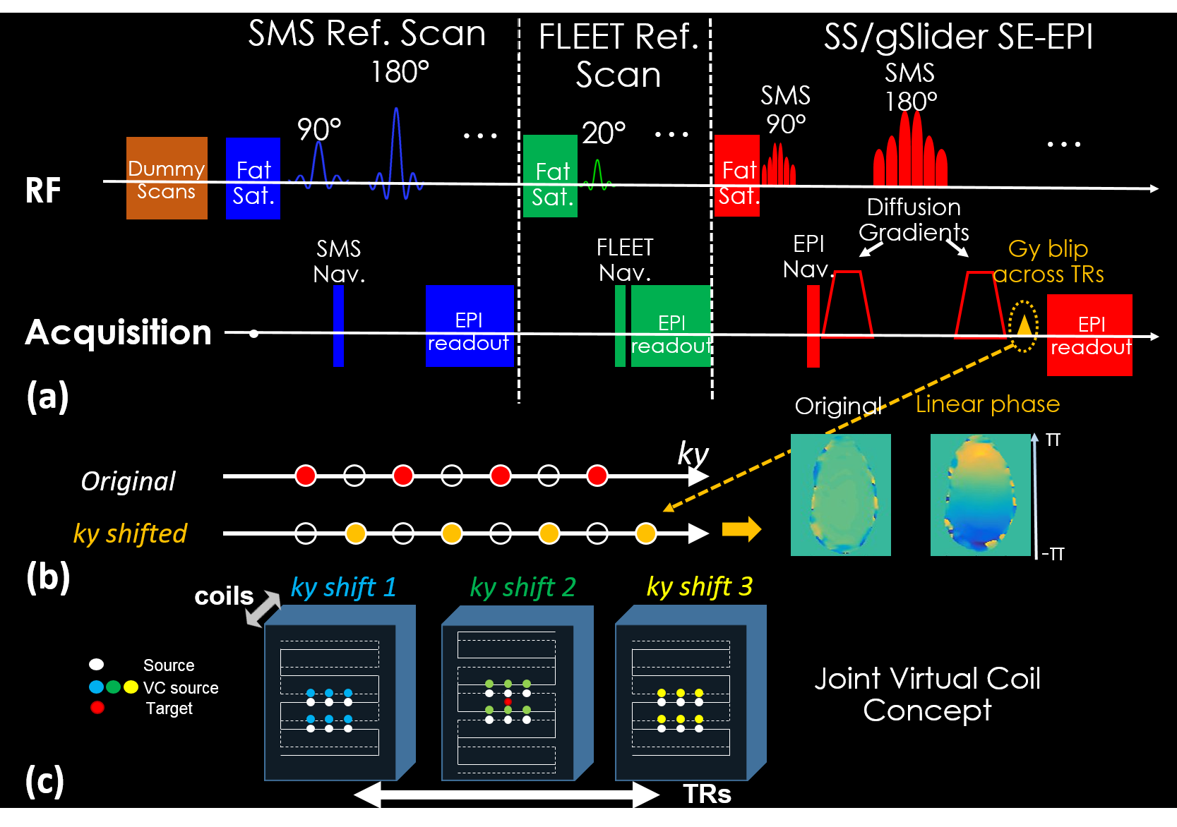
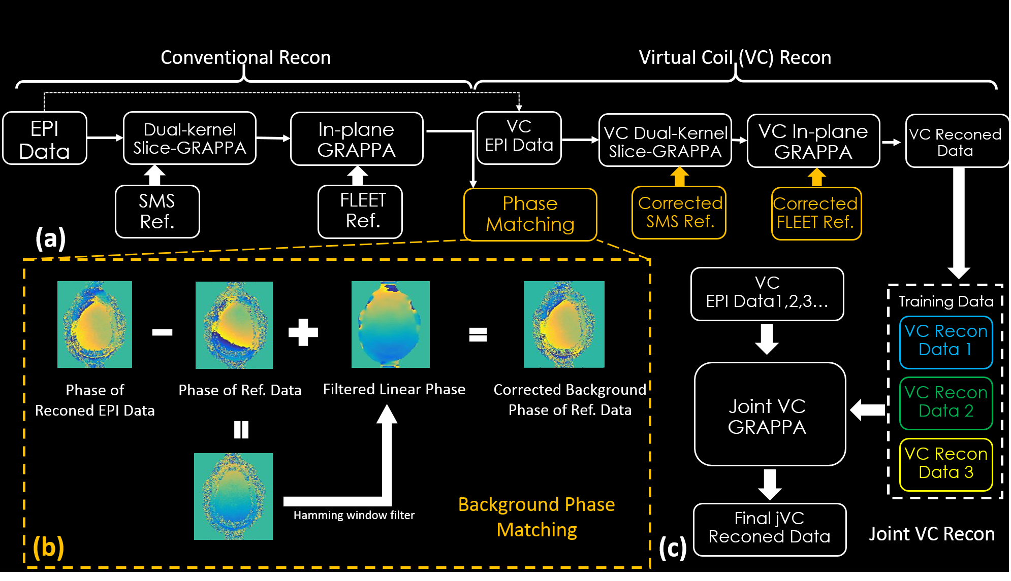
Fig.1a shows the sequence diagram, where different Gy prewinding-blips are added across TRs to generate differing linear image phases or ky shifts (Fig.1b). Fig.1c shows the benefits of such ky shifts in creating more unique k-space source points for VC and JVC reconstructions. Fig.2a shows the flowchart of VC reconstruction, where conventional reconstructed EPI data are used for the phase matching process, to add diffusion-direction-specific background phase into the training data of VC-GRAPPA (Fig.2b). The under-sampled raw data are then re-reconstructed using VC-GRAPPA, where a set of VC-GRAPPA kernel weights is created specifically for each diffusion weighted acquisition/direction. For jVC-GRAPPA reconstruction, the multi-kyshift VC-reconstructed data from different TRs are utilized as training data, and the under-sampled raw EPI data are then re-reconstructed again by the estimated weights.
Data: the following were acquired using a Siemens Prisma 3T Scanner:
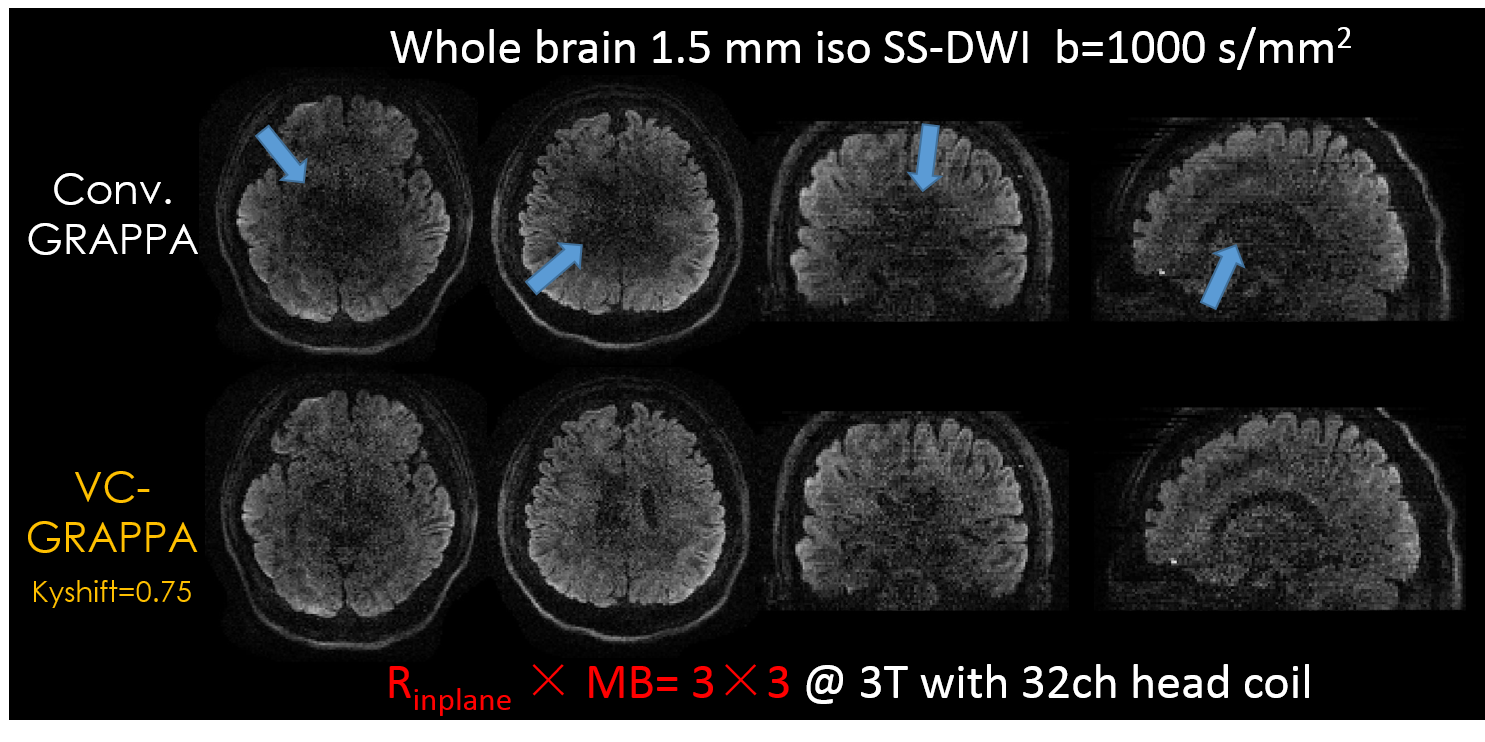
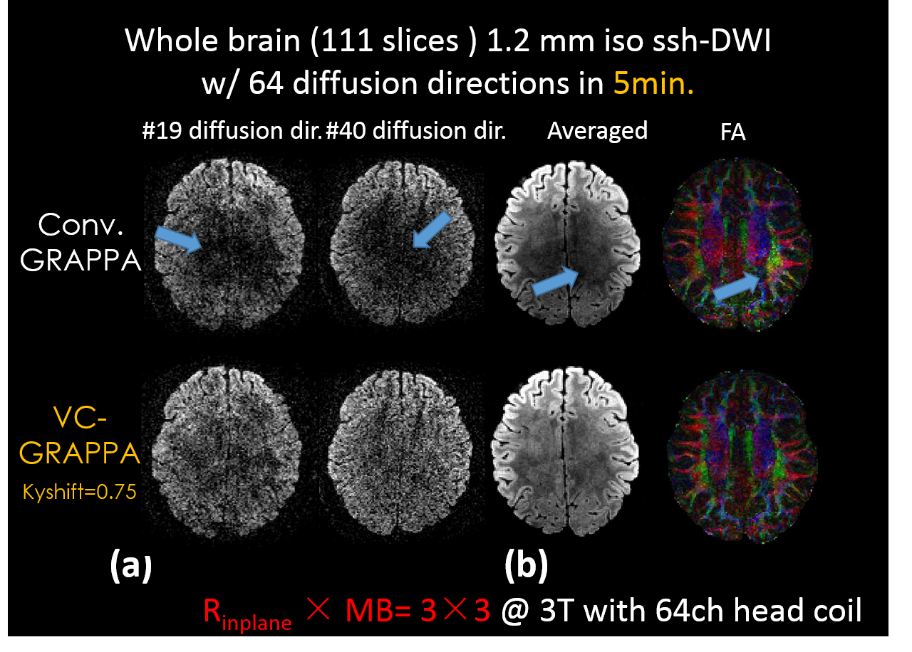
i) SS-EPI: 1.5mm and 1.2mm isotropic datasets, both with Rinplane MB=3x3, FOV: 230x230mm2, b=1000s/mm2, 64-directions, with constant 0.75 ky-shift across TRs. Total of 93 and 111 slices were acquired for whole-brain coverage at TE/TR/Tacq= 69ms/3.3s/4min and 78ms/4.4s/5min, respectively.
ii) gSlider-EPI: whole-brain 0.86mm isotropic, RinplanexMBxgSlider=4x2x5, FOV: 220x220x129mm2, b=1000s/mm2, 30-directions, Tacq12min. For each direction, 5 RF-encoded thin-slab volumes (4.3mm-slabs) are acquired sequentially and combined to reconstruct 0.86mm slices. Ky-shifts are looped by 1, 2 and 3 across TRs (RF-encodings).
Reconstructions: VC-GRAPPA was applied to SS-EPI, and both jVC- and VC-GRAPPA were applied to gSlider-EPI. For jVC, three consecutive RF-encodings with ky-shift of 1, 2 and 3 are jointly reconstructed. Reconstructions were compared to conventional-GRAPPA.
Results
Figure 3 and Fig. 4(a) show that VC-GRAPPA improves the quality of both 1.5 and 1.2mm isotropic datasets at high acceleration factors, where conventional reconstruction results in undesirable dark patches at the center of images. The blue arrows highlight image structures that were successfully recovered through VC reconstruction. Figure 4(b) show the 64-directions averaged-DWIs and colored-FA maps obtained from conventional and VC-GRAPPA reconstructions of the 1.2mm data. The results demonstrate that both averaged-DWI and FA-map were improved through VC-GRAPPA, with significant reduction in artifacts and g-factor noise for this 9-fold accelerated acquisition. The total acquisition time was only 5min, and longer scan across more directions could improve the SNR of this high-isotropic resolution acquisition further still.
Figure 5 shows that jVC-GRAPPA can be used to improve the reconstruction of gSlider data, where (a) shows the reconstructed RF-encoded thin-slabs (3 out of 5 shown) and (b) shows representative 2 of 5 resolved slices at 0.86 mm slice thickness. It can be observed that joint-reconstruction across slab-encoding improves reconstruction quality without smoothing away structural differences that exist across the slices within each slab.
Discussion
In this study, VC and jVC reconstructions were employed to achieved high-quality reconstructions at 8-9 fold acceleration in high resolution diffusion imaging. Complementary shifted-ky sampling strategy across TRs was used to better take advantage of smooth-phase and joint-information priors in the reconstruction. The proposed framework, which utilizes the conjugate phase information, can also be easily incorporated to SENSE(11) based single and multi-shot diffusion reconstructions (e.g., MUSE(12), low rank(13) and MUSSELS(14)), to effectively double the number of coil channels for reconstruction.Acknowledgements
This work was supported in part by NIH research grants: R01EB020613, R01EB019437, R24MH106096, P41EB015896, and the shared instrumentation grants: S10RR023401, S10RR019307, S10RR019254, S10RR023043References
1. Bammer R, Keeling SL, Augustin M, Pruessmann KP, Wolf R, Stollberger R, Hartung H-P, Fazekas F. Improved diffusion-weighted single-shot echo-planar imaging (EPI) in stroke using sensitivity encoding (SENSE). Magn. Reson. Med.2001;46:548–554.
2. Setsompop K, Cohen-Adad J, Gagoski BA, Raij T, Yendiki A, Keil B, Wedeen VJ, Wald LL. Improving diffusion MRI using simultaneous multi-slice echo planar imaging. Neuroimage 2012;63:569–580.
3. Blaimer M, Choli M, Jakob PM, Griswold MA, Breuer FA. Multiband phase-constrained parallel MRI. Magn. Reson. Med. 2013;69:974–980.
4. Blaimer M, Gutberlet M, Kellman P, Breuer FA, Köstler H, Griswold MA. Virtual coil concept for improved parallel MRI employing conjugate symmetric signals. Magn. Reson. Med.2009;61:93–102.
5. Blaimer M, Jakob PM, Breuer FA. Regularization method for phase-constrained parallel MRI. Magn. Reson. Med. 2014;72:166–171.
6. Kettinger AO, Kannengiesser SAR, Breuer FA, Vidnyanszky Z, Blaimer M. Controlling the object phase for g-factor reduction in phase-Constrained parallel MRI using spatially selective RF pulses. Magn. Reson. Med. 2017. doi: 10.1002/mrm.26890.
7. Huang F, Akao J, Vijayakumar S, Duensing GR, Limkeman M. k-t GRAPPA: Ak-space implementation for dynamic MRI with high reduction factor. Magn. Reson. Med. 2005;54:1172–1184.
8. Çukur T, Lustig M, Nishimura DG. Multiple-profile homogeneous image combination: Application to phase-cycled SSFP and multicoil imaging. Magn. Reson. Med. 2008;60:732–738.
9. Chen F, Shi X, Chen S, Johnson EM, Chen B, Ren G, Wei X, Wang S, Ying K. Accelerated model-based proton resonance frequency shift temperature mapping using echo-based GRAPPA reconstruction. Magn. Reson. Imaging 2015;33:240–245.
10. Setsompop K, Fan Q, Stockmann J, et al. High-resolution in vivo diffusion imaging of the human brain with generalized slice dithered enhanced resolution: Simultaneous multislice (gSlider-SMS). Magn. Reson. Med. 2017. doi: 10.1002/mrm.26653.
11. Pruessmann KP, Weiger M, Scheidegger MB, Boesiger P. SENSE: Sensitivity encoding for fast MRI. Magn. Reson. Med. 1999;42:952–962.
12. Chen N kuei, Guidon A, Chang HC, Song AW. A robust multi-shot scan strategy for high-resolution diffusion weighted MRI enabled by multiplexed sensitivity-encoding (MUSE). Neuroimage 2013;72:41–47.
13. Liao C, Chen Y, Cao X, Chen S, He H, Mani M, Jacob M, Magnotta V, Zhong J. Efficient parallel reconstruction for high resolution multishot spiral diffusion data with low rank constraint. Magn. Reson. Med.2017: 1359-1366.
14. Mani M, Jacob M, Kelley D, Magnotta V. Multi-shot sensitivity-encoded diffusion data recovery using structured low-rank matrix completion (MUSSELS). Magn. Reson. Med.2017;78:494–507.
Figures