0407
Development and Proof of Principle for Free Radical Imaging with the Novel Field-cycling in vivo DNP-MRI1Innovation Center for Medical Redox Navigation, Kyushu University, Fukuoka, Japan, 2Pharmaceutical Sciences, University of Shizuoka, Shizuoka, Japan, 3Osaka University, Suita, Japan, 4University of Yamanashi, Yamanashi, Japan, 5Kinjo Gakuin University, Nagoya, Japan, 6Fuji Electric Co., Ltd, Tokyo, Japan
Synopsis
In vivo DNP-MRI was reported as a new imaging method for free radical species in vivo1, and the field-cycling was utilized to overcome the large difference between electron and nuclear spin resonances2,3. Unfortunately the development of the clinical DNP-MRI has severe difficulties due to less applicability.
Here, we developed the novel field-cycling DNP-MRI for clinical trial by rotating the magnets for MRI (0.3T) and ESR (4.7mT), and the free radicals placed under the palm of volunteers were visualized, superimposed on the anatomical MRI image, and demonstrated the proof of principle for free radical imaging with the newly developed field-cycling DNP-MRI.
INTRODUCTION
In vivo DNP-MRI is a new imaging method for free radical species in vivo1, and its main advantage is the high spatiotemporal resolution, which rivals that of conventional MRI. Due to Overhauser effect, the proton interacted with free radical should be 330 times higher polarized than that of thermal equilibrium by inducing the electron spin resonance of free radical. The field-cycling in DNP-MRI is very effective to increase the magnetic field of MRI2,3, but the clinical trials of in vivo DNP-MRI have not been reported because of smaller observation space and less capabilities of the field-cycling system. Here, we demonstrate the development of a novel field-cycling DNP-MRI which enables us for clinical application, and the proof of principle for free radical imaging in human palm.METHODS
The stable free radical, Cmp and a raw chicken wing were purchased from Sigma Aldrich and the supermarket, respectively. MRI console, MRI and ESR power amplifiers were purchased from Japan Redox. Co. ltd. NMR coil in the solenoid configuration for transmission and receiving and field gradient coils were purchased from Neomax Co. ltd. The surface coil for ESR irradiation was prepared in-house as reported previously5. The DNP-MRI data were obtained as described in the figure captions, and analyzed using the Image J software package (http://rsb.info.nih.gov/ij/).RESULTS
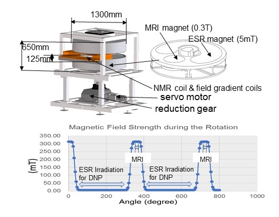
The field-cycling was performed by rotating the jacket composed with a permanent MRI magnet (310mT) and an arc-shaped permanent ESR magnet (4.7mT) at the maximum of 0.5rps with a servomotor. The field-cycling system was connected to a conventional MRI system.
Figure 1 demonstrates 3D-blueprints of the novel field-cycling DNP-MRI and the external magnetic fields B0 for EPR irradiation and MRI, which are cycling stably between 4.7mT and 310mT. The detection field has homogeneity better than 20ppm over 120x120x90mm volume and the stability within +/- 10ppm by controlling the temperature of magnets within 1.0 degree Celsius. In the field-cycling DNP-MRI, the NMR coil and the field gradient coils are fixed in the frame, and the movement of the external magnets causes the electromagnetic induction in the field gradient coils. The conditions of MRI were optimized to avoid the eddy current and the electromagnetic induction of the field gradient coils under the rotation of external magnets.
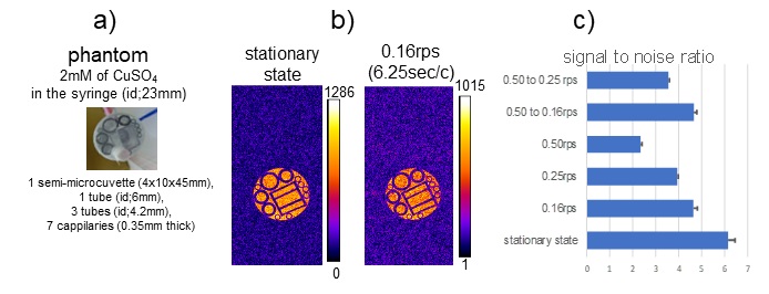
As shown in figure 2, the clear MRI image could be obtained under rotating the external magnets by adjusting exactly the timing of MRI pulse-sequence. The signal-to-noise ratio of the images under rotating the external magnets depended on the rotation-speed, and the rotation at 0.16 and 0.25 rps decreased 25% and 37% of the SN ratio from that of the stationary state, respectively.
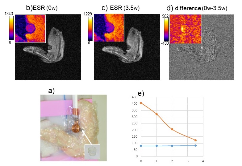
The small cup filled with 2mM of the stable free radical, Cmp was implanted at a raw chicken wing, and the images with and without ESR irradiation were obtained using the field-cycling DNP-MRI (figure 3). The ESR irradiation through the spiral surface coil clearly decreased the image intensity, the difference between the images with and without ESR irradiation depended linearly on the power of ESR irradiation and the image of free radical was obtained by subtracting the two images.
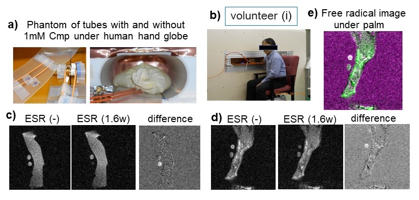
Next, we obtained the proof of principle for free radical imaging using the developed field-cycling DNP-MRI (Figure 4). The phantom consisted of 2 Agilent tubes containing either water or 1mM Cmp was located under the human hand globe (Figure 4b), and the images with and without ESR irradiation were obtained using in vivo DNP-MRI (Figure 4c). The subtraction of the image with ESR irradiation from that without irradiation gave a clear image of the free radical. As the first clinical trial of in vivo field-cycling DNP-MRI, the free radical located under the palm of healthy volunteers was imaged (Figure 4b, d). The heating by the ESR irradiation was measured to be less than 2 degree Celsius. Figure 4e demonstrates the free radical image located under the palm of healthy volunteer.
CONCLUSION
We developed the novel field-cycling DNP-MRI for clinical trial, and demonstrated the free radical imaging with less than 0.5 mm of the spatial resolution. The image of free radical placed under the palm of healthy volunteer was superimposed on the anatomical MRI image. Previously, we succeeded in the simultaneous images of radicals labeled with 14N and 15N nuclei by changing the ESR irradiation in DNP-MRI4, and then demonstrated the imaging of free radical intermediates produced from endogenous molecules, such as CoQ10 and FAD6. The intrinsic radical, melanin, might be also visualized. These facts indicate that the newly developed DNP-MRI could perform the functional/metabolic imaging by visualizing intrinsic radicals, redox intermediates and/or metal enzymes.Acknowledgements
We thank Mr. Ohkubo at Meiko Inc. Dr. Hyodo at Gifu University, and Dr. Naganuma at Japan Redox Inc. for carrying out the experiments. This work was jointly supported by the "Formulation of Advanced Collaborative Medical Innovation Center" as Special Coordination Funds for Promoting Science and Technology, Ministry of Education, Culture, Sports, Science and Technology, Japan and by KAKENHI Grant Numbers 16H05113 (H.U.) from the Japan Society for the Promotion of Science.References
1. Lurie DJ, Bussell DM, Bell LH, et al. Proton-electron double magnetic resonance imaging of free radical solutions. J. Magn. Reson. 1988;76:366-370.
2. Lurie DJ, Hutchison JMS, Bell LH., et al. Field-cycled proton electron double-resonance imaging of free-radicals in large aqueous samples. J. Magn. Reson. 1989;84(2):431-437.
3. Lurie DJ, Davies GR, Foster MA, et al. Field-cycled PEDRI imaging of free radicals with detection at 450mT. Magn Reson Imaging. 2005;23;175-181.
4. Utsumi H, Yamada K, Ichikawa K, et al. Simultaneous molecular imaging of redox reactions monitored by Overhauser-enhanced MRI with 14N- and 15N-labeled nitroxyl radicals. Proc Natl Acad Sci U S A, 2006;103(5):1463-1468.
5. Matsumoto S, Yamada K, Hirata H, et al. Advantageous application of a surface coil to EPR irradiation in Overhauser-enhanced MRI. Magn Reson Med 2007;57: 806-811.
6. Hyodo F, Ito S, Yasukawa K, et al. Simultaneous and spectroscopic redox molecular imaging of multiple free radical intermediates using dynamic nuclear polarization-magnetic resonance imaging. Anal Chem, 2014;86(15):7234-7238.
Figures