0392
Cortical feedback to superficial layers of V1 contains predictive scene information.1Institute of Neuroscience & Psychology, University of Glasgow, Glasgow, United Kingdom, 2Faculty of Psychology and Neuroscience, Maastricht University, Maastricht, Netherlands
Synopsis
A central characteristic of brain function is the ability to merge sensory input with internal representations of the world, but relatively little is known about cortical feedback channels that facilitate internal representations. We blocked feedforward input to subsections of human primary visual cortex by occluding one quarter of the visual field while participants viewed 384 real-world scenes. Using high-resolution 7T fMRI, we show that superficial layers of V1 exhibit predictive response properties unique from those associated with V1 feedforward processing. Our findings suggest that feedback to superficial layers of V1 provides neurons with contextual information not available via feedforward input.
Introduction
A central characteristic of brain function is the ability to merge sensory input with internal representations of the world1. Many experiments have furthered our understanding of the features that modulate early sensory areas through feedforward channels, but relatively little is known about the feature space that drives cortical feedback channels. Accessing and describing these internal feedback information channels will provide information about the structure of internal representations and is central to fully understanding neural computations2.
To study feedback, one must disentangle feedforward and non-feedforward sources of input to an area, which involves independent stimulation or inactivation of feedback and feedforward pathways. This can be achieved by pairing single- or multiunit recordings with electrical stimulation, pharmacological intervention, cooling or optogenetics3-4, but these methodologies are generally too invasive for studying the healthy human brain. A non-invasive strategy to measure feedback is to homogenize feedforward input using visual occlusion while recording with functional MRI. Feedback influences wide-spread dendritic activity, but does not necessarily lead to spiking activity in cells1. Functional MRI is therefore particularly powerful for studying feedback because it is sensitive to energy-consumption associated with dendritic spatially-specific activity5.
Here, we asked whether feedforward sensory input and predictive feedback signals could be read out from different layers of cortex, and what types of information these two inputs to early visual cortical neurons contain. By utilizing an occlusion paradigm in combination with recordings from high-resolution fMRI, we compared depth-specific V1 responses to sensory- and prediction-based computational models and to high-level scene category information.
Methods
We blocked feedforward input to subsections of human retinotopic visual cortex by occluding one quarter of the visual field6-7. Participants viewed 384 real-world scenes (192 occluded and 192 non-occluded scenes) in 5 scanning sessions while we recorded V1 responses using high-resolution 7T fMRI. Data were acquired using the CMRR multi-band accelerated 2D Gradient Echo EPI sequence8 (resolution: 0.80mm isotropic; number of slices: 56; slice spacing: 0mm; TR: 2000ms; TE: 25ms; multi-band factor: 2; iPAT factor: 3; flip angle: 75º).
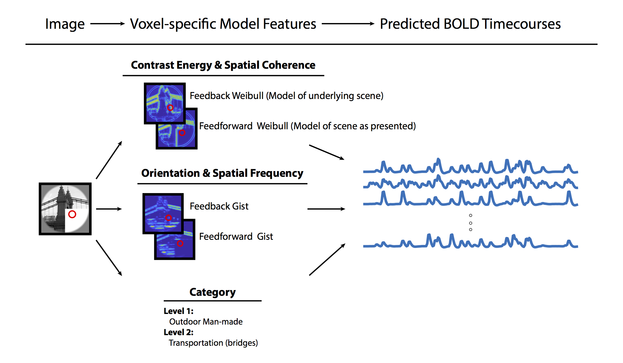
For each V1 voxel, we calculated a population receptive field9 and defined voxel-specific feedforward and feedback models with varying levels of computational complexity (Figure 1). Model information included contrast energy and spatial coherence (Weibull model10), orientation and spatial frequency (Gist model11), and high-level category information (SUN database hierarchy12). V1 neurons in the lower-right scene quadrant received meaningful sensory input during non-occluded scene presentations, but not during occluded scene presentations. Therefore, feedforward models were defined as responses related to scenes as they were presented. Feedback models were defined as responses related to scenes as they would be predicted (i.e. responding as if the scene was not occluded). Responses from each of these models were convolved with a hemodynamic response function to create model time courses. We fitted model time courses to fMRI data and calculated the unique variance explained by models using semi-partial correlation statistics. We defined a voxel’s tuning to feedforward and feedback signals as the ratio of unique variance explained by sensory feedforward and predictive feedback models.
Results
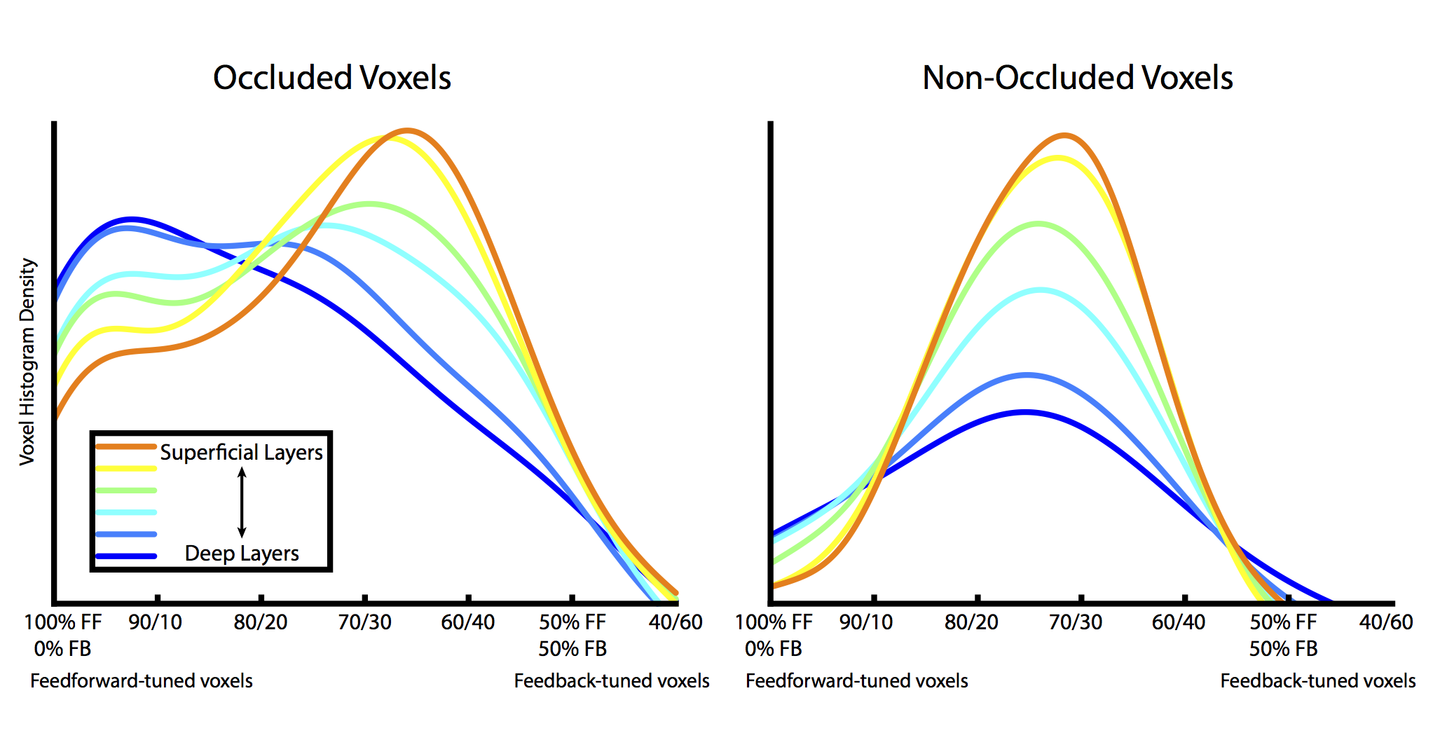
Results from cortical depth-specific voxel tuning analyses are shown in Figure 2. For six evenly-spaced cortical depths (between 10% and 90% of the total cortical thickness), we show the probability density of voxel tuning to feedforward and feedback models. Voxels in deep layers of cortex are more likely to be heavily tuned to feedforward signals. However, tuning shifts toward feedback signals in superficial layers, where many feedback connections terminate in cortex1. Interestingly, these depth-dependent tuning properties are only observable in occluded voxels (where feedforward inputs are blocked), as voxels in non-occluded areas have similar tuning through all cortical depths (Figure 2, right side).Conclusion
Superficial layers of V1 exhibit predictive (and higher-level) response properties unique from the orientation and spatial frequency properties typically associated with V1 feedforward processing. Our findings suggest that feedback connections terminating in superficial layers provide V1 neurons with contextual information not available via localized feedforward input.Acknowledgements
This work was supported by the European Research Council (ERC StG 2012_311751-BrainReadFBPredCode) and the Human Brain Project (EU grant 604102), both awarded to L.M.References
1. Larkum M. A cellular mechanism for cortical associations: an organizing principle for the cerebral cortex. Trends in Neuroscience, 2013;36(3):141-151.
2. Petro LS, Muckli L. The laminar integration of sensory inputs with feedback signals in human cortex. Brain and Cognition, 2016;112:54-57.
3. Muckli L, Petro LS. Network interactions: non-geniculate input to V1. Current Opinion in Neurobiology, 2013;23:195–201.
4. Petro LS, Vizioli L, Muckli L. Contributions of cortical feedback to sensory processing in primary visual cortex. Frontiers in Psychology, 2014;5:1223.
5. Logothetis NK. The ins and outs of fmri signals. Nat Neurosci. 2007;10(10):1230–2.
6. Muckli L, De Martino F, Vizioli L, et al. Contextual feedback to superficial layers of V1. Current Biology. 2015;25(20):2690-5.
7. Smith FW, Muckli L. Nonstimulated early visual areas carry information about surrounding context. Proceedings of the National Academy of Sciences USA, 2010;107:20099-103.
8. Moeller S, Yacoub E, Olman CA, et al. Multiband multislice GE-EPI at 7 tesla, with 16-fold acceleration using partial parallel imaging with application to high spatial and temporal whole-brain fMRI. Magn Reson Med. 2010;63(5):1144-1153.
9. Dumoulin SO, Wandell BA. Population receptive field estimates in human visual cortex. Neuroimage, 2008;39:647-660.
10. Groen I, Ghebreab S, Prins H, et al. From image statistics to scene gist: evoked neural activity reveals transition from low-level natural image structure to scene category. J Neurosci, 2013;33(48):18814–24.
11. Oliva A, Torralba A. Modeling the shape of the scene: A holistic representation of the spatial envelope. Int. J. Comput. Vision, 2001;42(3):145–175.
12. Xiao J, Hays J, Ehinger KA, et al. Sun database: Large-scale scene recognition from abbey to zoo. In CVPR, pages 3485–3492. IEEE Computer Society, 2010.
Figures