0386
Parallel Transmit Excitation Pulses for Shuttered Echo Planar Imaging1Biomedical Engineering, Vanderbilt University, Nashville, TN, United States, 2Institute of Imaging Science, Vanderbilt University, Nashville, TN, United States, 3Radiology, Vanderbilt University, Nashville, TN, United States
Synopsis
A parallel transmission based shutter excitation is proposed for multishot EPI that overcomes its sensitivity to motion and dynamic phase changes between shots. The g-factor performance, flip angle error, and SAR are characterized as a function of the number of transmit coils, and compared to ideal single-channel excitation.
Introduction
Echo planar imaging (EPI) is the most widely used pulse sequence for rapid functional, diffusion, and perfusion imaging, and has been the focus of recent developments to increase its speed and spatial resolution [1]. However, increased resolution requires longer readouts which extend echo times and reduce functional contrast and SNR at 7 Tesla, while increasing geometric distortions and blurring. Multishot EPI is a classic method to increase spatial resolution without increasing readout durations, but is sensitive to motion and dynamic phase changes between shots. To address this, ‘in-plane multiband’ or ‘shuttered EPI’ has recently been proposed and validated in 3T breast diffusion-weighted imaging [2]. The method works by imaging a set of spatially disjoint shutters in each shot, and then separately reconstructing and motion-correcting those images and combining them using a phase-insensitive image combination. However, its application to high-resolution neuroimaging is limited by the long excitation pulse durations required to simultaneously excite a slice and a set of in-plane shutters. Here we describe and validate an approach to shorten the pulse durations using parallel transmission at 7 Tesla.Method
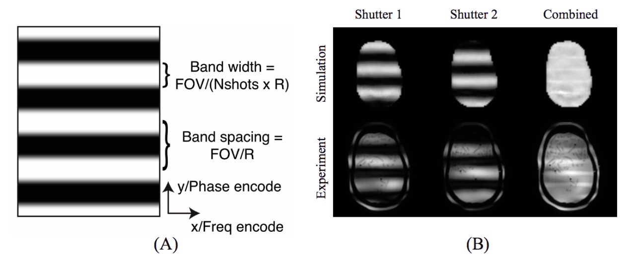
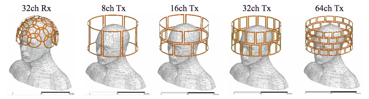
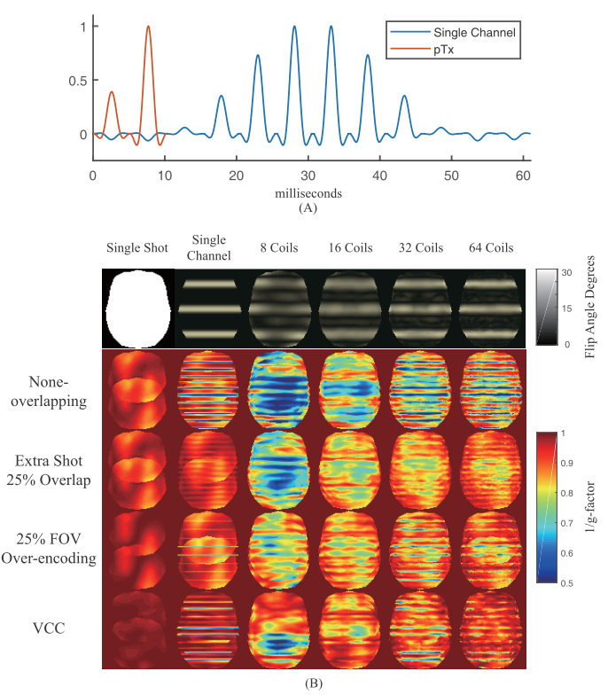
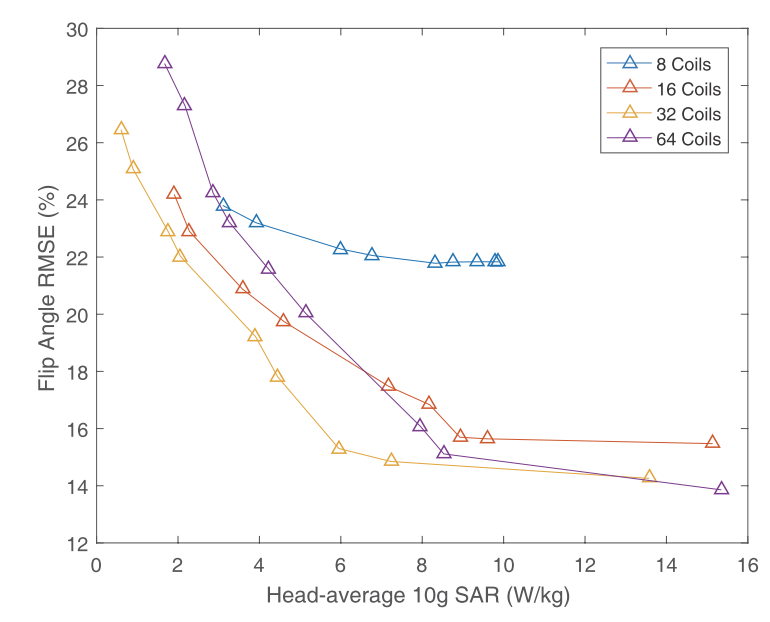
Slice-selective shutter excitation pulses were designed using the small-tip parallel transmit (pTx) spokes pulse design method of Ref. 3, which is an iterative algorithm that alternately updates RF pulse weights, target phase patterns, and x-y gradient blip areas between slice-selective subpulses. The in-plane desired excitation pattern was set to a shutter flip angle pattern (Fig 1a), and the RF pulses were designed to minimize flip angle error while regularizing for low head-average 10g SAR. Studies were performed to validate the pulses and evaluate their performance. First, an experiment was performed on a 7T MRI scanner (Philips Healthcare, Best, Netherlands) on a 3D-printed human head phantom [4] with 8 channel transmit and 32 channel receive head coils. MRCodeTool (MRCode BV, Netherlands) was used for B0 shimming, B1+ map processing, and RF pulse interfacing. Pulses were designed to excite two complementary patterns with 4 shutters across a 22.4 cm FOV, for a 2 shot shuttered EPI acquisition with acceleration factor R = 4, shutter time-bandwidth-product 3, Fig 1a. The pulses had 5 spokes, 4.8 ms duration and excited 3 mm-thick slices with a 1 degree flip angle Their patterns were imaged with a gradient-recalled echo sequence with TE/TR = 4.4/10 ms. Next, a simulation study was performed to evaluate shuttered EPI g-factor maps with different numbers of transmit coils, using 2-spoke pulses. Four transmit arrays and a 32-channel receive array were simulated using HFSS (Ansys Inc, Canonsburg, PA) (Fig 2) with a 3D human model. Multiple strategies were also compared to mitigate g-factor losses between the shutters, which included acquiring an extra shot with all shutter patterns spaced closer together, 25% FOV-oversampling in each shot, and virtual conjugate coil (VCC) reconstruction [5]. The pulses’ SARs were calculated based on a high-resolution 7T gradient echo fMRI sequence protocol (90 flip angle, 2 RF spokes, 4s TR, 100 slices, 0.6 mm slice thickness) and were compared between the transmit coil arrays and across a range of average 10g SAR regularization parameters in the pulse design.Results
The predicted and achieved experimental 2-shot patterns excited by the pTx pulse are shown in Fig 1b. The target shutters were excited as expected, with 2.8 cm widths and 5.6 cm spacings. Fig 3a shows that a two-spoke pTx shutter pulse for a 1 mm slice thickness is 6x shorter than a single channel pulse that excites the same nominal pattern. Fig 3b illustrates the various methods that mitigate g-factor losses between the shutters, and their effectiveness improves with an increasing number of transmit channels. Fig 4 shows the relationship between excitation flip angle error and regularized 10g SAR in the brain. The excitation error and SAR are both reduced by increasing the number of transmit coils, up to 32 coils. Overall, while 64 coils gives the best 1/g maps, 32 channels achieves the best tradeoff between excitation error and SAR.Discussion & Conclusion
pTx-based shutter excitation pulses for shuttered multishot EPI were proposed and characterized in terms of their g-factor performance compared to ideal single-channel excitation, and as a function of the number of transmit coils. A large number of transmit channels enables short pulse durations with low SAR, while maintaining the desired shutter patterns. Different strategies were demonstrated to mitigate g-factor losses between the shutters. A many-coil pTx array can be driven with array-compressed pTx [6] to minimize the number of power amplifiers needed for these excitations. The proposed shutter excitation can also be combined with simultaneous multislice excitation [7], and can extended to large-tip-angle pTx pulses for refocusing in spin echo sequences [8].Acknowledgements
Funding by NIH R01 EB 016695.References
[1] D A Feinberg, S Moeller, S M Smith, E Auerbach, S Ramanna, M F Glasser, K L Miller, K Ugurbil, and E Yacoub. Multiplexed echo planar imaging for sub-second whole brain fMRI and fast diffusion imaging. PLoS One, 5(12):e15710, 2010.
[2] Taviani, V., Alley, M. T., Banerjee, S., Nishimura, D. G., Daniel, B. L., Vasanawala, S. S. and Hargreaves, B. A. (2017), High-resolution diffusion-weighted imaging of the breast with multiband 2D radiofrequency pulses and a generalized parallel imaging reconstruction. Magn. Reson. Med., 77: 209–220. doi:10.1002/mrm.26110.
[3] Grissom, W. A., Khalighi, M.-M., Sacolick, L. I., Rutt, B. K. and Vogel, M. W. (2012), Small-tip-angle spokes pulse design using interleaved greedy and local optimization methods. Magn Reson Med, 68: 1553–1562. doi:10.1002/mrm.24165.
[4] Khan, A. F., Drozd, J. J., Moreland, R. K., Ta, R. M., Borrie, M. J., Bartha, R. and and the Alzheimer's Disease Neuroimaging Initiative (2012), A novel MRI-compatible brain ventricle phantom for validation of segmentation and volumetry methods. J. Magn. Reson. Imaging, 36: 476–482. doi:10.1002/jmri.23612
[5] Kettinger, A. O., Kannengiesser, S. A. R., Breuer, F. A., Vidnyanszky, Z. and Blaimer, M. (2017), Controlling the object phase for g-factor reduction in phase-Constrained parallel MRI using spatially selective RF pulses. Magn. Reson. Med.. doi:10.1002/mrm.26890.
[6] Cao, Z., Yan, X. and Grissom, W. A. (2016), Array-compressed parallel transmit pulse design. Magn. Reson. Med., 76: 1158–1169. doi:10.1002/mrm.26020.
[7] Sharma, A., Bammer, R., Stenger, V. A. and Grissom, W. A. (2015), Low peak power multiband spokes pulses for B1+ inhomogeneity-compensated simultaneous multislice excitation in high field MRI. Magn. Reson. Med., 74: 747–755. doi:10.1002/mrm.25455.
[8] Cao, Z., Donahue, M. J., Ma, J. and Grissom, W. A. (2016), Joint design of large-tip-angle parallel RF pulses and blipped gradient trajectories. Magn. Reson. Med., 75: 1198–1208. doi:10.1002/mrm.25739.
Figures