0381
Fast imaging with ultrahigh isotropic resolution using partition-encoded simultaneous multi-slab (PRISM)1BRIC, UNC at Chapel Hill, Chapel Hill, NC, United States
Synopsis
Fast imaging with ultrahigh isotropic resolution has gained widespread interests but is technically challenging. The simultaneous multi-slice (SMS) and 3D imaging can hardly achieve high isotropic resolution without compromising temporal resolution or imaging contrast that needs long TR. A novel method, Partition-encoded Simultaneous Multi-slab (PRISM) imaging, was proposed to mitigate the physical constraints of thin slice excitation by applying partition encoding onto simultaneous multi-slab acquisition and provide ultrahigh isotropic resolution while maintaining the acceleration capability and TR flexibility. Using the PRISM technique, whole-brain fMRI with spatial resolution of 1mm isotropic and a temporal resolution of 2 seconds has been achieved.
Introduction
Fast MR imaging with ultrahigh isotropic resolution is desirable in many studies, such as the structural/functional architecture of cortical layers1-2 or within small subcortical nuclei3-4. The volumetric excitation of 3D imaging5-6 provides improved SNR efficiency and acceleration capability, and the slice thickness can be extremely thin and well-defined using partition encoding. However, the rapid volumetric excitation makes 3D imaging not suitable for the acquisitions that require a long repetition time (TR), such as diffusion imaging. On the other hand, simultaneous multislice (SMS) imaging excites multiple slices simultaneously7-8, reducing the acquisition time by the SMS factor and offering the flexibility of choosing a desired TR by setting the SMS factor properly. Nevertheless, the spatial resolution of SMS imaging along slice direction is limited owing to the physical constraints of slice excitation. The recently-proposed method, Generalized Slice Dithered Enhanced Resolution (gSlider), addresses this issue by RF encoding within the slab9. However, RF encoding is relatively more sensitive to field inhomogeniety and the regularization used during reconstruction results in spatial blurring9. The novel method, proposed herein, named Partition-encoded Simultaneous Multi-slab (PRISM), breaks the limit of excitation slice thickness and allows a higher slice acceleration rate than SMS factor based on the finding of a novel k-space sampling pattern for spatially noncontiguous slabs.Method
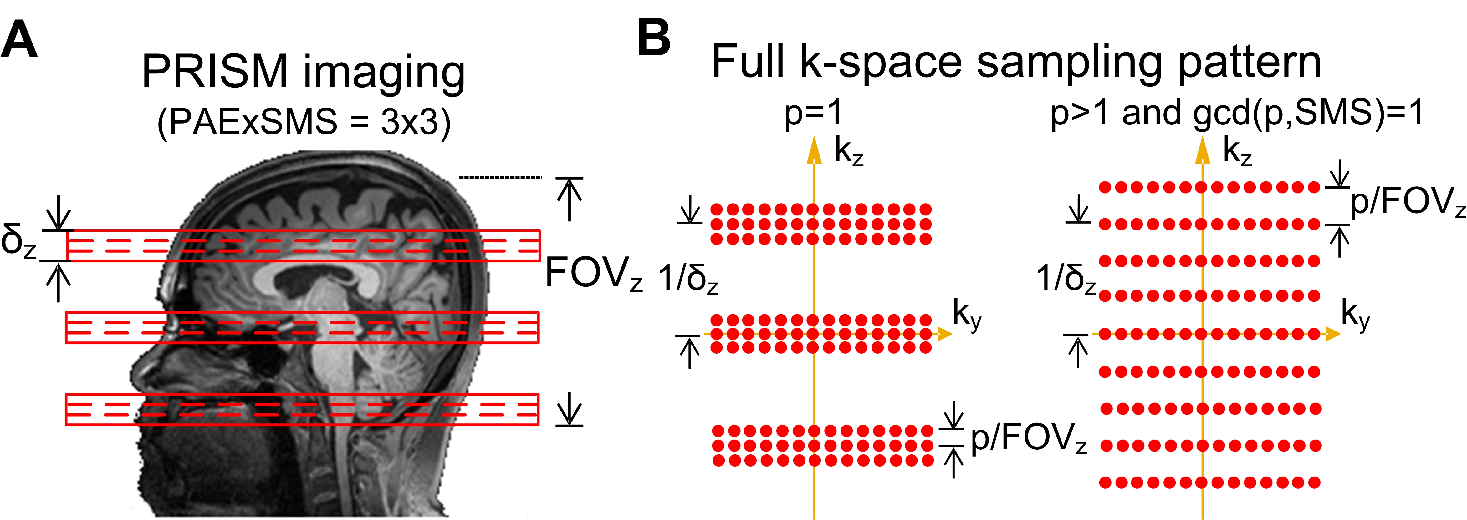
The proposed k-space sampling patterns of PRISM imaging are illustrated in Figure 1. Assuming a SMS factor of 3 and the number of partition encoding (PAE) of 3 (see Figure 1A), the sampling pattern would include three k-space bands and each band consist of three k-planes. The total number of data points in 3D k-space for PRISM is the same as that of SMS approach and therefore the temporal resolution is not compromised. The k-space distance between two adjacent bands is the reciprocal of the slab thickness. The k-space distance between adjacent planes within a band is p/FOVz, where p is a natural number. The orthogonality of PRISM encoding will be fulfilled as long as the k-space distance between bands is 1/δz and the distance between adjacent planes within a band is p/FOVz, where p is coprime to the SMS factor. With the k-space sampling pattern of p>1, the slice acceleration rate higher than SMS factor becomes feasible because the maximal PAE blip size is reduced.Results
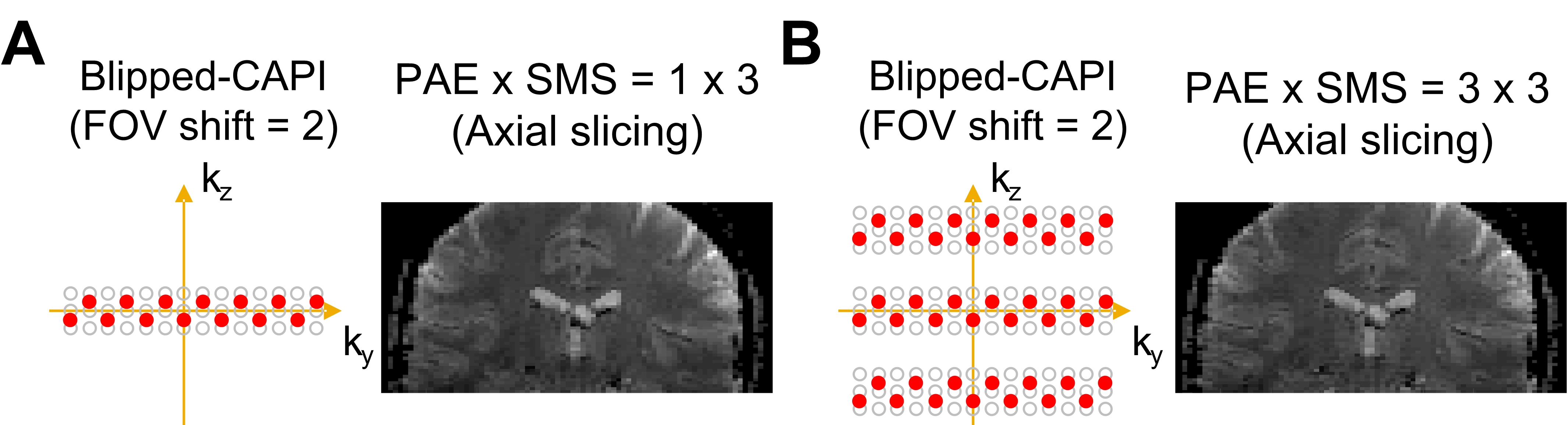
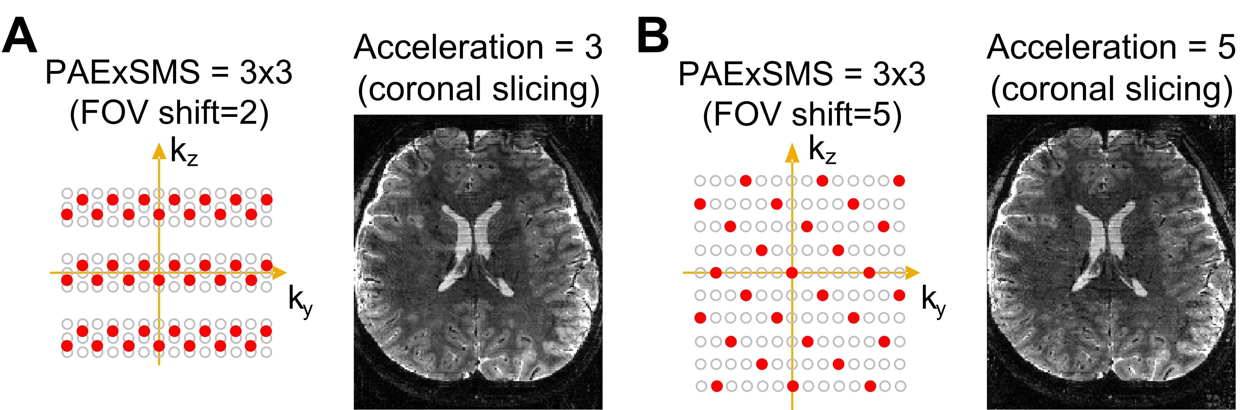
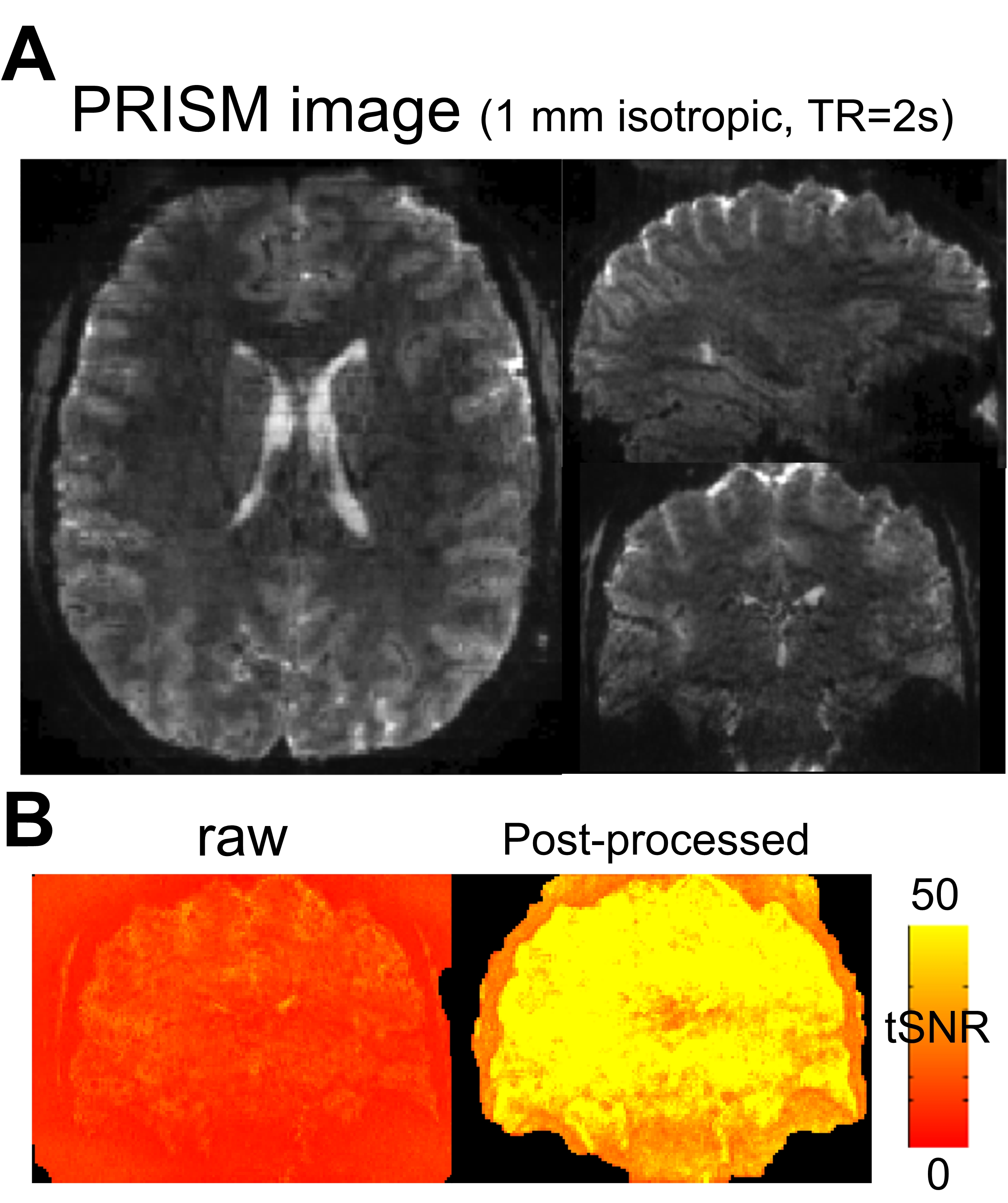
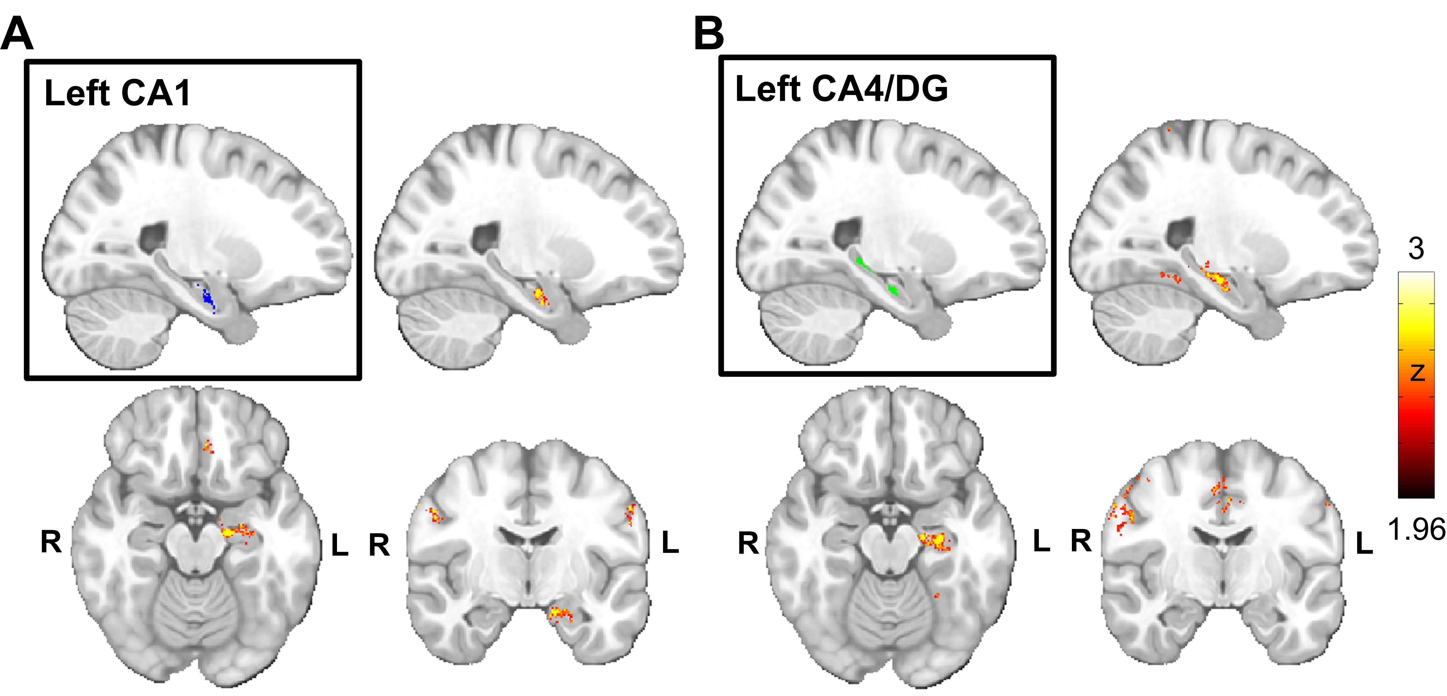
The left panels in Figure 2A and 2B illustrated the sampling patterns of blipped-CAIPI technique in order to reduce the g-factor penalty. The empirical SMS image with a slice thickness of 1.2 mm was shown in Figure 2A as the reference. Figure 2B showed the acquired PRISM image with the slab thickness of 3.6 mm and the number of PAE=3 (slice thickness = 1.2mm). The normalized mean-square-error (MSE) between SMS and PRISM images was 1.1%. Additionally, the simulation results in Figure 3A and 3B demonstrated the feasibility that the slice acceleration rate could be higher than SMS factor. The right panel in Figure 3A showed the eSPIRiT10-reconstructed PRISM image from the accelerated data of which both the SMS factor and acceleration rate were 3. Based on the sampling pattern illustrated in the right panel of Figure 1B, the slice acceleration rate of 5 can be achieved by employing the sampling pattern shown in the left panel of Figure 3B and a comparable image quality (normalized MSE=1.0%) was demonstrated in the right panel. For fMRI, we have achieved whole-brain acquisition with 1 mm isotropic resolution in 2 seconds at 7T, as shown in Figure 4A. The temporal SNR (tSNR) was improved from ~18 to ~74 by post-processing (see Figure 4B). The achievement enabled the study of cortical-hippocampal subfield interaction based on resting-state functional connectivity (rsFC). We recruited 8 healthy adults with age of 31.8±6.6 years old. The connectivity patterns associated with CA1 and with CA4/DG were shown to be distinct from each other (see Figure 5A and 5B).Discussion
Our novel PRISM technique will open a new avenue for high spatiotemporal-resolution imaging by the following features: 1) break the constraint of thin slice excitation while maintaining the flexibility of TR; 2) provide a higher acceleration rate than SMS factor without increasing the specific absorption rate (SAR). The second feature is particularly beneficial for diffusion imaging of which the SAR is generally high. Compared to 3D imaging, the PRISM imaging is also less vulnerable to head motion because of shorter encoding time. Finally, whole-brain fMRI acquisition with spatial resolution of 1mm isotropic in 2 seconds has been achieved using PRISM. The application of PRISM on the study of cortical-hippocampal subfield connectivity was also demonstrated.Acknowledgements
No acknowledgement found.References
1. Koopmans, P.J., M. Barth, and D.G. Norris, Layer-specific BOLD activation in human V1. Hum Brain Mapp, 2010. 31(9): p. 1297-304.
2. Polimeni, J.R., et al., Laminar analysis of 7T BOLD using an imposed spatial activation pattern in human V1. Neuroimage, 2010. 52(4): p. 1334-46.
3. Satpute, A.B., et al., Identification of discrete functional subregions of the human periaqueductal gray. Proc Natl Acad Sci U S A, 2013. 110(42): p. 17101-6.
4. De Martino, F., et al., Spatial organization of frequency preference and selectivity in the human inferior colliculus. Nat Commun, 2013. 4: p. 1386.
5. Poser, B.A., et al., Three dimensional echo-planar imaging at 7 Tesla. Neuroimage, 2010. 51(1): p. 261-6.
6. Poser, B.A. and D.G. Norris, Investigating the benefits of multi-echo EPI for fMRI at 7 T. Neuroimage, 2009. 45(4): p. 1162-72.
7. Weaver, J.B., Simultaneous multislice acquisition of MR images. Magn Reson Med, 1988. 8(3): p. 275-84.
8. Paley, M.N., et al., Simultaneous parallel inclined readout image technique. Magn Reson Imaging, 2006. 24(5): p. 557-62.
9. Setsompop, K., et al., High-resolution in vivo diffusion imaging of the human brain with generalized slice dithered enhanced resolution: Simultaneous multislice (gSlider-SMS). Magn Reson Med, 2017.
10. Uecker, M., et al., ESPIRiT--an eigenvalue approach to autocalibrating parallel MRI: where SENSE meets GRAPPA. Magn Reson Med, 2014. 71(3): p. 990-1001.
Figures