0379
Three Vencs for the Price of Two: Efficient Multi-Venc Phase-Contrast MRI for Improved Velocity Dynamic Range1Department of Radiology, Northwestern University, Chicago, IL, United States, 2Department of Biomedical Engineering, Northwestern University, Evanston, IL, United States, 3Cardiovascular MR R&D, Siemens Medical Solutions USA, Inc., Chicago, IL, United States, 4Philips Healthcare, Gainesville, FL, United States
Synopsis
Single-venc 4D flow MRI is inherently limited by the need to set a velocity encoding sensitivity (venc), where velocity(v)>venc results in velocity aliasing and v<<venc results in elevated noise. Thus, we propose a novel multi-venc encoding scheme for reconstruction of a triple-venc 4D flow dataset from our previously described 7TR dual-venc sequence. This triple-venc dataset can be used to improve velocity unwrapping without increasing scan time. The aim of this study was to systematically evaluate the utility of 7TR triple-venc velocity encoding to improve velocity dynamic range and further decrease velocity noise beyond the capabilities of current dual-venc methods.
Purpose
Comprehensive evaluation of cardiovascular disease often requires measurement across a wide velocity dynamic range (VDR) to quantify high velocity jets (400 cm/s) near adjacent low circulation zones (10 cm/s). However, evaluation by conventional single-venc 4D flow MRI is inherently limited by the need to set a velocity encoding sensitivity (venc), where velocity(v)>venc results in velocity aliasing and v<<venc results in elevated noise. To address this limitation, we previously developed a dual-venc 4D flow MRI sequence, using a shared reference scan followed by two 3D acquisitions in 7 repetition times (TR).1,2 The conventional dual-venc reconstruction used the non-aliased high-venc data to unwrap the low-venc data with a higher velocity-to-noise ratio (VNR). However, current dual-venc unwrapping algorithms are often limited by a high-to-low venc ratio of ~2:1 to avoid problematic multiple phase wraps. Specifically, Zwart et al. found that while decreasing the low-venc increases the VNR, it in turn increases the v to venc ratio, increasing noise sensitivity and limiting the reconstruction algorithm.3 Multi-venc encodings have shown utility in improving VDR at the expense of further increased scan or reconstruction time.4,5
Thus, we propose a novel multi-venc encoding scheme for reconstruction of a triple-venc 4D flow dataset from our previously described 7TR dual-venc sequence. This triple-venc dataset can be used to improve velocity unwrapping without increasing scan time. The aim of this study was to evaluate the utility of a prototype 7TR triple-venc velocity encoding implementation to improve the VDR and VNR beyond the capabilities of current dual-venc methods, using a highest-to-lowest venc ratio of 3:1 and a bi-conditional unwrapping algorithm.
Methods
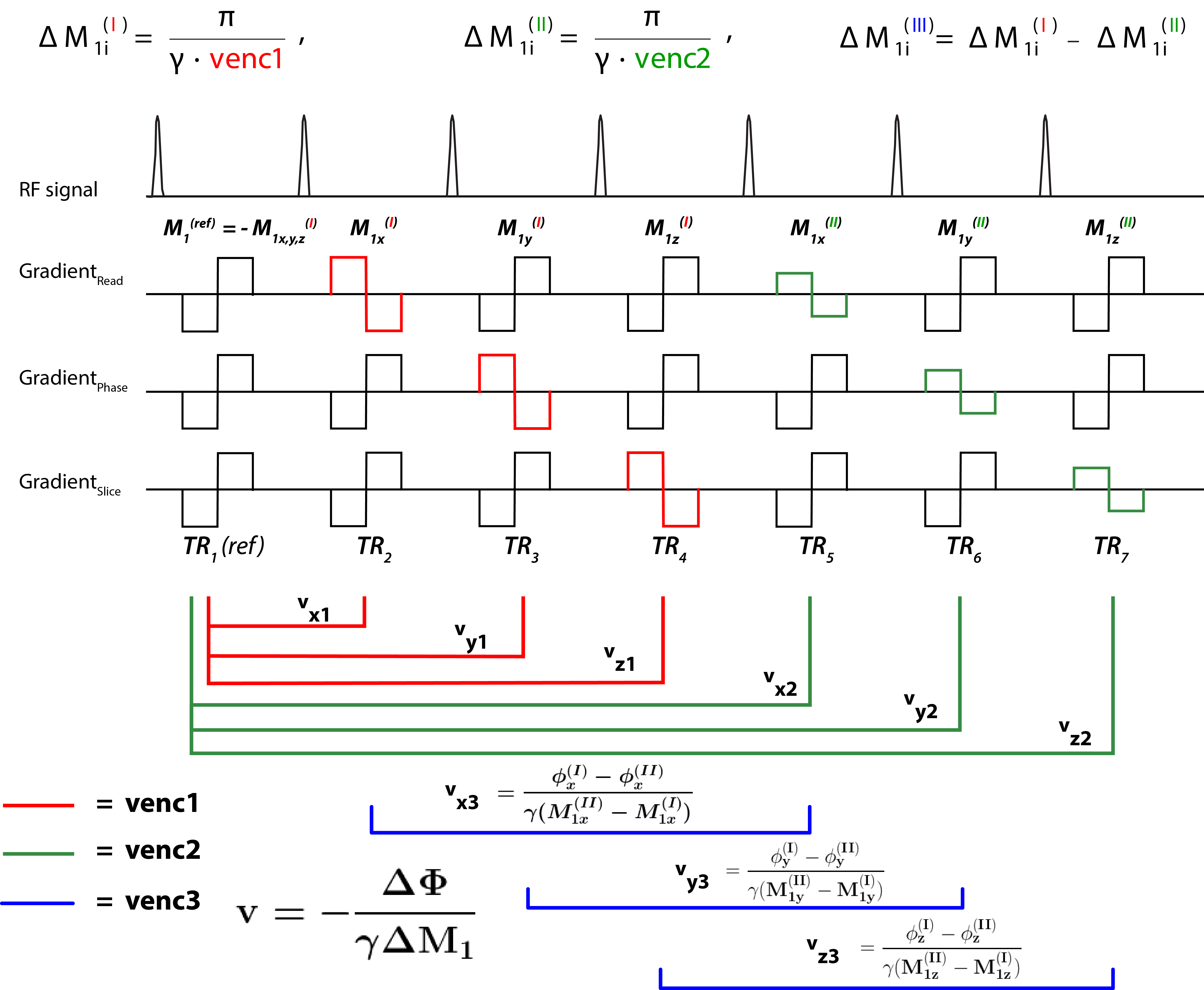
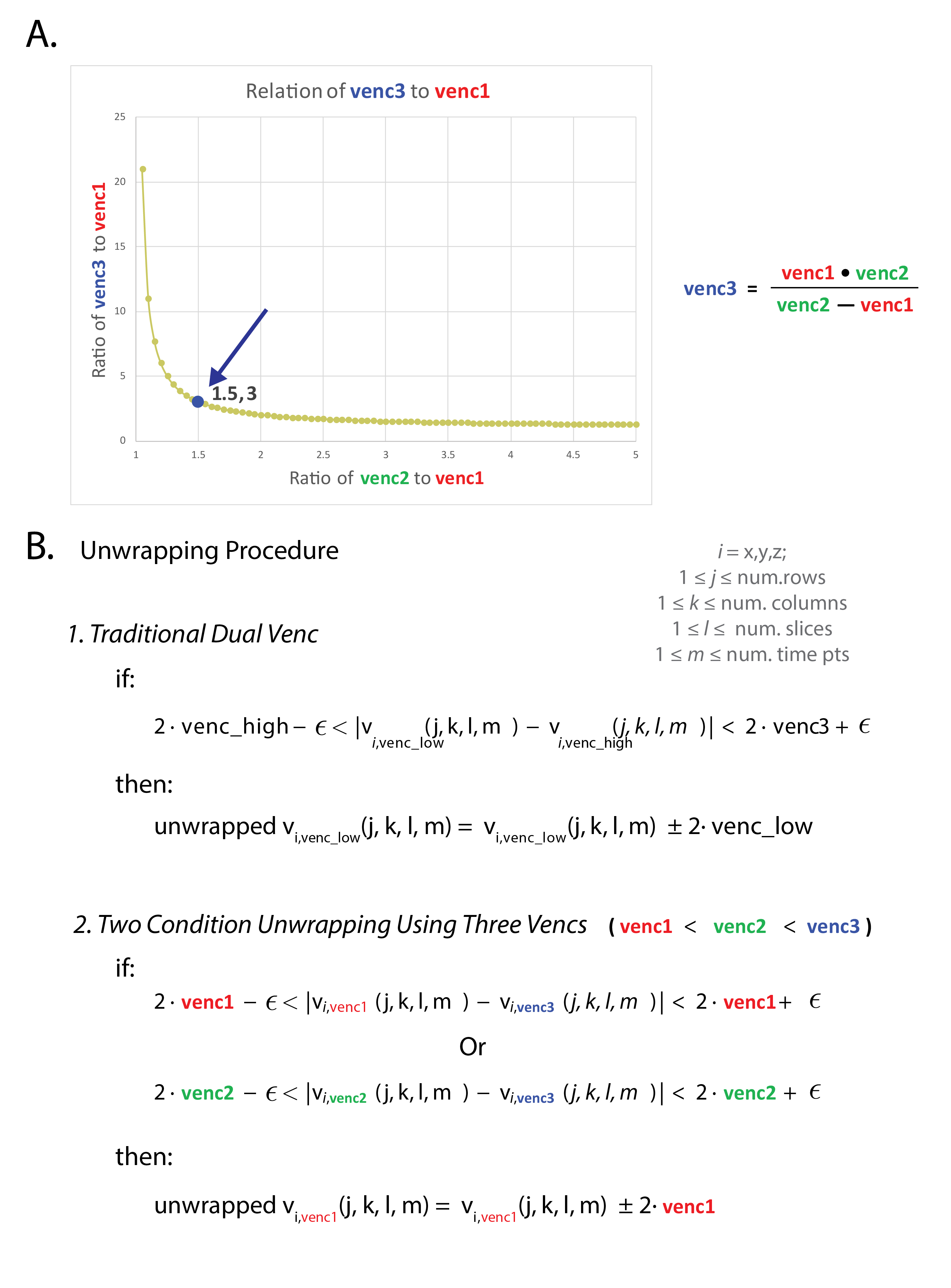
Intrinsic multi-venc encoding: Time-resolved velocities were calculated for the three vencs as shown in Figure 1. Multi-venc reconstruction employed a shared reference scan to calculate flow images with venc1 (lowest venc, reference scan in TR1 subtracted from TR2-4, red) and venc2 (TR5-7-TR1, green). An additional set of phase-difference images utilizing 6 TRs (TR2-7) yielded venc3 (highest venc, blue). The desired change in first-order gradient moment for venc1 (I), ΔM1i(I), i= x,y,z, was equally distributed between the reference scan and subsequent venc1 scans , with venc2 (II) first moments calculated based on the same reference. The resulting venc3 (III) depended on the change in first moment between the venc1 and venc2 scans, $$$venc3_i=\frac{\pi}{\gamma(M_{1i}^{(I)}-M_{1i}^{(II)})} $$$. This new scheme enables selection of variable ratios of the three vencs (Fig. 2A), which can increase VDR, and thus further improve VNR.
Multi-venc reconstruction: Data from phase-difference images for all 3 vencs were used to un-alias the venc1 scan to generate a single, unwrapped dataset with the favorable VNR of the lowest venc. Using a venc3:venc2:venc1 ratio of 3:1.5:1, for a given voxel, if |venc1,2-venc3| was within the 2*venc1,2±ε boundary (Figure 2B2), venc1 was considered aliased. Multi-venc phase-difference reconstruction including Maxwell correction for all 3 vencs was implemented in the scanner's online reconstruction pipeline. Offline processing included eddy current correction.
In-vitro flow phantom validation: Multi-venc 4D flow MRI was tested in-vitro using a pulsatile flow pump and a 3D-printed aorta with an 80% coarctation in the descending aorta. All phantom data were acquired with the following multi-venc sequence parameters: venc1/venc2/venc3=110/165/330 cm/s, TR/TE=5.0/2.6 ms, FA=15°, resolution=(2.4 mm)3. A 4TR single-venc (SV) scan was used for comparison with: venc=330 cm/s, TR/TE=4.7/2.2 ms, FA=15°, resolution=(2.4 mm)3.
In-vivo application: Multi-venc 4D flow MRI was evaluated in-vivo in 4 healthy volunteers (all male, age=26, 37, 56, 75 y.o., venc1/venc2/venc3 = 50/75/150 cm/s, TR/TE=5.5/3.1 ms, FA=7°, typical resolution = 2.4×2.4×2.7 mm3). One volunteer had an additional SV scan (venc=150 cm/s, TR/TE=4.9/2.5 ms, FA=7°, resolution=2.4×2.4×2.6 mm3). All scans were acquired on a 1.5T MAGNETOM Aera system (Siemens Healthcare, Erlangen, Germany) with k-t GRAPPA, R=5.
Results
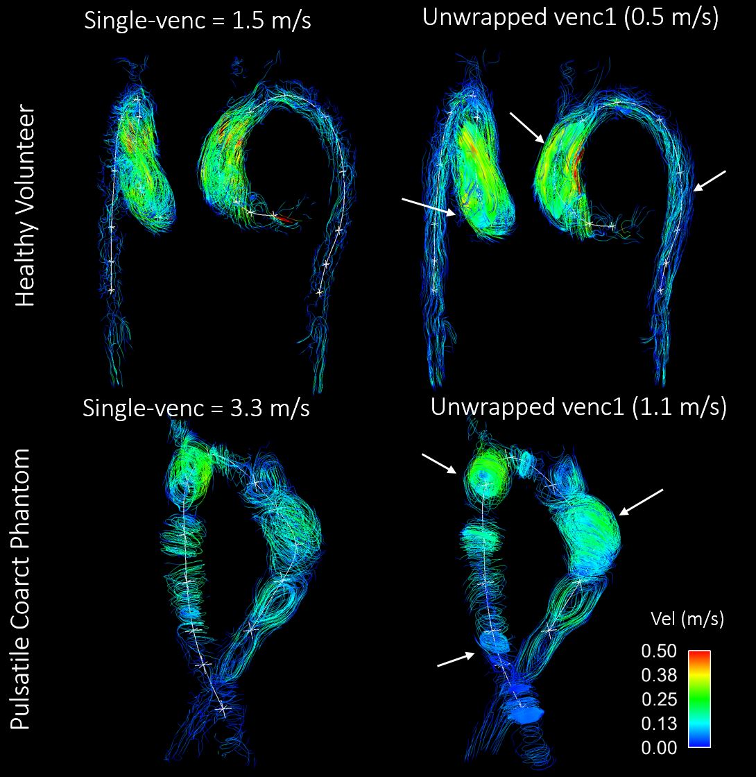
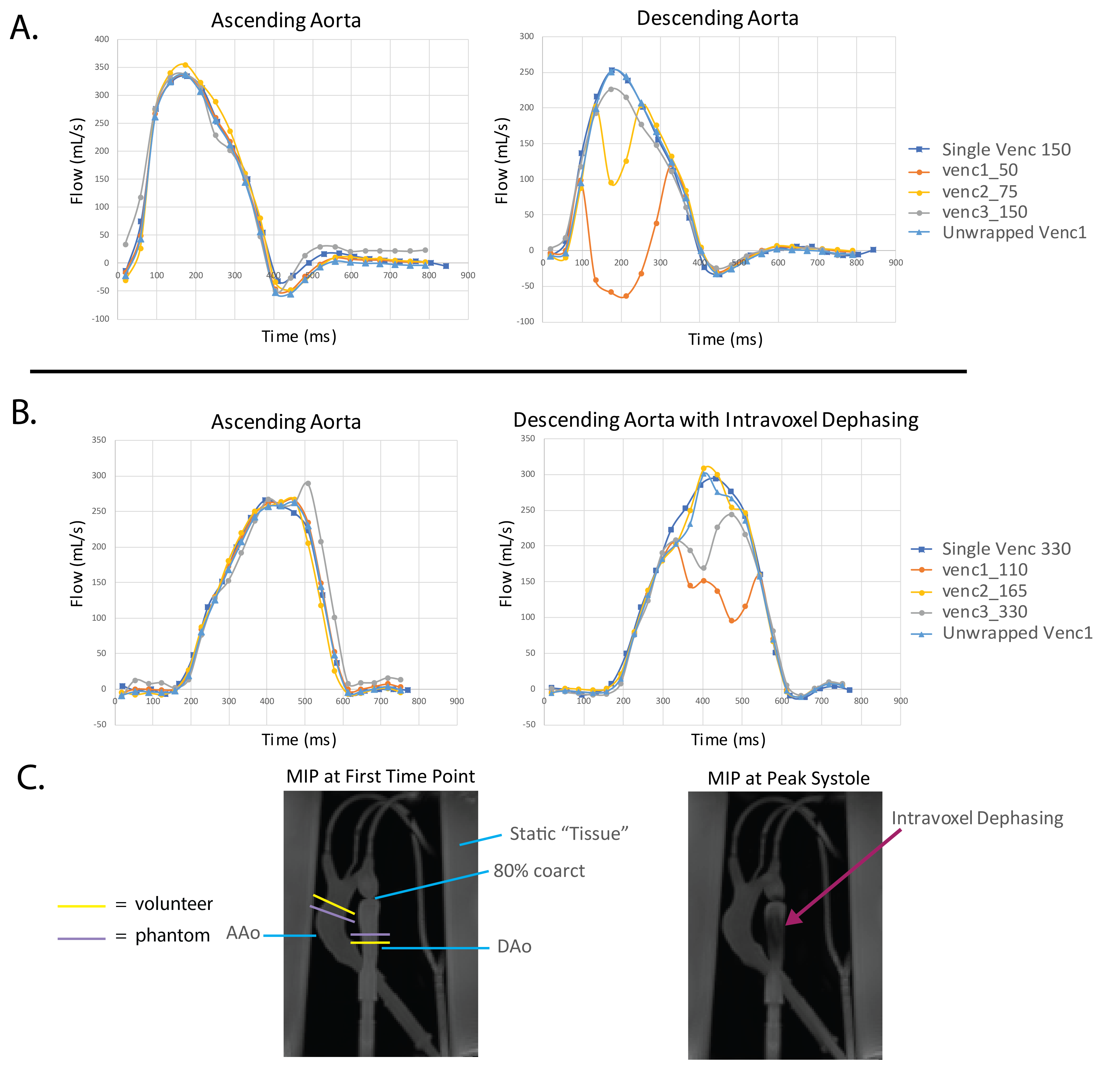
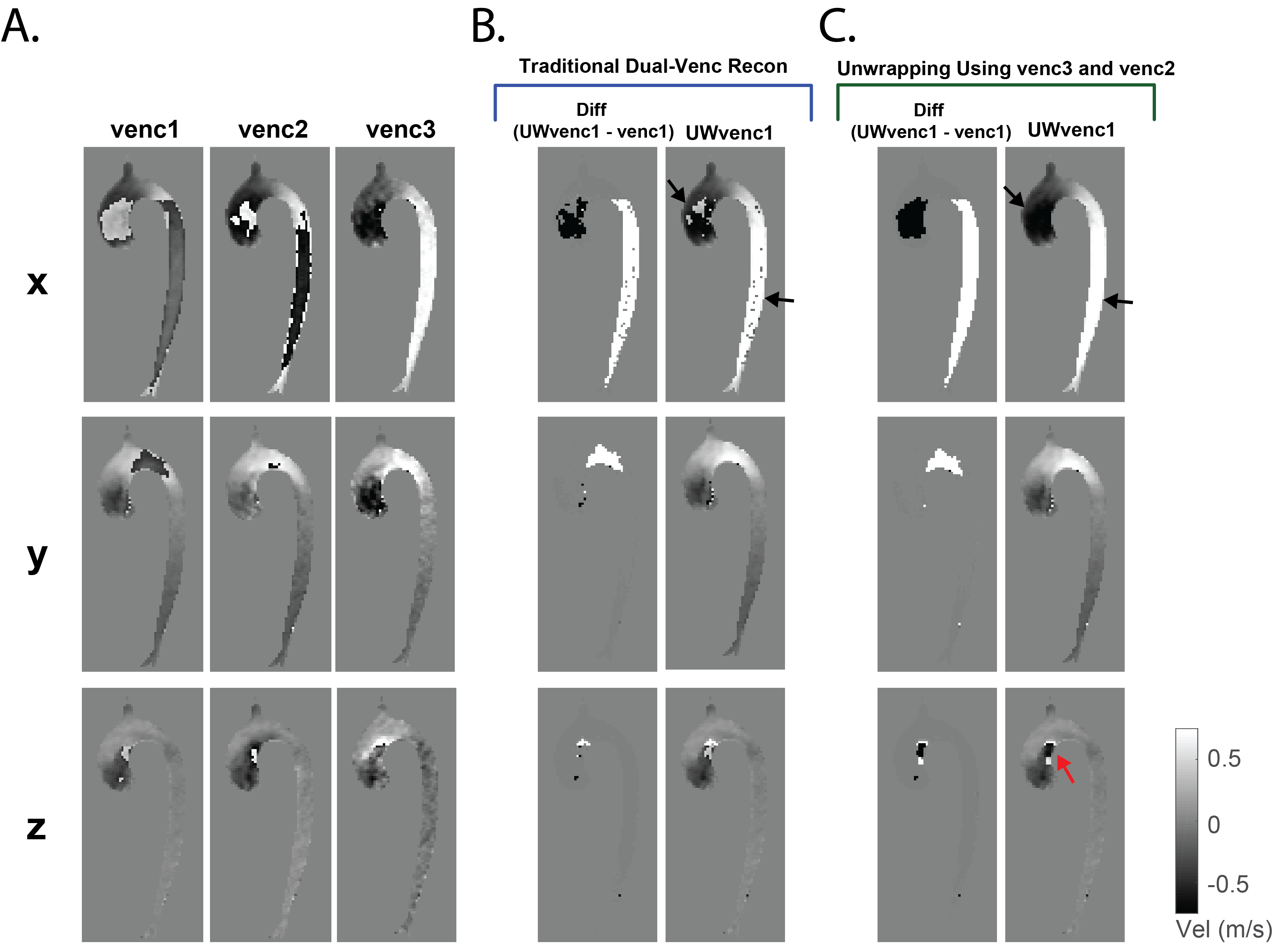
Multi-venc reconstruction and unwrapping were performed successfully in all datasets. Compared to SV 4D flow MRI, multi-venc encoding provided increased VNR (153% and 165% in the phantom and one volunteer, respectively) and improved streamline integrity (Figure 3). Flow waveforms in Figure 4 depict successful multi-venc unwrapping in areas of predictable flow, however artifacts due to intravoxel dephasing could not be recovered. Figure 5 shows improved multi-venc unwrapping compared to dual-venc unwrapping, however, we still noticed some missed or incorrectly unwrapped voxels (red arrow).Discussion and Conclusions
The proposed efficient multi-venc encoding scheme has the potential to extend VDR beyond a traditional dual-venc algorithm. Future investigations will include an improved unwrapping algorithm that considers voxel-by-voxel velocities in all three vencs simultaneously (i.e. model fitting), comparison to a 7TR DV acquisition with venc2=venc3 and traditional unwrapping, as well as further investigations of the temporal blurring and correlative noise effects of venc3 using a simpler phantom.Acknowledgements
Grant support by NIH F30HL137279, NIH R21 AG055954, and AHA 16SDG30420005.References
1. Schnell, S., et al., Improved assessment of aortic hemodynamics by kt accelerated dual-venc 4D flow MRI in pediatric patients. JCMR, 2016. 18(1): p. 1.
2. Schnell, S., et al., Accelerated dual-venc 4D flow MRI for neurovascular applications. JMRI, 2017. 46(1): p. 102-114.
3. Zwart, N.R. and J.G. Pipe, Multidirectional high‐moment encoding in phase contrast MRI. MRM, 2013. 69(6): p. 1553-1563.
4. Binter, C., et al., Bayesian multipoint velocity encoding for concurrent flow and turbulence mapping. MRM, 2013. 69(5): p. 1337-1345.
5. Knobloch, V., et al., Mapping mean and fluctuating velocities by Bayesian multipoint MR velocity encoding‐validation against 3D particle tracking velocimetry. MRM, 2014. 71(4): p. 1405-1415.
Figures