0367
Cardiac Single Breath-hold Balanced SSFP Cine ‘Watermark’ provides Cardiac Function via Magnitude and 2D Myocardial Strain via Phase1MRI Research Center, Auburn University, Auburn, AL, United States, 2Siemens Healthineers, Malvern, PA, United States
Synopsis
Cardiac MRI myocardial tagging enables quantification of myocardial strain. However, tagging remains limited to a research context due to the time-intensive analysis and need to run multiple sequences. CMR sequences must be fast and efficient. We previously developed a FLASH-based cine sequence called Cine ‘Watermark’ (CWM) that acquires normal cine magnitude images plus ‘hidden’ (via phase) cardiac strain data for calculating myocardial strain. Here we present a Balanced SSFP (bSSFP) version of CWM that presents improved SNR and scan efficiency. The bSSFP CWM is demonstrated in human subjects at 3T and its performance is compared to conventional Grid-Tagging MRI.
Purpose
Cardiac MRI (CMR) myocardial tagging enables quantification of myocardial strain throughout the cardiac cycle1, 2. However tagging remains limited to a research context due to the time-intensive analysis and need to run multiple sequences. CMR sequences must provide maximum diagnostic information with minimum operator effort, time and cost3. Previously we developed a FLASH-based CMR cine sequence, called Cine Watermark (CWM), with a ‘hidden’ added capability to acquire cardiac strain data while requiring no extra operator effort4. The CWM acquires normal cine magnitude images plus spatial-modulated tagging added only into the image phase (‘watermark’) allowing quantitative cine myocardial strain calculation. Here we present a Balanced SSFP (bSSFP) version of CWM that presents improved SNR and scan efficiency. The bSSFP CWM is demonstrated in human subjects at 3T and its performance is compared to conventional Grid-Tagging MRI.Methods
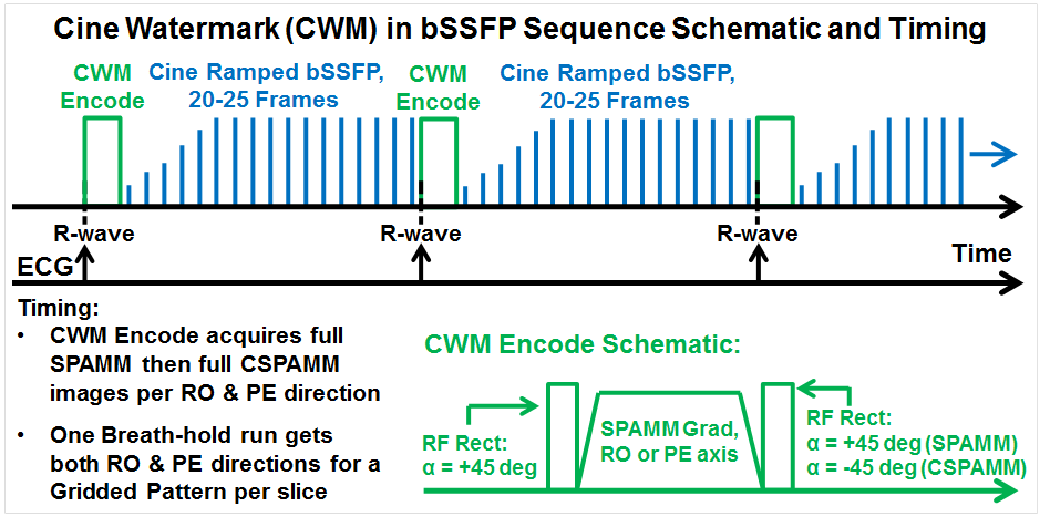
Fig 1 illustrates the bSSFP CWM sequence that runs continuously as steady-state with ECG-trigger to acquire data on every heartbeat. A classic “1-1” saturation SPAMM was applied with each non-selective rectangular RF pulse flip angle = 45°. The cine bSSFP readout was customized with ramped start-up flip angle for fast approach to steady-state and at end of cardiac R-R cycle can incorporate either α/2 magnetization restore method, or just apply spoil gradients before the next triggered SPAMM encode. Within one 25-second breath-hold, four CWM acquisitions are performed that include cine SPAMM and CSPAMM in both readout (RO) and phase encode (PE) axes directions. These RO and PE direction sets provide orthogonal line-tag sets and are combined for a grid-tag set. An optional secondary 8-second breath-hold scan acquired a phase reference image for improved background phase consistency. For performance comparison, a standard Grid-Tagging with cine FLASH readout sequence was used with as similar imaging parameters as possible on the same image slices. Imaging parameters included for both bSSFP CWM and Standard Grid-Tagging: FOV = 256x224 mm, matrix = 128x112, Slice thickness = 8 mm, pixel size = 2x2 mm, Bandwidth = 977 Hz/pixel, GRAPPA acceleration = x2, cine frames = 20, Averages = 1, SPAMM line spacing = 8 mm; bSSFP CWM only: flip angle = 30°; standard Grid-Tagging only: flip angle = 12°. All image and strain analyses were performed offline using customized Matlab programs (Mathworks, Natick, MA) to include: 1) correct the SPAMM and CSPAMM background phase with the reference scan phase, 2) calculate complex image sum and difference (SPAMM±CSPAMM) objects, 3) calculate the magnitude of the complex sum (SPAMM+CSPAMM) to produce normal cine images with no tagging, 4) calculate the phase of the complex difference (SPAMM-CSPAMM) to produce high contrast tagging-only images, and 5) apply Discrete Model Free (DMF)5 strain analysis to process the tagged images and calculate cardiac left ventricular (LV), short-axis (SA), six-sector, circumferential strain (Ecc) graphs. Segmentation lines drawn onto magnitude images were used to automatically segment corresponding phase images. Six healthy human subjects, 19-30 yo, 3 female, with informed consent, were scanned in a 3T Verio scanner (Siemens, Erlangen, Germany) with a 32-chan anterior/posterior RF coil array (Invivo, Gainesville, Florida). Scan slice locations included one long-axis (LA) 4-chamber and 1-3 mid-LV SA views.Results
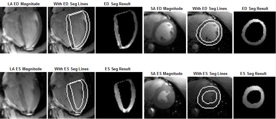
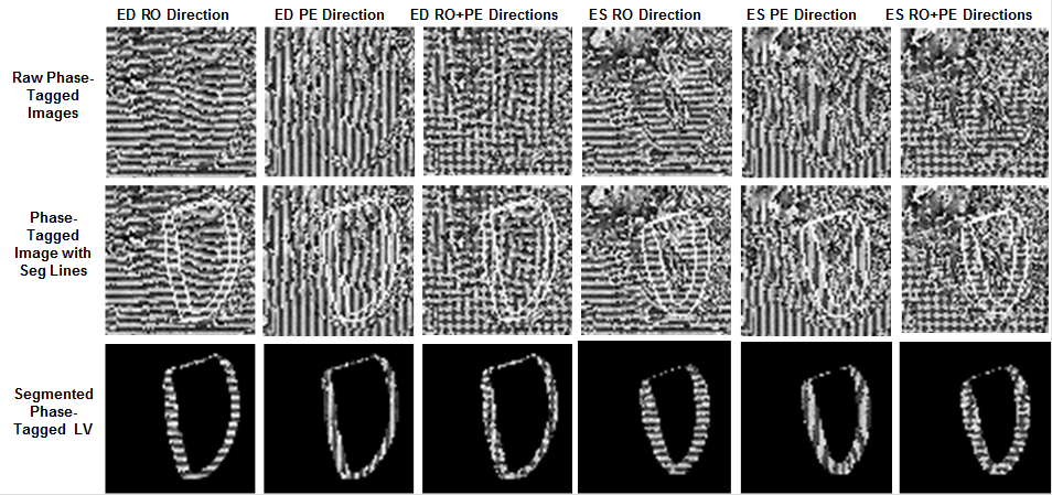
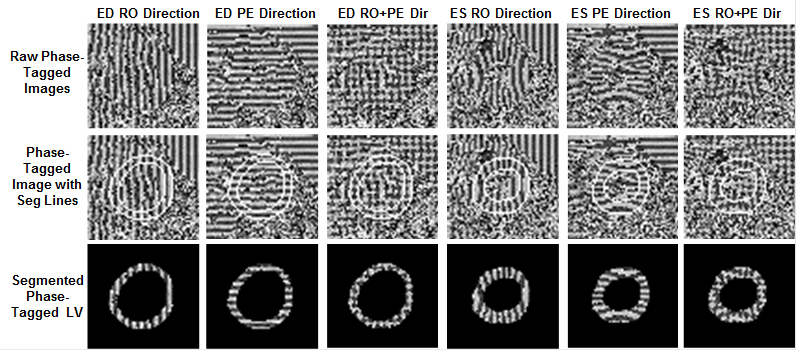
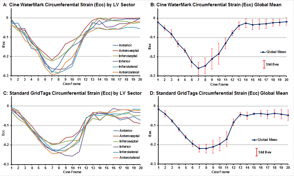
Within 25-second breath-holds, all CWM subjects provided cine magnitude and spatial-modulated phase image sets suitable for strain analysis. Fig 2 presents representative CWM LA and SA magnitude cine frames at end-diastole (ED) and end-systole (ES) time-points with subsequent LV segmentation. Fig 3 presents LA phase-tagged results at ED and ES with spatial-modulation applied in the RO, PE and combined RO+PE (grid) directions. All show strong tag contrast. The segmented LV images show clear displacement and bending of the phase-tagging patterns at ES. Similar to Fig 3, Fig 4 presents SA phase-tagged results that also show strong contrast and clear displacement and bending of the phase-tagging patterns at ES. Fig 5 presents representative SA non-filtered/non-smoothed Ecc strain results with good agreement and minimal offset bias between bSSFP CWM and standard Grid-Tagging. All sectors show nominal strains with consistent trajectories. Some CWM scans presented T1 fading with noisy tagging in the late cardiac cycle frames.Discussion
The CWM can acquire cardiac cine and phase-tagged cine images in a single breath-hold scan. The high SNR from bSSFP and x2 GRAPPA acceleration were essential for CWM to achieve this capability. The bSSFP ramped flip angle greatly reduced artifacts. The Ecc strain results generally agree with the previous literature2.Conclusions
We developed an accelerated bSSFP Cine ‘Watermark’ sequence with an integrated hidden capability to quantify myocardial strain. Future work is needed to reduce T1 fading effects.Acknowledgements
Special thanks for assistance by Dr. Martha Forloines, Mr. Julio Yanes, and Ms. Lily Strassberg.References
1. Axel L, Dougherty L. “MR imaging of motion with spatial modulation of magnetization”, Radiology 1989;171:841–9.
2. Edvardsen T, et al. “Quantitative Assessment of Intrinsic Regional Myocardial Deformation…”, Circulation 2002;106:50-56.
3. Edelstein WA, et al. “MRI: Time Is Dose—and Money and Versatility”, J Am Coll Radiol 2010;7(8): 650–2.
4. Beyers RJ, et al. “Accelerated Cardiac Cine ‘Watermark’ MRI Provides Cardiac Function via Magnitude Cine and 2D Myocardial Strain Via Spatially Modulated Phase”, Proc. Intl. Soc. Mag. Reson. Med. 25, 2017.
5. Denney TS Jr, McVeigh ER. “Model-free reconstruction of three-dimensional myocardial strain from planar tagged MR images”, J Magn Reson Imaging 1997;7(5):799-810.
Figures