0350
30 brain MRI exams in 1 hour using a multi-contrast EPI sequence1Department of Neuroradiology, Karolinska University Hospital, Stockholm, Sweden, 2Department of Clinical Neuroscience, Karolinska Institutet, Stockholm, Sweden, 3MR Applied Science Laboratory Europe, GE Healthcare, Stockholm, Sweden
Synopsis
An in-house developed multi-contrast EPI sequence producing six MR contrasts (T1-FLAIR, T2-w, DWI, ADC, T2*-w, and T2-FLAIR) was used as a fast brain MRI protocol. Seven healthy volunteers were scanned repeatedly to investigate how many brain MRI exams that can be performed over the course of one hour. With a net scan time of one minute and an additional minute for table movements and switch of subjects, it was possible to perform 30 brain MRI examinations in 1 h.
Introduction
In clinical brain CT imaging, the actual scan is completed in a few seconds making the examination time essentially only dependent on the patient setup and management, often lasting around 10 min. For MRI, the time taking the patient on and off the table is usually short in comparison to the scan protocol of a clinical brain MRI exam. We have recently presented a multi-contrast EPI sequence1 capable of producing six MR contrasts (T1-FLAIR, T2-w, DWI, ADC, T2*-w, and T2-FLAIR) with full brain coverage in about one minute. These contrasts correspond well to what is needed in a brain trauma MRI protocol2, although it remains to be seen for which patient categories confident clinical diagnoses can be made. With an MRI exam time around one minute, the patient setup time now becomes large in comparison, similar to the CT case. In this study, we therefore asked the question: What is the upper limit of the number of brain MRI exams that can be performed in one hour on a clinical 3T system?Methods
Seven healthy volunteers were recruited to be scanned with our in-house developed multi-contrast EPI sequence on a GE MR750w system (GE Healthcare, WI, USA) using an 8-channel RF coil (Invivo Corp., FL, USA). The volunteers were scanned repeatedly with minimal delay between the examinations over the course of 1 h. Each examination was registered as a separate patient as in a clinical setting. The scan protocol consisted of only the multi-contrast EPI sequence with axial slices at predefined slice locations relative to the landmark position. Hence, no localizer scan or graphical prescription of slice locations was performed. The scan parameters were: FOV = 24x24 cm, matrix 180x180, GRAPPA R = 3, 30 5 mm axial slices with FOV center 30 mm anterior of the isocenter. The net scan time, excluding prescan, was 0:55 min (cf. Protocol B in Ref 1). A video camera was used to monitor the times required between four steps in the examination procedure. The four steps (or timestamps) were, in order: 1) switch of subjects, 2) landmark (setting origin) on subject, 3) subject's head at the isocenter of the magnet, 4) scan end. Between steps 1 and 2, the foam mattress and headrest paper were swapped between subjects to simulate the necessary hygiene measures in a clinical environment.Results
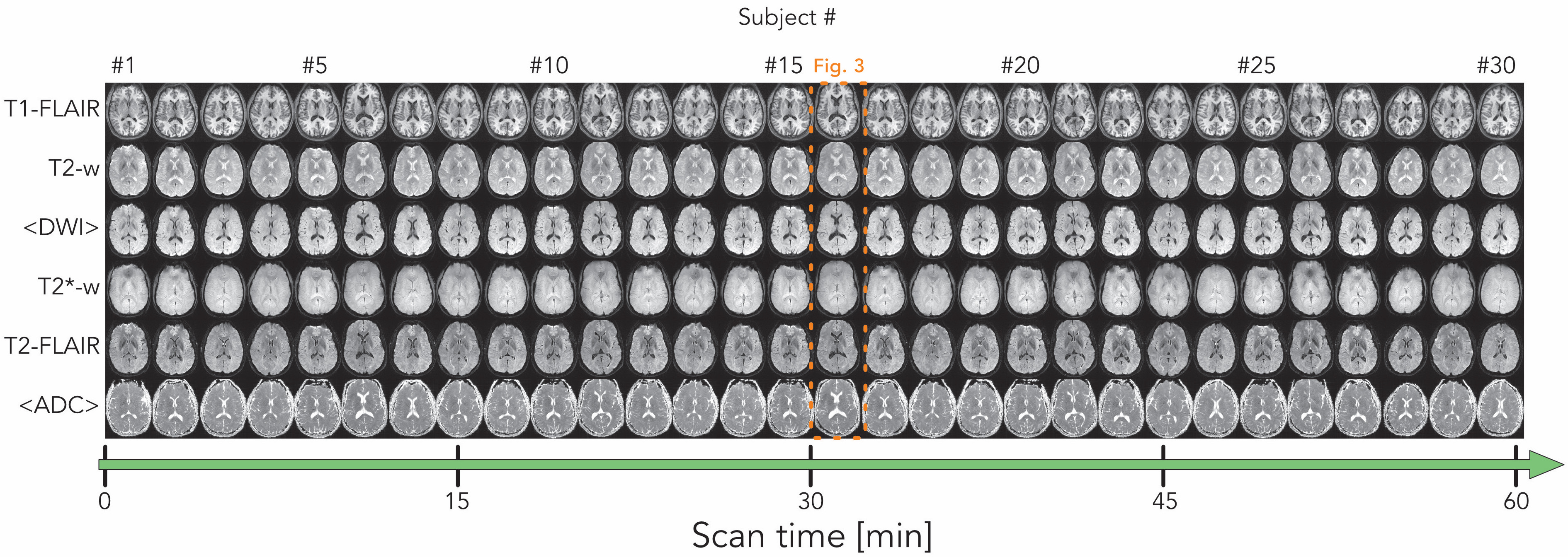
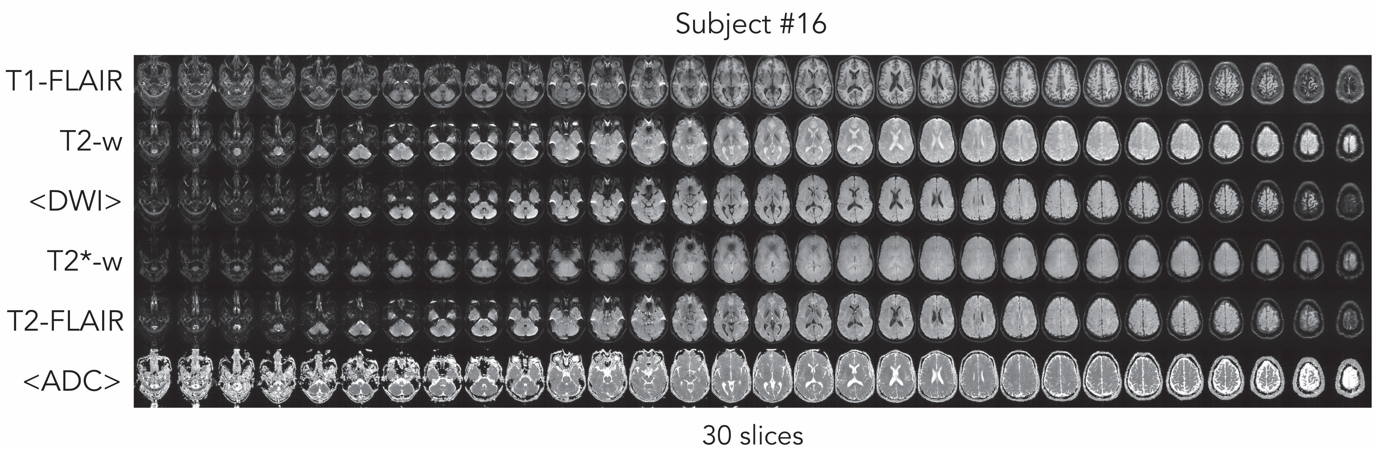
Fig. 1a provides an overview of the four timestamps in each MR exam, between which times were measured. Fig. 1b shows that it was possible to fit 30 subjects during the course of 1 h. Each bar consists of the four measured times for the corresponding subject. Taking the mean and one standard deviation across the 30 subjects, the average total scan time (including prescan and user interface delays) was 79 ± 5 [s] (Fig. 1c, yellow bar). Adding table movement, mattress swap, patient switching and landmark time, the average examination time per subject became 2:03 ± 0:06 min. Fig. 2 shows the six MR contrasts for one slice of each of the 30 subjects. Five of the seven subjects were repeated 5-6 times, which can be identified in the figure. Despite no localizer or manual slice prescription and high throughput, it was possible to reproduce slice orientations with good left-right symmetry, slice angulation and FOV centering, with some variation in the head-feet direction. The images in Fig. 2 have been cropped by the same amount for all examinations. All slices for subject #16 are shown in Fig. 3 to show all data acquired per exam.Discussion
We have shown that it is possible to perform brain MRI examinations of 30 healthy volunteers in one 1 h, with the six most common MR contrasts that are of particular interest in e.g. brain trauma2. On these trained subjects, the scan time and non-scan time contributed to one minute each, the latter also involving patient registration and change of mattress/head cover. In a real clinical scenario, the non-scan time will likely be at least 1-2 min longer, even for patients with full mobility. Nevertheless, provided the multicontrast EPI data will show to be reliable for diagnosis in the future, MR exams like this would be mostly limited to patient handling and almost as fast as CT.Acknowledgements
No acknowledgement found.References
1. Skare, S. et al. A 1-minute full brain MR exam using a multicontrast EPI sequence. Magn. Reson. Med. (2017). doi:10.1002/mrm.26974
2. Di Muzio, B. Brain trauma protocol (MRI). Radiopaedia Available at: https://radiopaedia.org/articles/brain-trauma-protocol-mri. (Accessed: 29th May 2017)
Figures
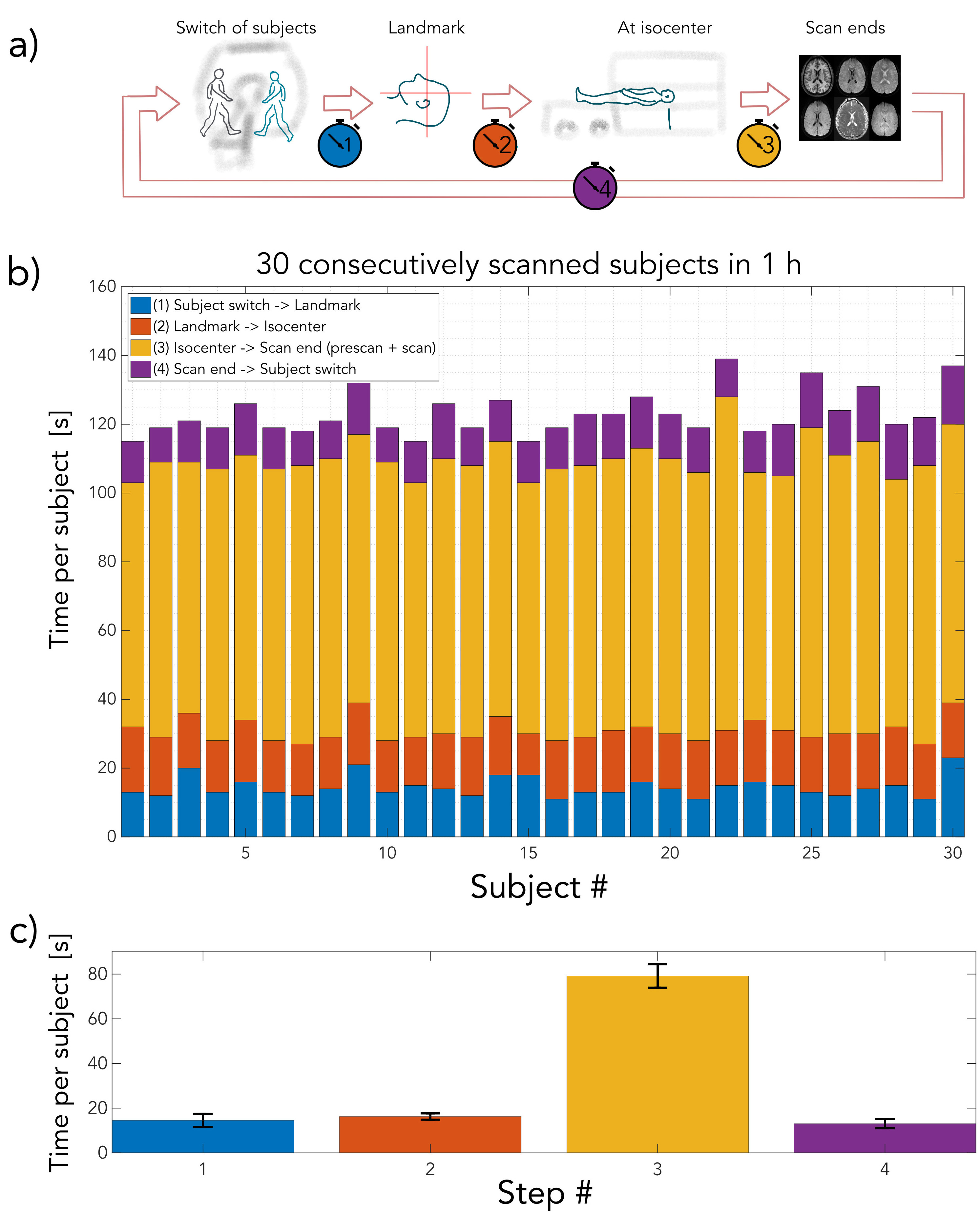
a) Time measured between four timestamps: Switching of subjects, setting landmark on subject, subject at the scanner's isocenter, end of scan. b) Times required for each exam for the 30 subjects, divided into four steps. c) Mean ± SD across subjects for the times between 1-2, 2-3, 3-4, 4-1. The mean scan time incl. prescan was 1:19 min and the average time between the switch of subjects was 2:03 min