0345
Accelerated 3D 1 mm isotropic T2w-Imaging of the Prostate in less than 3 min1School of Biomedical Engineering and Imaging Sciences, King's College London, London, United Kingdom, 2Siemens Healthcare Limited, Frimley, United Kingdom, 3Cancer Imaging, King's College London, London, United Kingdom
Synopsis
Three dimensional (3D), isotropic T2-weighted imaging of the prostate requires a long acquisition time. Here we propose a 3D T2-prepared multi-shot bSSFP acquisition with a variable density undersampled trajectory and TV-SENSE reconstruction. Results from a healthy volunteer study demonstrate that 3D 1 mm isotropic resolution T2-weighted images of the prostate can be acquired in 2 min 45 s, with image quality that is comparable to the clinical standard turbo spin echo sequences but only takes 1/3 of the acquisition time.
Purpose
Prostate cancer is the most common male cancer1. The current clinical multiparametric MRI protocol for prostate cancer detection consists of a 2D multi-slice T2-weighted (T2w) Turbo Spin Echo (TSE) sequence, 2D multi-slice diffusion-weighted single shot echo-planar (SS-EPI) sequence, and a dynamic contrast enhanced 3D T1-weighted Dixon spoiled gradient recalled (VIBE) sequence, allowing for the detection of focal lesions for confirmatory targeted biopsy.
Three dimensional (3D), isotropic high-resolution T2w imaging enables the visualization of the prostate in multiple arbitrarily angulated anatomical planes, thereby potentially improving sensitivity for cancer detection, particularly within the transition zone, enabling more precise image-guided biopsy and ultimately improving surgical planning or focal treatment.
However, 3D T2w TSE imaging has been hampered by long acquisition times, and is susceptible to motion and blurring artifacts from the long echo train sampling. Here we propose to combine a T2-prepared2,3 multi-shot 3D balanced steady state free precession (bSSFP) sequence with a Cartesian trajectory with spiral profile order (CASPR)4 with a variable density undersampling and Total Variation regularized SENSE (TV-SENSE) reconstruction5 to enable fast 3D isotropic T2w-imaging of the prostate.
Methods
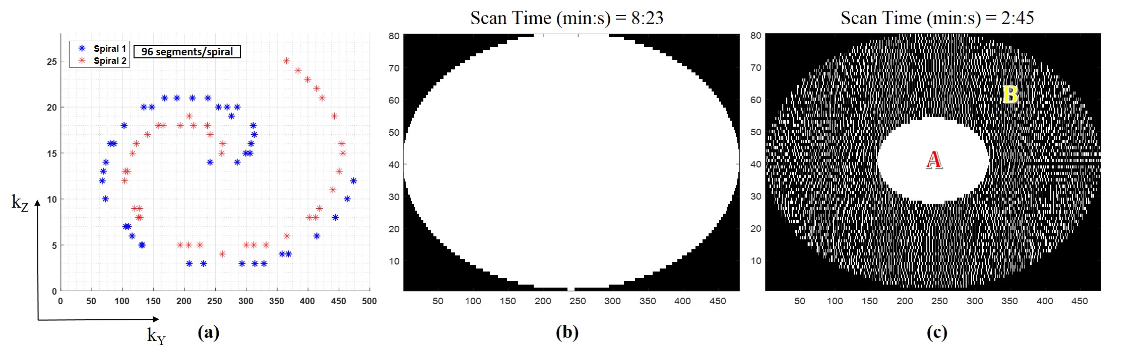
This study was approved by the local institutional review board. Following informed consent, five healthy male subjects were scanned on a 3T PET-MR scanner (Biograph mMR, Siemens Healthcare, Erlangen, Germany). MR measurements were obtained using a prototype 3D segmented balanced steady state free precession (bSSFP) acquisition preceded by a T2-preparation module2,3 with duration of 90 ms (TR = 1600 ms). Imaging parameters included: transversal plane, 304x304x60 matrix, 1x1x1 mm3 resolution, flip angle (FA) = 57o, bSSFP TR/TE = 4/2 ms. The acquisition was three-fold (3x) prospectively accelerated using a variable density CASPR trajectory (Figure 1(c) with a fully-sampled centre A and a four-fold undersampled periphery B). The fully-sampled and accelerated datasets were reconstructed offline in MATLAB (Mathworks, Natick, MA) using regularized TV-SENSE reconstruction described in5, which involved minimizing the following cost function:
$$$ \hat{I}=arg\space min_I\left\{\parallel EI-K\parallel^2_2+\space \lambda_S\parallel \triangledown_SI\parallel_1 \right\} $$$
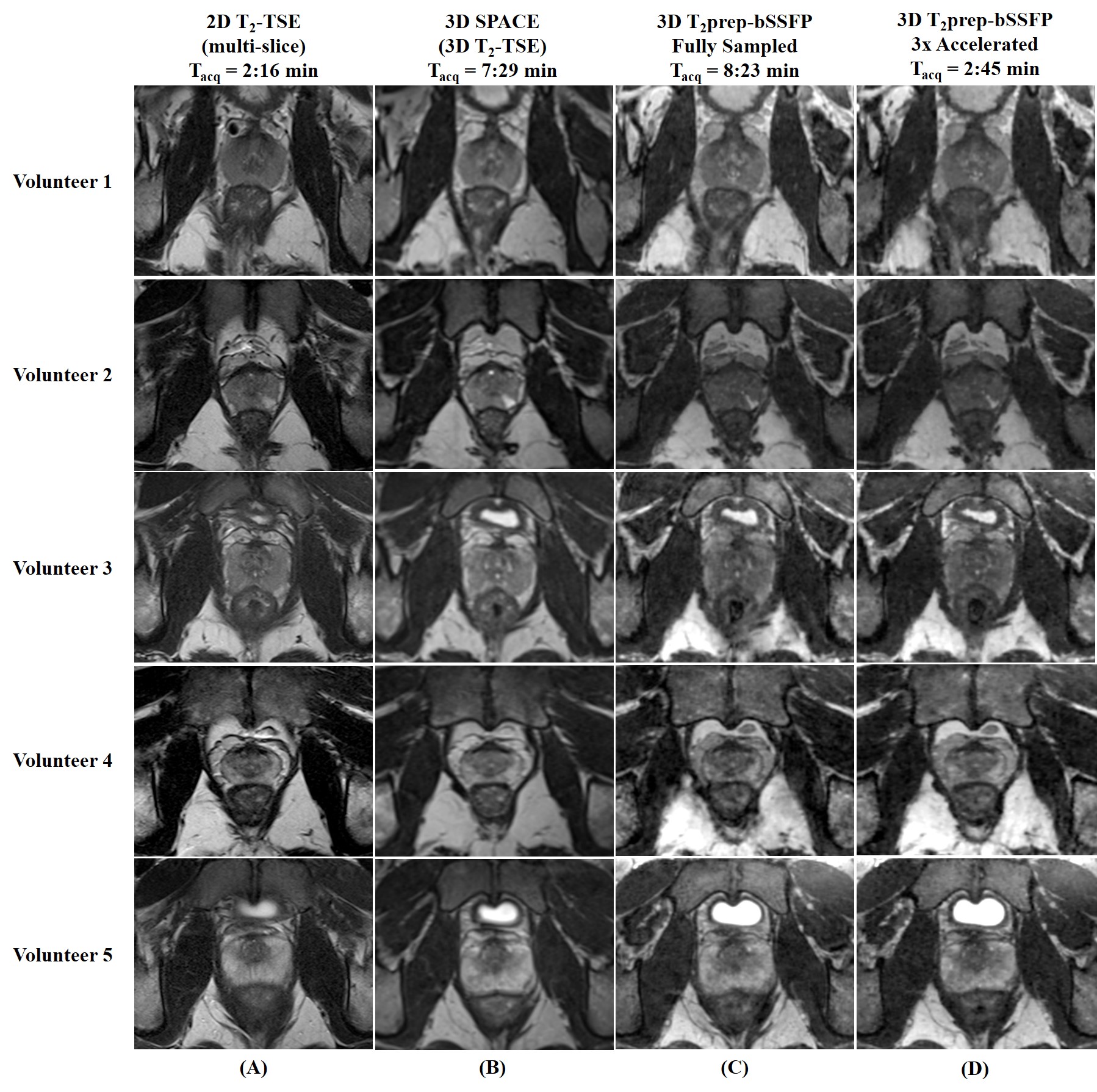
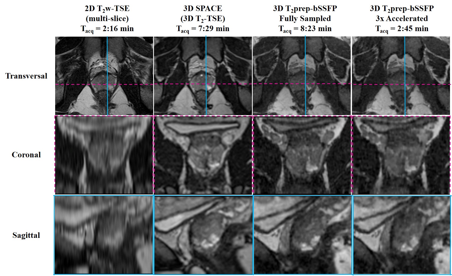
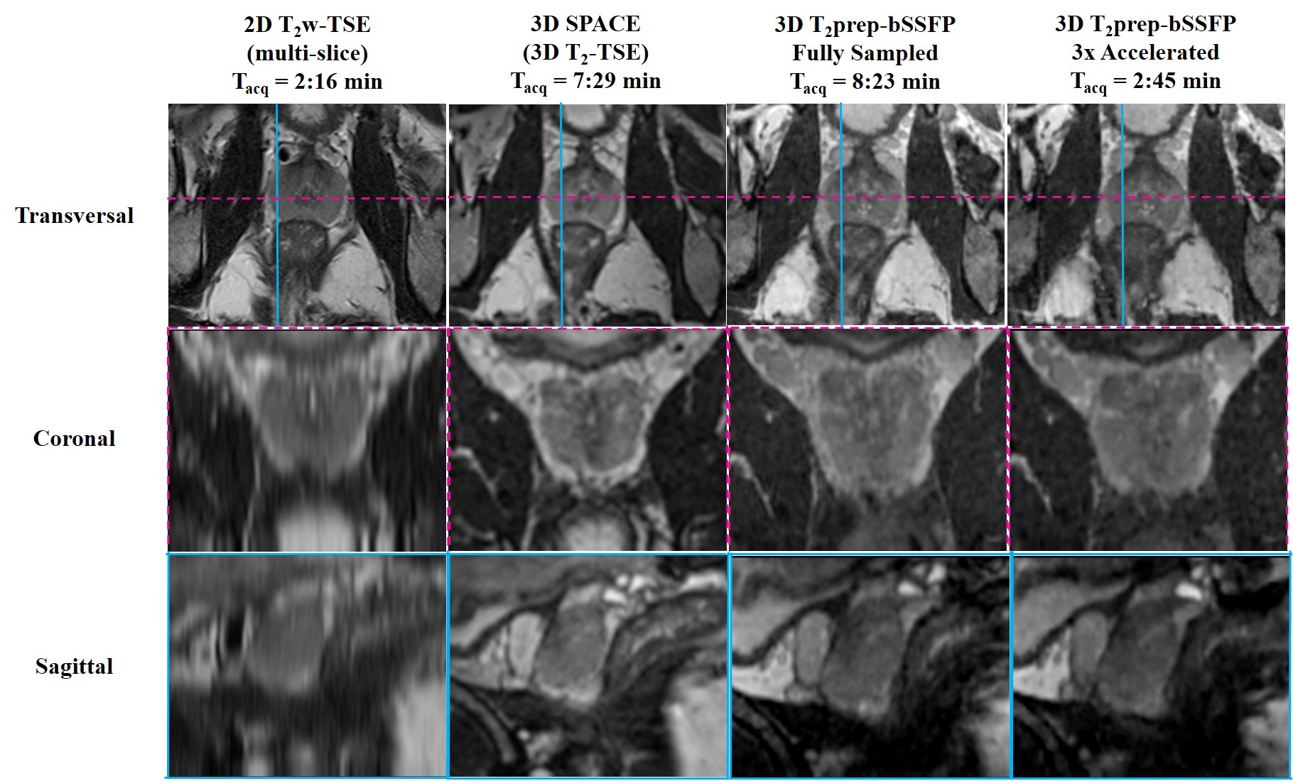
Where, I is the reconstructed image, E is the SENSE encoding operator, K is the undersampled k-space, $$$ \triangledown_S $$$ is the first order finite difference operator, and $$$ \lambda_S $$$ is the regularization parameter. The accelerated reconstructions were compared with the fully-sampled reference in terms of acquisition time (Tacq), apparent signal to noise ratio (SNR), SNReff (SNR/√Tacq), and visually remaining undersampling artifacts. Image quality of the accelerated images was compared to (i) the clinical standard multi-slice transversal 2D T2w TSE and (ii) 3D SPACE (Siemens, 3D T2w fast spin echo) sequences with matched imaging parameters except the following: (i) 2D T2w TSE – 0.6x0.8x3 mm3 resolution, TR/TE = 6470/89 ms, FA = 150o (ii) 3D SPACE – TR/TE = 1700/101 ms, FA = 135o. The scan time (min:s) for the multi-slice transversal 2D T2w TSE was 2:16, while that for the 3D SPACE sequence was 7:29. The 3D fully-sampled T2prep-bSSFP acquisition required a scan time of 8:23, whereas the 3x prospectively accelerated T2prep-bSSFP acquisition required only 2:45.
Results & Discussion
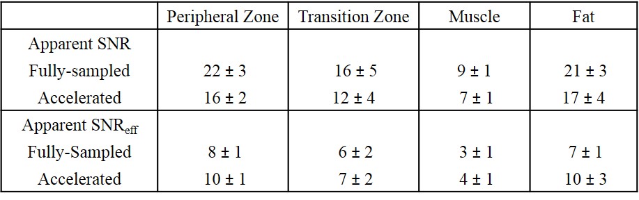
Figure 1 shows the Cartesian CASPR trajectory that is centric in the ky-kz plane, and the fully-sampled and 3x accelerated masks. Figure 2 shows a single matched transversal slice for five healthy volunteers scanned using the multi-slice 2D T2w TSE, 3D SPACE, 3D fully-sampled T2prep-bSSFP, and 3D accelerated T2prep-bSSFP sequences. The accelerated T2prep-bSSFP images display similar image quality to the fully-sampled reference, as indicated by the mean apparent SNR (n = 5 subjects) over different regions summarized in Table 1. Furthermore, the fast 3D T2prep-bSSFP reconstructions have image quality comparable to the clinical standard high resolution 2D T2w TSE, and appear visually sharper than the 1 mm3 isotropic 3D SPACE images (Figure 2). Figures 3 and 4 illustrate a single matched transversal slice for two representative healthy subjects from the multi-slice 2D T2w TSE, 3D SPACE, 3D fully-sampled and accelerated T2prep-bSSFP reconstructions, reformatted into the sagittal and coronal planes. The accelerated T2prep-bSSFP acquisitions appear to have no visually remaining artifacts from the TV-SENSE reconstruction along all the three dimensions (Figures 3 and 4), and display comparable image quality with the fully-sampled data.Conclusion
We have demonstrated the feasibility of 3D T2w imaging in the prostate with 1 mm isotropic resolution in 2 min 45 s, by combining 3D multi-shot T2prep-bSSFP acquisitions with a variable density accelerated trajectory and TV-SENSE reconstruction. This facilitates high-resolution isotropic prostate imaging in a clinically feasible scan time.Acknowledgements
We acknowledge funding from The King’s Health Partners Research and Development Challenge Fund, TOHETI, NIHR BRC, GSTT/KCL BRC, CRUK/EPSRC Cancer Centre, and Siemens Healthineers. This work was supported by the Wellcome EPSRC Centre for Medical Engineering at King's College London (WT 203148/Z/16/Z).References
1. R. Siegel, J. Ma, Z. Zou, and A. Jemal, “Cancer Statistics, 2014”, CA Cancer J Clin, 64(1), 9 – 29, 2014.
2. R. M. Botnar, M. Stuber, P. G. Danias, K. V. Kissinger, and W. J. Manning, “Improved coronary artery definition with T2-weighted, free-breathing, three-dimensional coronary MRA”, Circulation, 99, 3139-3148, 1999.
3. J. H. Brittain, B. S. Hu, G. A. Wright, C. H. Meyer, A. Macovski, and D. G. Nishimura, “Coronary Angiography with Magnetization-Prepared T2 Contrast”, Magn Reson Med, 33, 689-696, 1995.
4. C. Prieto, M. Doneva, M. Usman, M. Henningsson, G. Greil, T. Schaeffter, and R. M. Botnar, “Highly Efficient Respiratory Motion Compensated Free-Breathing Coronary MRA Using Golden-Step Cartesian Acquisition”, J Magn Reson Imaging, 41, 738-746, 2015.
5. G. Cruz, D. Atkinson, C. Buerger, T. Schaeffter, and C. Prieto, "Accelerated Motion Corrected Three-Dimensional Abdominal MRI Using Total Variation Regularized SENSE Reconstruction", Magn Reson Med, 75, 1484-98, April 2016.
Figures