0344
Reduced distortion in prostate DWI by using split echo type TSE-DWI (SPLICE) with MultiVane acquisition1Philips Japan, Shinagawa, Tokyo, Japan, 2Department of Radiology, Gifu University Hospital, Gifu, Japan, 3Philips Healthcare, Shinagawa, Tokyo, Japan
Synopsis
To reduce image distortion in prostate DWI, split-echo type TSE-DWI (SPLICE) was combined with MultiVane (current Philips implementation of PROPELLER) acquisition, named MV-SPLICE. To avoid non-CPMG artifacts in SPLICE, the spin echoes and stimulated echoes were separated by the unbalanced readout gradient and acquired in separate MultiVane k-space for separate reconstruction. ADCs and SNRs in transition zone and peripheral zone, and distortion for DWI and T2W images in the anterior-posterior direction of prostate diameter for MV-SPLICE were compared to conventional EPI and MultiVane TSE-DWI (MV-ALSOP). We demonstrated that MV-SPLICE is insensitive to distortion and can provide comparable ADC measurement.
INTRODUCTION
Prostate diffusion weighted image (DWI) has gained more attention for prostate cancer detection1. Most studies employ single-shot echo-planar imaging (ssh-EPI) but EPI is sensitive to field inhomogeneity and suffers from distortion. TSE (turbo spin echo) DWI has been developed for less sensitivity to field inhomogeneity, due to the refocusing pulses2.
A challenge of TSE-DWI is that TSE violates the Carr-Purcell-Meiboom-Gill (CPMG) conditions. Imperfect refocusing pulses generate spin echoes (SE) and stimulated echoes (STE) and cause destructive interference. To solve this, two approaches have been proposed. One is the Alsop method, which eliminates non-CPMG component3. The second one is SPLICE (split-echo acquisition of FSE signals)4, where SE and STE are separated and acquired in separate k-spaces. Alsop method has less blurring but suffers from low signal-to-noise ratio (SNR) because of discarding half of the signal. On the contrary, SPLICE has higher SNR but suffers from blurring due to long echo-train-length (ETL). Therefore, to reduce ETL by using multi-shot acquisition, we have combined SPLICE and MultiVane (MV), named MV-SPLICE. MultiVane is current Philips implementation of PROPELLER5 (periodically-rotated-overlapping-parallel-lines-with-enhanced-reconstruction). The aim of this study is to investigate clinical usefulness of MV-SPLICE in prostate DWI with b-value of 1000 s/mm2 in volunteers, compared to EPI and MultiVane TSE-DWI (MV-ALSOP).
METHODS
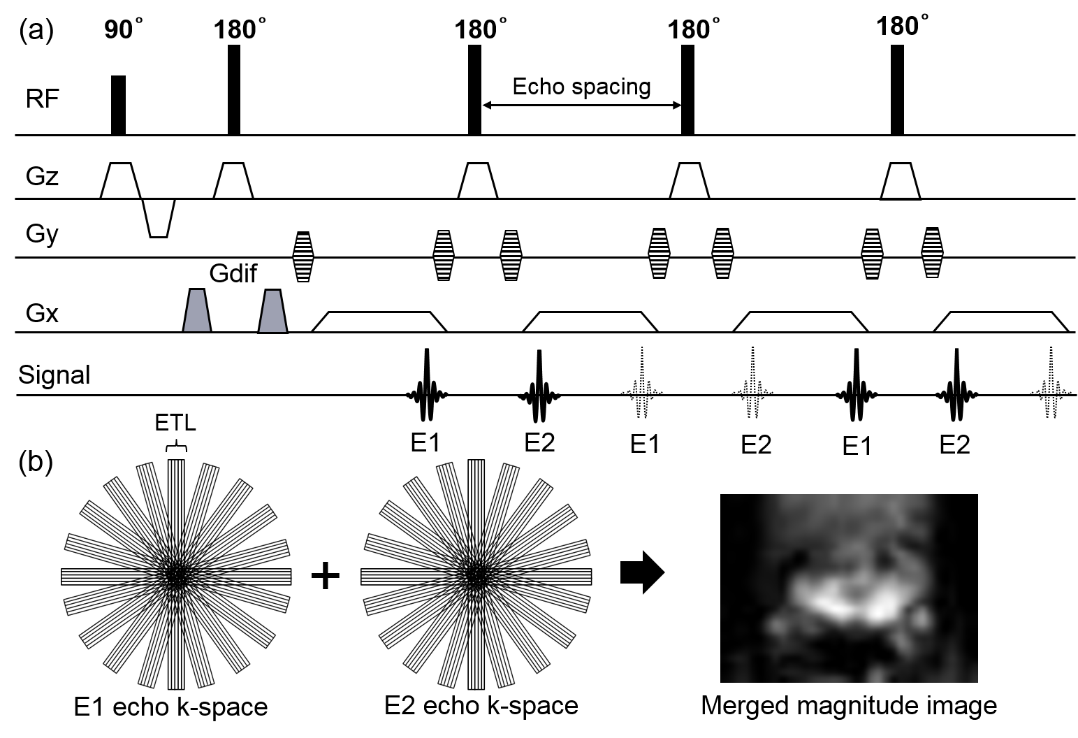
MV-SPLICE scheme: To avoid non-CPMG artifacts, SE (solid lines) and STE (dashed lines) were separated by unbalanced readout gradient (Fig. 1a). Diffusion gradients (shaded area) were applied along the readout direction. Odd (E1) and even (E2) echoes were acquired in separate MultiVane k-space for separate reconstruction. These magnitude images were merged to form the final image.
Subject and equipment: The sequences were implemented on a 3.0-T MR scanner (Philips, Ingenia R5.3). Five healthy volunteers were examined after obtaining informed consent required by hospital review board. EPI, MV-ALSOP, and MV-SPLICE were acquired for image comparison.
Sequence parameters: Axial single-shot EPI, MV-ALSOP, and MV-SPLICE were obtained. For evaluating distortion as mentioned below, axial T2W images were obtained. Sequence parameters are summarized in Table 1.
Validation of MV-SPLICE in brain: To validate whether MV-SPLICE has SNR adequate for apparent diffusion coefficient (ADC) analysis, ADC analysis in white matter (WM) and gray matter (GM) was performed. Calculated ADC maps were generated by using b=0 and b=1000 s/mm2 DWI images. Circular region-of-interests (ROIs) were placed on genu of corpus callosum (GCC) for WM and left and right caudate nucleus head (CNH) for GM. The average ADCs of GCC and CNH were compared among three sequences.
Assessment of ADC and SNR in prostate: Calculated ADC maps were generated by using b=0 and b=1000 s/mm2 DWI images. ROIs were placed on left and right transition zones (TZ), left and right peripheral zones (PZ). The average ADCs of TZ and PZ were compared among three sequences. SNRs in TZ or PZ with b-value of 1000 s/mm2 were calculated as SNR = SAve/SSD, where SAve is the average signal intensity in TZ or PZ, and SSD is mean SD in obturatorius internus muscle. The average SNRs were compared between MV-ALSOP and MV-SPLICE.
Evaluation of distortion in DWI images: Distortion for DWI and T2W images in the anterior-posterior direction was quantified as Distortion = diameter DWI – diameter T2W, where diameter DWI and diameter T2W were diameter in DWI and T2W respectively. The average distortion was compared among three sequences.
Statistical analysis: For each analysis, data are presented as means ± SD. To adjust P values in multiple testing, three groups Holm method was used. For SNR analysis, paired t-test was used. P values less than 0.01 was considered significant.
RESULTS
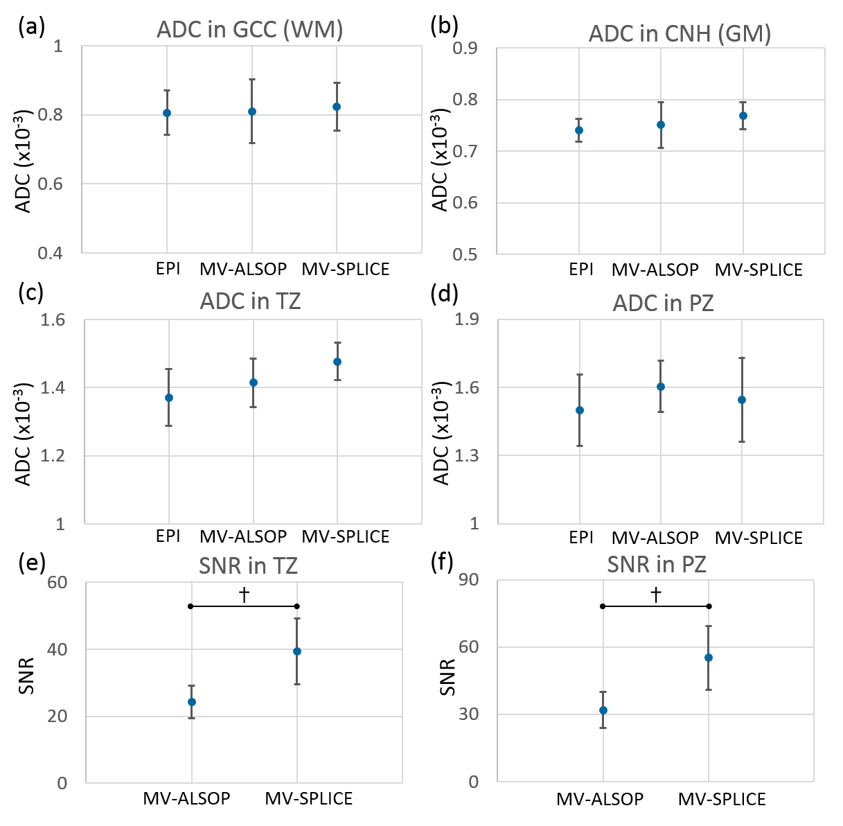
For validation of MV-SPLICE in brain, ADCs of GCC and CNH are shown in Fig. 2a,b. ADCs of GCC and CNH were not significantly different among three sequences and comparable to reference6-7.
ADCs of TZ and PZ were not significantly different among three sequences and comparable to reference8 (Fig. 2c,d). SNRs of TZ and PZ for MV-SPLICE were higher than those of MV-ALSOP; TZ (p<0.0001), PZ (p<0.00001), as shown in Fig. 2e,f.
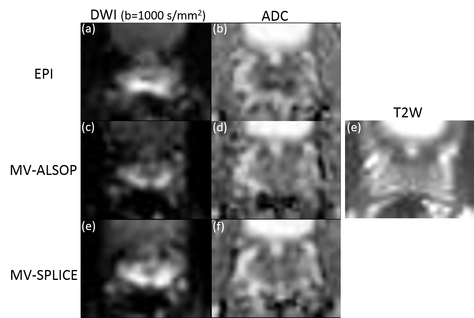
Representative DWI and ADC images for three sequences are shown in Figs. 3. Some distortion of the prostate shape can be observed only in EPI (Fig. 3a). Measured distortion of the prostate in the A-P direction is shown in Fig. 4.
Distortion of EPI image was larger than that of MV-ALSOP and MV-SPLICE; MV-ALSOP (p=0.0048), MV-SPLICE (p=0.0048).
CONCLUSION
We have demonstrated the use of MV-SPLICE for prostate DWI. The results indicate that MV-SPLICE is insensitive to distortion and may provide better diagnosis for prostate cancer compared to conventional EPI and TSE-DWI sequence.Acknowledgements
No acknowledgement found.References
1. Tamada T et al. Diffusion-weighted MRI and its role in prostate cancer. NMR Biomed. 2014;27:25-38
2. Norris DG et al. On the application of ultra-fast rare experiments. Magn Reson Med. 1992;27:142-164
3. Alsop DC. Phase insensitive preparation of single-shot RARE: Application to diffusion imaging in humans. Magn Reson Med.1997;38:527-533
4. Schick F. SPLICE: Sub-second diffusion-sensitive MR imaging using a modified fast spin-echo acquisition mode. Magn Reson Med.1997;38:638-644,
5. Pipe JG et at. Multishot diffusion-weighted FSE using PROPELLER MRI. Magn Reson Med. 2002;47:42-52
6. Osamu A et al. Normal aging in the central nervous system: quantitative MR diffusion-tensor analysis. Neurobiol Aging. 2002;23:433-441,
7 Helenius J et al. Diffusion-Weighted MR Imaging in Normal Human Brains in Various Age Groups. AJNR Am J Neuroradiol. 2002;23:194-199
8. Wang X et al. High-b-value diffusion-weighted MRI for the detection of prostate cancer at 3 T. Clin Radiol. 2014;69:1165-1170
Figures