0326
Zero Time of Echo imaging with an Adiabatic Fat Suppression Pulse at 7T1GE Healthcare, Pisa, Italy, 2Imago7, Pisa, Italy, 3IRCCS Stella Maris, Pisa, Italy, 4GE Healthcare, Munich, Germany, 5GE Healthcare, Waukesha, WI, United States, 6University of Pisa, Pisa, Italy, 7King's College London, London, United Kingdom
Synopsis
We present results using a Zero Time of Echo sequence ("Silent") with an Adiabatic Fat Suppression Pulse (ASPIR). As well as providing fat suppression that is relatively robust to field inhomogeneities at 7T, ASPIR-Silent also generates images with an additional off-resonant contrast. We show this contrast derives primarily from Magnetisation Transfer effects, and give an in vivo example.
Introduction
Silent, a 3D-radial “Zero Time of Echo” (ZTE) sequence, acquires 7T images with proton density or T1-weighted contrast1. Fat suppression can be applied before the Silent imaging module of typically 32-512 spokes (“segment”) using a standard chemically-selective pulse and crusher-gradient combination.
The fat signal has a blurring effect in radial sequences, but B0 and transmit B1 inhomogeneities at 7T limit the efficacy of conventional fat saturation pulses. STIR relies on T1 rather than frequency to suppress fat, reducing the effect of B0 inhomogeneities at the cost of adverse T1 weighting. Spectral Partial Inversion Recovery sequences2 are as effective as STIR but perturb water signal much less. Adiabatic SPIR (ASPIR, SPAIR) pulses apply the fat-selective inversion in an adiabatic manner3, reducing dependence on B1 homogeneity4. Such approaches have been used at 3T in breast and liver imaging. However, adiabatic pulses rely on off-resonant and spin-locking approaches to nutate the spins3, and at the offsets required for 7T, these effects may not be negligible.
We applied an adiabatic fat suppression pulse (ASPIR) before each Silent segment. This suppressed the fat signal but introduced an additional image contrast attributable to Magnetisation Transfer.
Methods
ZTE images were acquired with and without ASPIR fat suppression on a General Electric MR 950 7T scanner (gradients=50mT/m, slew=200T/m/s; Nova Medical birdcage transmission coil, Nova Medical 32-channel receive array coil) in the head. The ASPIR pulse was applied offset from the imaging frequency by about 1500Hz; it was a hyperbolic secant pulse (duration=40ms, amplitude=0.1G, bandwidth=2000Hz).A delay of approximately 100ms between the ASPIR inversion and the centre of the Silent segment meant the average signal from fat was close to zero. Image parameters: receive bandwidth=±31.25kHz, TR=130ms, spokes/segment=64, TE=16us, Flip Angle=2°, Field of View=18cm, matrix=128x128x128, voxel size=1.4mm cube, scan time=48s.
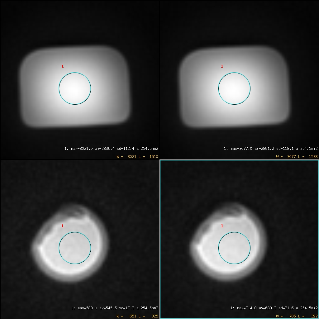
Phantom experiments were performed on a hard-boiled egg and on a bottle of Manganese Chloride (MnCl2). Regions of interest were measured in the two phantoms in the Silent images without (Su) and with (Ss) ASPIR preparation (Su).
Results
Fig 1 shows ROI placement in the phantom images.
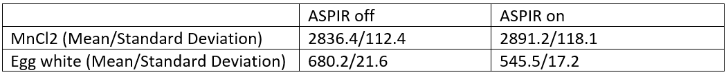
Fig 2 tabulates the mean and standard deviation of the ROI measurements in the phantom images
The Magnetisation Transfer Ratio (MTR), (Su-Ss)/Su, for the ROI in this egg white was calculated as 19.8% (percentage units). The observed change in the ROI of the MnCl2 phantom was small compared to the standard deviations.
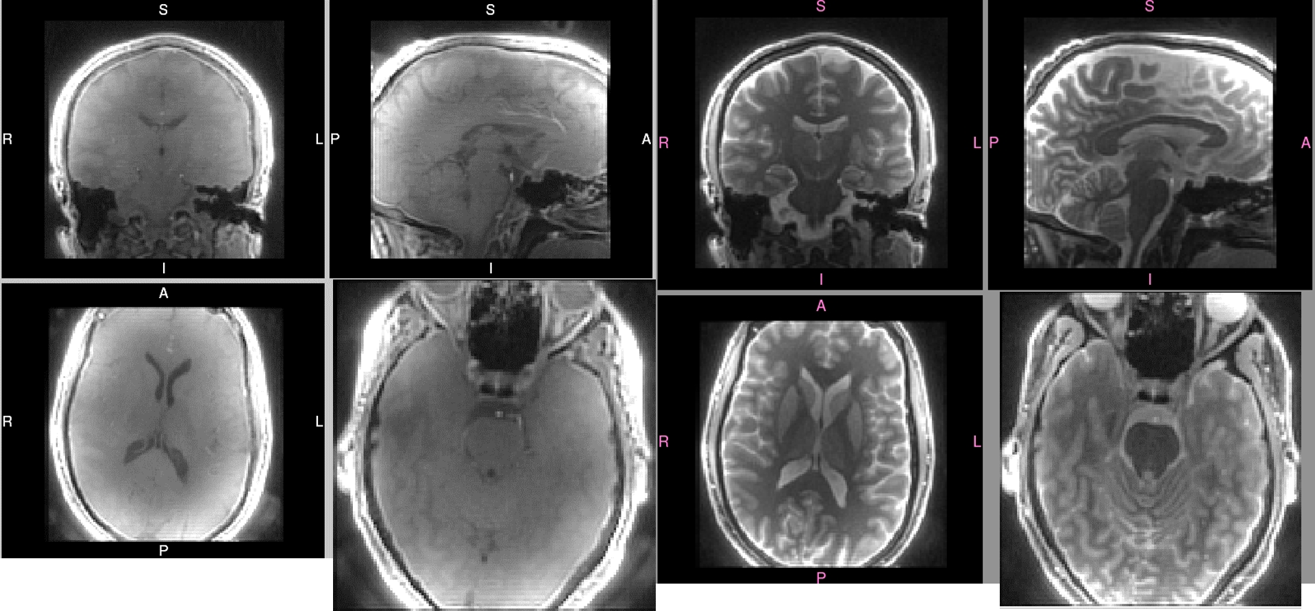
Fig 3 shows Silent images taken without and with ASPIR preparation in the head. Fat suppression is good in the ASPIR-prepared image, removing signal from the fatty sheaths surrounding the optic nerves.
Discussion
The phantom experiments help separate two possible effects of the ASPIR pulse: T1-rho and Magnetisation Transfer.
1) The ASPIR pulse parameters (duration=40ms, amplitude=0.1G) resemble those used in T1-rho-weighted imaging5. During the ASPIR fat inversion, the adiabatic pulse spin-locks the fat magnetisation to an extent determined by the pulse shape, and local transmit B1. The water component may also experience a spin-locking effect, even if it undergoes zero NET nutation during the pulse.
2) MTR imaging experiments use an offset of 1000-2000Hz, which is considered to induce a genuine MT effect6. The ASPIR offset (1500Hz) will, according to a standard MT model7, saturate short-T2 components which will transfer their magnetisation to the "liquid" observed pool.
Others8 showed by simulation and experiment that a Gaussian off-resonant pulse applied with offset 1500Hz produces very little T1-rho weighting in an MnCl2 phantom. We observed no significant change in our MnCl2 phantom when the ASPIR pulse was applied, which suggests the pulse is not introducing significant T1-rho weighting.The egg white signal is attenuated by the ASPIR pulse. MnCl2 exhibits no MT, but the denatured protein in egg white shows an MT effect9. A full simulation of the MT and T1-rho effects of the ASPIR pulse is beyond the scope of this current work, but such models are being developed10. We conclude that the contrast we have observed in ASPIR-Silent images arises mostly from Magnetisation Transfer effects.
Conclusions
Silent images prepared with an ASPIR pulse provide a contrast weighted by magnetisation transfer, which could be of clinical interest in many areas. The fortuitous "Buy One, Get One Free" dual effect of fat suppression and magnetisation transfer is welcome at 7T, where SAR limitations often occur. Others have previously investigated UTE Magnetisation Transfer sequences11. ASPIR-prepared Silent offers an intriguing step forward in the ability to combine the indirect observation of substances with short T2 via the MT effect with their direct observation using the Zero Time of Echo property of Silent.Acknowledgements
No acknowledgement found.References
1) Costagli M, Symms MR, Angeli L et al. Assessment of Silent T1-weighted head imaging at 7 T. Eur Radiol. (2016) 26(6):1879-88
2) Kaldoudi E, Williams SCR, Barker GJ, Tofts PS. A Chemical Shift Selective Inversion Recovery Sequence for Fat-Suppressed MRI: Theory and Experimental Validation. Magnetic Resonance Imaging (1993) 11: 341-355
3) Garwood M, DelaBarre L. The return of the frequency sweep: designing adiabatic pulses for contemporary NMR. J Magn Reson. (2001) 153(2):155-77.
4) Iyama Y, et al. Fat Suppressed Contrast-Enhanced T1-Weighted Dynamic Magnetic Resonance Imaging at 3T: Comparison of Image Quality Between Spectrally Adiabatic Iversion Recovery and the Multiecho Dixon Technique in Imaging of the Prostate. J Comput Assist Tomogr (2017) 41: 382–387
5) Villanueva-Meyer JE, Barajas RF, Mabray MC et al. Differentiation of brain tumor-related edema based on 3D T1rho imaging. European Journal of Radiology (2017) 91: 88-92
6) Bagary MS, Hutton SB, Symms MR et al. Structural neural networks subserving oculomotor function in first-episode schizophrenia. Biol Psychiatry (2004) 56(9):620-7
7) Sled JG, Pike GB. Quantitative imaging of magnetization transfer exchange and relaxation properties in vivo using MRI. Magn Reson Med. (2001) 46(5):923-31.
8) Moran PR, Hamilton CA. Near-resonance spin-lock contrast. MRI (1995) 13(6):837-846.
9) Finelli DA, Hurst GC, Frank HA et al. Analysis of magnetization transfer effects on T1-weighted spin-echo scans using a simple tissue phantom simulating gadolinium-enhanced brain lesions. J Magn Reson Imaging. (1997) 7(4):731-8.
10) Malik SJ, Teixeira RPAG, Hajnal JV. Extended Phase Graph formalism for systems with Magnetization Transfer and Chemical Exchange. Submitted for publication; retrieved 5th Nov 2017 from https://arxiv.org/ftp/arxiv/papers/1709/1709.00832.pdf
11) Springer F, Martirosian P, Machann J et al. Magnetization Transfer Contrast Imaging in Bovine and Human Cortical Bone Applying an Ultrashort Echo Time Sequence at 3 Tesla. Magnetic Resonance in Medicine (2009) 61:1040–1048
Figures