0316
An Optimized Single-shot Sequence for Fast T2w Imaging of the Brain1Electrical and Computer Engineering, University of Arizona, Tucson, AZ, United States, 2Medical Imaging, University of Arizona, Tucson, AZ, United States, 3Biomedical Engineering, University of Arizona, Tucson, AZ, United States
Synopsis
T2-weighted imaging of the brain using single shot sequences such as HASTE suffer from reduced spatial resolution, blurring artifacts and decreased conspicuity of small lesions. We present an analytic framework to design the refocusing flip angles for the HASTE sequence tailored for brain imaging. The flip angles are optimized to minimize SAR and blurring, and maximize SNR. The utility of this sequence is demonstrated by incorporating it in a brain tumor protocol and comparing its performance to conventional T2w Turbo Spin Echo in 21 patients.
Introduction
T2-weighted (T2w) imaging protocols play an important role in neuroimaging for the diagnosis of various pathologies. Turbo Spin Echo (TSE / FSE) has been the “workhorse” sequence for 2D T2w imaging, despite the longer acquisition times and vulnerability to artifacts from subject motion. While single-shot alternatives such as SSFSE / HASTE have been explored in the past for their motion robustnes1-4, they suffer from reduced spatial resolution, blurring and decreased lesion conspicuity. As a result, single-shot sequences are used only in pediatric imaging applications5,6. Recently, an empirical approach was described7 to optimize the refocusing flip angles for SSFSE. In this work, we reformulated it in an optimization framework to develop a HASTE-VFA sequence tailored for brain imaging. The refocusing flip angle train of HASTE-VFA was optimized to minimize SAR and blurring, and maximize SNR. We demonstrate the utility of this sequence by incorporating it in a brain tumor protocol and comparing its performance to conventional T2w TSE in 21 patients.Methods
Flip Angle Design:
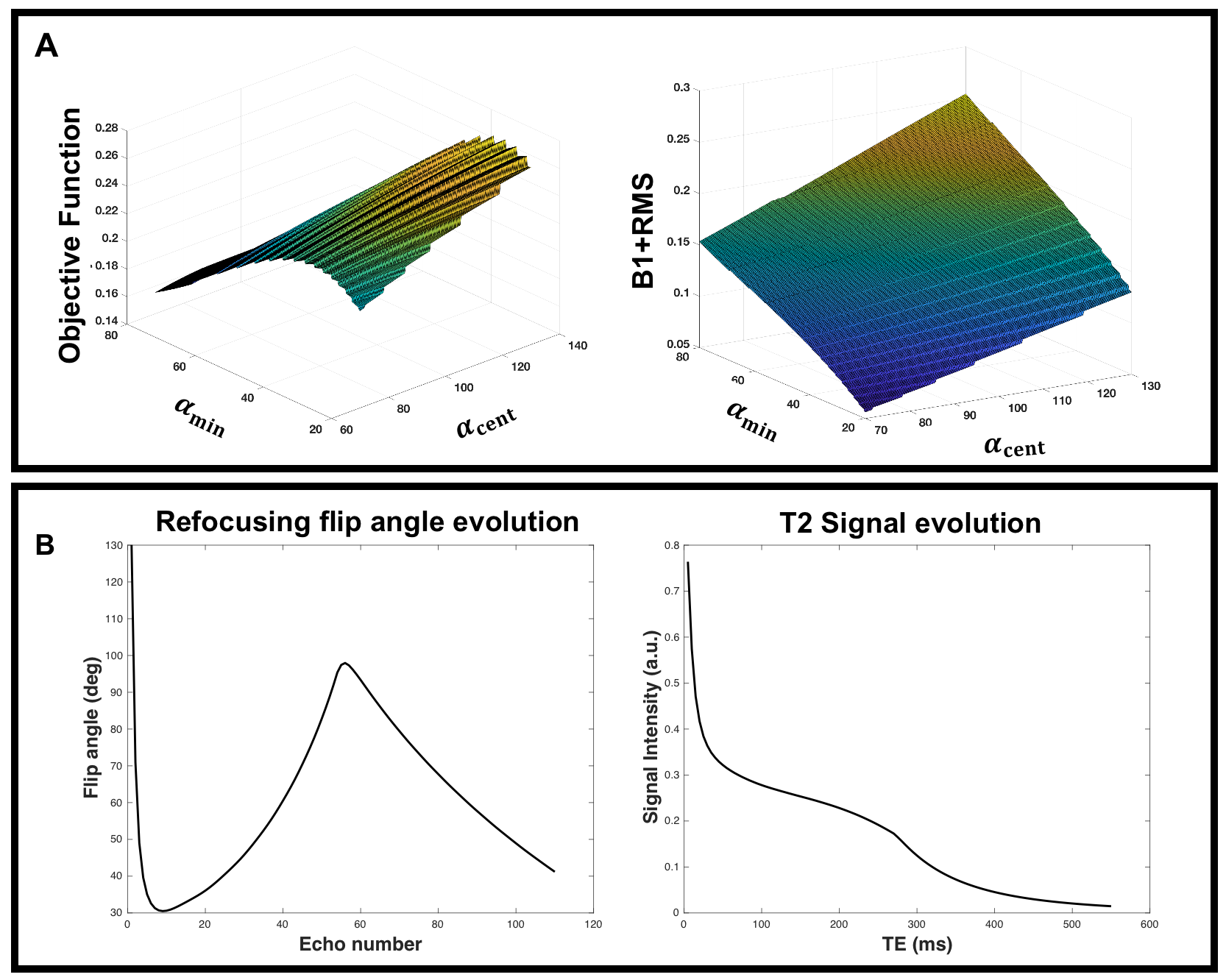
A commercial Siemens HASTE sequence was modified to incorporate the refocusing flip angle modulation scheme proposed by Busse et al8. In this scheme, the flip angles are parametrized by four control angles $$$\vec{\alpha} = [\alpha_{min},\alpha_{cent},\alpha_{end},\alpha_{max}]$$$. These control angles were optimized to achieve the following: (1) maximize the PSF peak and minimize the mean of the outer edge of PSF to improve the spatial resolution (2) maximize the signal at the central echo to maximize the SNR (3) minimize the SAR of the sequence to decrease the imaging TR which is usually SAR limited. In addition, constraints were added to limit the choice of $$$\alpha_{min}$$$ as it has been shown9-11 to be proportional to signal dephasing due to motion. Since the signal evolution is insensitive to $$$\alpha_{end}$$$, it was fixed at 45° and based on8 $$$\alpha_{max}$$$ was set to 130°.
Representing the problem as a multi-objective optimization:
$$\max_{\alpha_{min},\alpha_{cent}} psf_{max}(\alpha_{min},\alpha_{cent}) \& \min_{\alpha_{min},\alpha_{cent}} psf_{outer}(\alpha_{min},\alpha_{cent}) \& \max_{\alpha_{min},\alpha_{cent}} sig_{cent}(\alpha_{min},\alpha_{cent})\\ \text{s.t. } \text{ sar }(\alpha_{min},\alpha_{cent}) < \eta;\\ \alpha_{min} > \delta$$
The objective function can be reformulated by the $$$\epsilon$$$-constraint method11:
$$\max_{\alpha_{min},\alpha_{cent}} psf_{max}(\alpha_{min},\alpha_{cent}) - psf_{outer}(\alpha_{min},\alpha_{cent}) \\ \text{ s.t. } sig_{cent}(\alpha_{min},\alpha_{cent}) < \tau; \\ \text{ sar }(\alpha_{min},\alpha_{cent}) < \eta; \\\alpha_{min} > \delta$$
The optimization problem was solved using a genetic algorithm implemented in MATLAB. A two-dimensional surface plot of the objective function is shown in Figure 1A showing dependence on the individual parameters $$$\alpha_{min}$$$ and $$$\alpha_{cent}$$$. The flip angle evolution corresponding to the optimal solution along with the T2 decay curve is shown in Figure 1B.
In vivo Imaging:
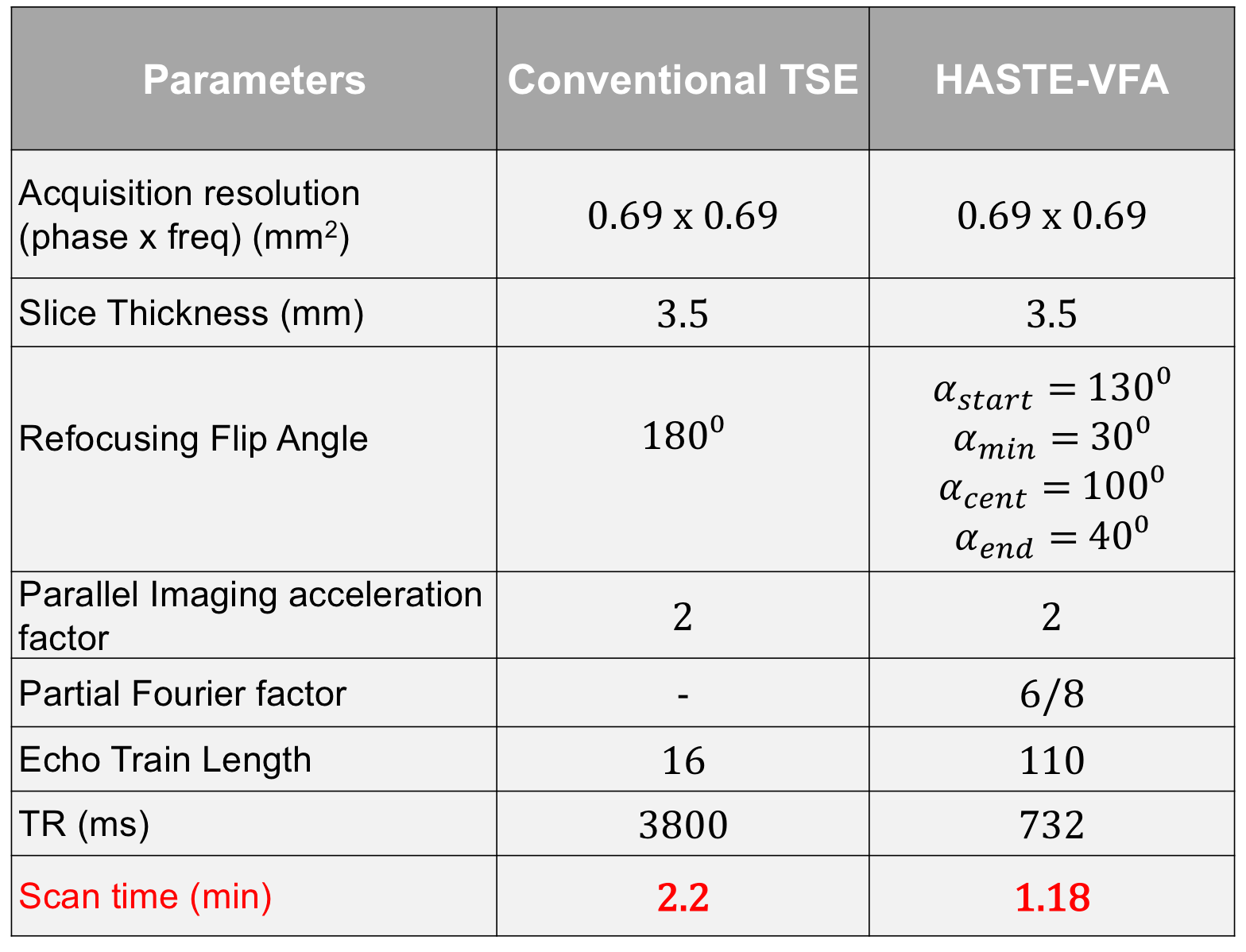
The optimized sequence was implemented and tested on a Siemens 3T scanner. The sequence was added to clinical brain tumor protocol at our institution as an addition to the routine T2w TSE sequence. With the approval of the Institutional Review Board and with informed consent, data was acquired from 21 patients. The sequence parameters are shown in Table 1.
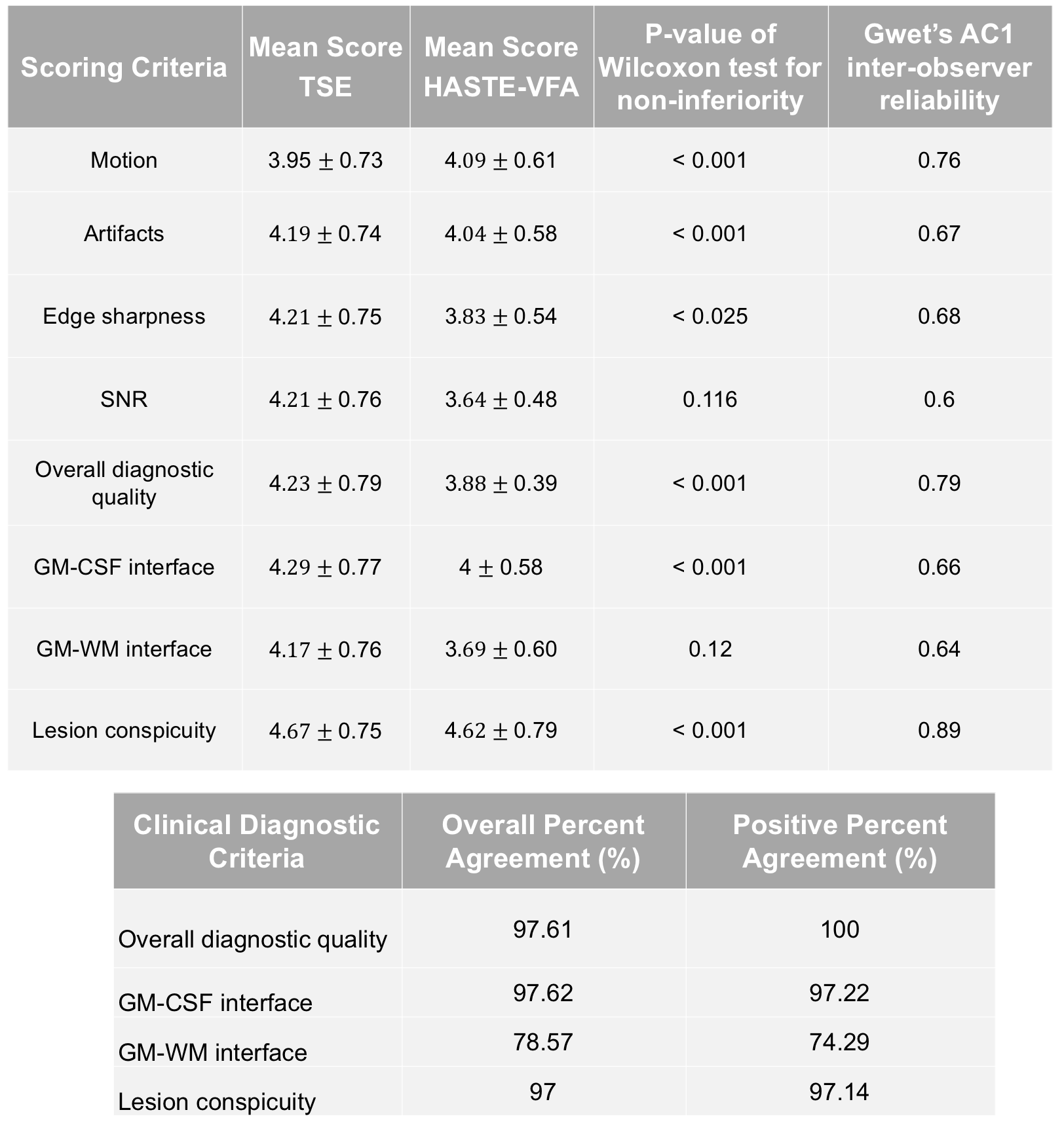
The image quality of the HASTE-VFA and the TSE sequences were assessed by two experienced neuro-radiologists in a blinded fashion, with a 2-week interval to avoid recall bias. Images were graded on a scale of 1-5 and scores were assigned based on the criteria listed in Table 2. Non-inferiority analysis between HASTE-VFA and TSE was done using a one-sided Wilcoxon signed rank test with a non-inferiority margin $$$\Delta$$$=0.5, $$$\alpha$$$=0.025 and Gwet’s AC1 metric was computed to measure the inter-observer variability.
Results
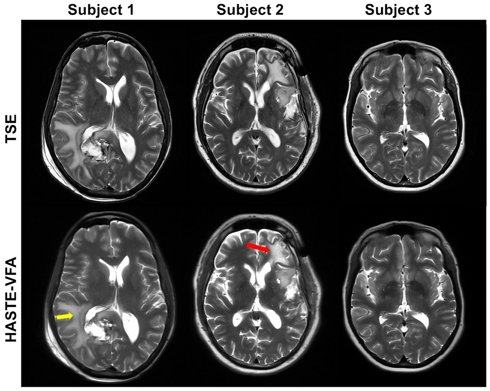
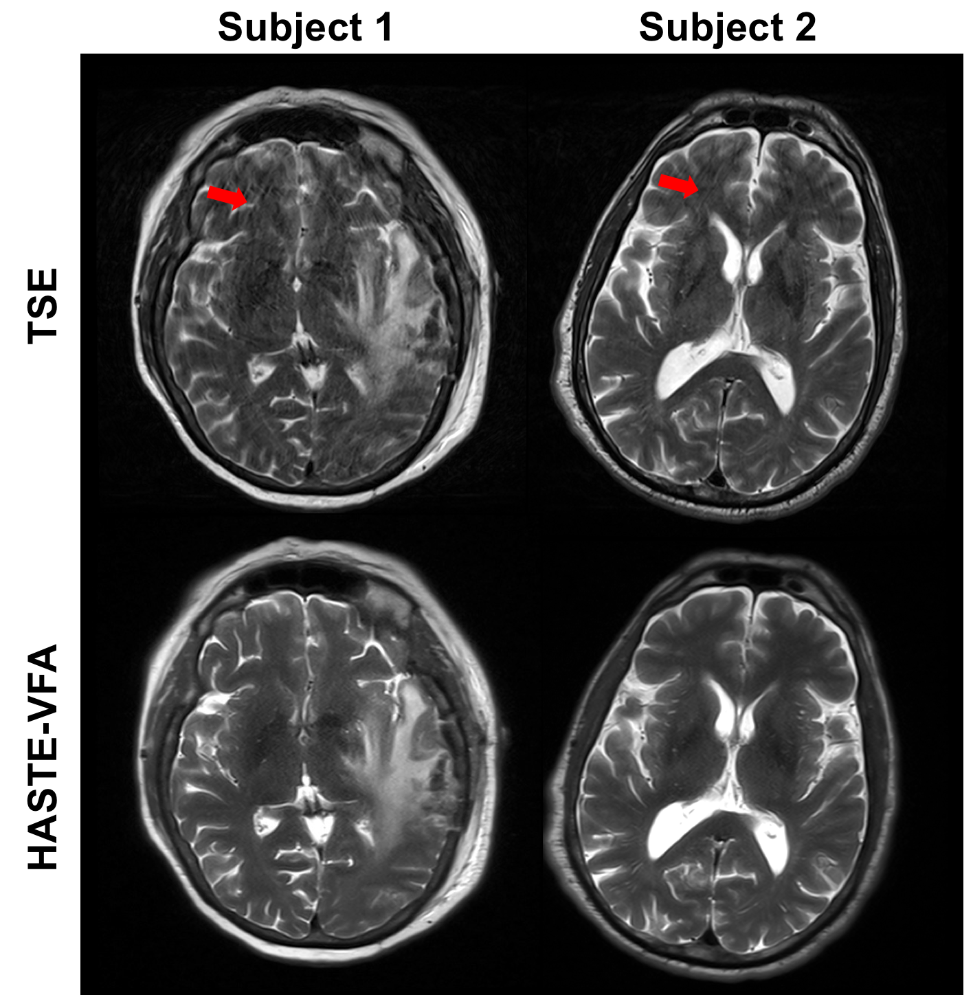
Representative axial images of the brain for 3 subjects are shown in Figure 2. Note that HASTE-VFA generates images at comparable contrast and SNR to the TSE. Figure 3 shows images from 2 subjects where the TSE scan exhibited severe motion artifacts. The proposed sequence is more robust because of the single shot acquisition and shorter scan time, whilst maintaining image quality.
The mean scores for the various criteria are shown in Table 2 along with the p-value from the Wilcoxon test. The median difference in scores between the two sequences for was significantly (p-value < 0.025) less than the non-inferiority margin ($$$\Delta$$$=0.5) for all criteria except SNR and GM-WM conspicuity, implying that HASTE-VFA is not inferior to conventional TSE. The inter-observer reliability between the readers indicate a moderate to strong agreement for all criteria. The high positive percent agreement and overall agreement further affirm the diagnostic utility of HASTE-VFA.
Conclusion
We have developed an optimization framework to design variable refocusing flip angle trains for HASTE sequences and have demonstrated its utility in brain imaging. HASTE-VFA generates images of comparable contrast and image quality as conventional TSE and being a 2x faster scan is more robust to subject motion related artifacts.Acknowledgements
No acknowledgement found.References
1. Filippi, M, Rocca, M.A, Wiessmann, M, Mennea, S, Cercignani, M, Yousry, T.A, Sormani, M.P, Comi, G. A comparison of MR imaging with fast-FLAIR, HASTE-FLAIR, and EPI-FLAIR sequences in the assessment of patients with multiple sclerosis. AJNR Am J Neuroradiol. 1999;20:1931–1938
2. Ge, Y., Korogi, Y., Sugahara, T. et al. Neuroradiology (2001) 43: 1046
3. Sugahara T, Korogi Y, Hirai T et al. Comparison of HASTE and segmented-HASTE sequences with a T2-weighted fast spin-echo sequence in the screening evaluation of the brain. Am J Roentgenol. 1997 Nov;169(5):1401-10.
4. Patel MR, Klufas RA, Alberico RA, Edelman RR. Half-fourier acquisition single-shot turbo spin-echo (HASTE) MR: comparison with fast spin-echo MR in diseases of the brain. Am J Neuroradiol. 1997 Oct;18(9):1635-40.
5. Penzkofer AK, et al. MR imaging of the brain in pediatric patients: diagnostic value of HASTE sequences. AJR Am J Roentgenol. 2002;179(2):509–14.
6. Patel DM, Tubbs RS, Pate G, et al. Fast-sequence MRI studies for surveillance imaging in pediatric hydrocephalus. J Neurosurg Pediatr. 2014;13:440–447.
7. Loening AM, Saranathan M, et al. Increased Speed and Image Quality in Single-Shot Fast Spin Echo Imaging Via Variable Refocusing Flip Angles. JMRI 2015 42 : 1747-1758
8. Busse RF, Brau A, Vu A, et al. Effects of refocusing flip angle modulation and view ordering in 3D fast spin echo. Magn Reson Med 2008; 60:640–649
9. Busse RF. Flow Sensitivity of CPMG Sequences with Variable Flip Refocusing and Implications for CSF Signal Uniformity in 3D-FSE Imaging. In: Proc 14th Annual Meeting ISMRM, Berlin; 2006
10. Madhuranthakam AJ, Busse RF, Brittain JH, Rofsky NM, Alsop DC. Sensitivity of low flip angle SSFSE of the abdomen to cardiac motion. In: Proc 15th Annual Meeting ISMRM, Berlin; 2007 (abstract 2523).
11. Litwiller DV, Holmes JH, Saranathan M, et al. Sensitivity of modulated refocusing flip angle single-shot fast spin echo to impulsive cardiac-like motion. In: Proc 22nd Annual Meeting ISMRM, Milan; 2014
12. Miettinen, KM. Nonlinear multi-objective optimization. International series in operations research & management science. Kluwer Academic Publishers, Boston. 1998
Figures