0314
Silent T2* Encoding using ZTE Combined with Gradient-Echo Burst (BURZTE)Rolf F Schulte1, Guido Buonincontri2, Mauro Costagli2, Anne Menini3, Florian Wiesinger1, and Ana Beatriz Solana1
1GE Healthcare, Munich, Germany, 2IMAGO7 Foundation, Pisa, Italy, 3GE Healthcare, Menlo Park, CA, United States
Synopsis
ZTE image encoding was combined with burst imaging by reversing segments of 3D radial spokes both in time and amplitude. This recalls gradient echoes for the individual spokes. Multiple burst echoes can be acquired by repeating the trajectories. This “burzte” pulse sequence encodes T2* in a silent manner.
Introduction
Burst imaging enables relatively rapid and silent encoding in MRI [1,2] via encoding multiple k-space lines with a train of short and evenly-spaced RF block pulses. Another silent acquisition method is ZTE [3,4,5], where spatial information is encoded with 3D radial trajectories, ramping the gradients already before the RF pulse with slow, hence silent switching from one spoke to the next one. In this work, ZTE is combined with gradient-echo burst encoding for silent T2* weighted imaging and mapping with reduced susceptibility artefacts. This is similar to an approach called Looping Star, where gradient-echoes are generated after ZTE excitation by creating k-space loops [6].Methods
After a block of standard ZTE encoding with multiple 3D radial k-space spokes, the direction of traversing k-space is reversed, thus recalling gradient echoes for each spoke/excitation (Fig.1). This was implemented into a ZTE sequence by reversing gradients and acquisition, switching off the RF pulses during the burst acquisition part and repeating theses gradient trajectories to collect multiple echoes. The pulse sequence, named T2*-BURZTE (Fig.2), was implemented on a GE MR750w scanner. Data is reconstructed automatically on the scanner using standard 3D gridding and FFT. Quantitative T2* maps were extracted by fitting the images of three echo times to an exponential decay (omitting even acquisition trains). Quantitative susceptibility maps (QSM) were generated by feeding the phase of the second burst echo (TE=24.4ms) to a conventional post-processing pipeline that included phase unwrapping and background field removal [7]. In vivo feasibility was demonstrated by imaging the brain of healthy volunteers.Results and Discussion
It is possible to acquire high-quality T2* weighted brain images in a silent manner. The image quality of even acquisitions, in particular train two (first burst echo), is impaired, most likely due to inconsistent echo-times from reversing the gradients. Fitting multiple echoes to an exponential decay enables extraction of quantitative T2* maps (Fig.3), while the phase can be used for generating QSM maps (Fig.4). It is also possible to acquire gradient-echoes of a full isotropic 3D volume in a few seconds by reducing the number of spokes (here by half) and by increasing the voxel size to (3mm)3. Further scan time reductions (to enable applications such as fMRI) are possible by reducing sampling further and combining BURZTE with advanced reconstructions, such as parallel imaging. The SNR-limitations of burst [2] are alleviated by a full 3D acquisition. T2*-BURZTE does not require calibration scans and additional artefact reduction methods, as for instance needed for EPI.Conclusion
T2*-BURZTE enables silent and rapid acquisition of gradient recalled burst echoes. Clinical applicability is facilitated by implementing support for arbitrary parameter selection into the pulse sequence (including off-isocentre modulation) and image reconstruction directly on the scanner.Acknowledgements
No acknowledgement found.References
- Burst imaging. Hennig J, Hodapp M. MAGMA. 1993; 1:39-48.
- Burst imaging—Can it ever be useful in the clinic? Doran SJ, Bourgeois ME, Leach MO. Concepts Magn Reson Part A. 2005; 26A(1):11-34.
- Fast imaging in liquids and solids with the back-projection low angle shot (BLAST) technique. Hafner S. Magn Reson Imaging 1994;12:1047-1051.
- Ultra-fast imaging using low flip angles and fids. Madio DP, Lowe IJ. Magn Reson Med 1995;34:525–529.
- Density of organic matrix of native mineralized bone measured by water- and fat-suppressed proton projection MRI. Wu Y, Ackerman JL, Chesler DA, Graham L, Wang Y, Glimcher MJ. Magn Reson Med. 2003;50:59-68.
- Looping Star: A Novel, Self-Refocusing Zero TE Imaging Strategy. Solana AB, Menini A, Wiesinger F. ISMRM 2016, #104.
- The impact of white matter fiber orientation in single-acquisition quantitative susceptibility mapping. Lancione M, Tosetti M, Donatelli G, Cosottini M, Costagli M. NMR in Biomedicine 2017;e3798.
Figures
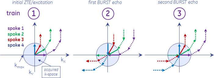
Fig. 1: k-space view of ZTE-burst encoding (4 spokes per segment). The
encoding of spokes 1-3 is stored in the higher-frequency parts of k-space (acquisition
train 1). When reversing the gradients in time and amplitude (and switching off
RF), the same k-space pathway is traversed backwards (acquisition train 2).
This leads to a first burst gradient echo inside the kmax circle, where TE per
spoke is different. When reversing again the gradients, a second BURST gradient
echo is acquired where TE per spokes is equal. Multiple burst gradient-echoes
can be collected by repeating trains 2 and 3.
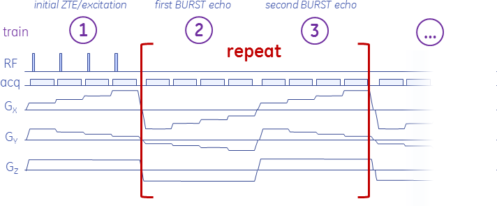
Fig. 2: Sequence diagram for T2*-BURZTE. Gradients are reversed in time and
amplitude to recall burst gradient echoes (Fig.1). Repeating 2 and 3 enables
recalling multiple echoes. Different spokes of the 3D radial k-space are
encoded in different segments. Because k-space contains only high-frequency
components after the odd trains, it is possible to start directly with the next
segment (recovery time=0ms) without the need for crusher gradients.
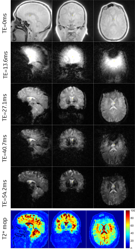
Fig. 3: In vivo
2mm-resolution brain scan. The ZTE/echo images were used to fit a quantitative
T2* map (bottom row).
(FOV=(20cm)3, resolution=(2mm)3,
matrix size=1003, flip angle=3°, 5 acquisition trains, BW=±31.25kHz, acquisition
duration=2:53min).
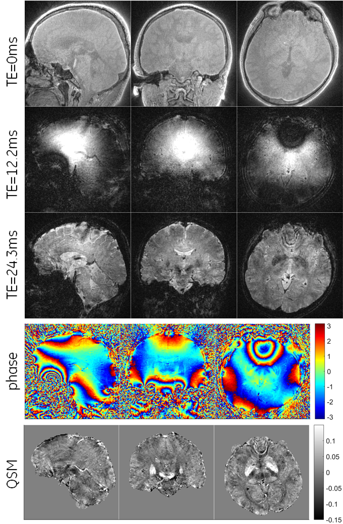
Fig. 4: In vivo
1mm-resolution brain scan with three acquisitions. Phase and QSM maps are shown
in the bottom rows.
(FOV=(19.2cm)3, resolution=(1mm)3,
matrix size=1923, flip angle=3°, 3 acquisition trains, BW=±31.25kHz, acquisition
duration=11:24min).
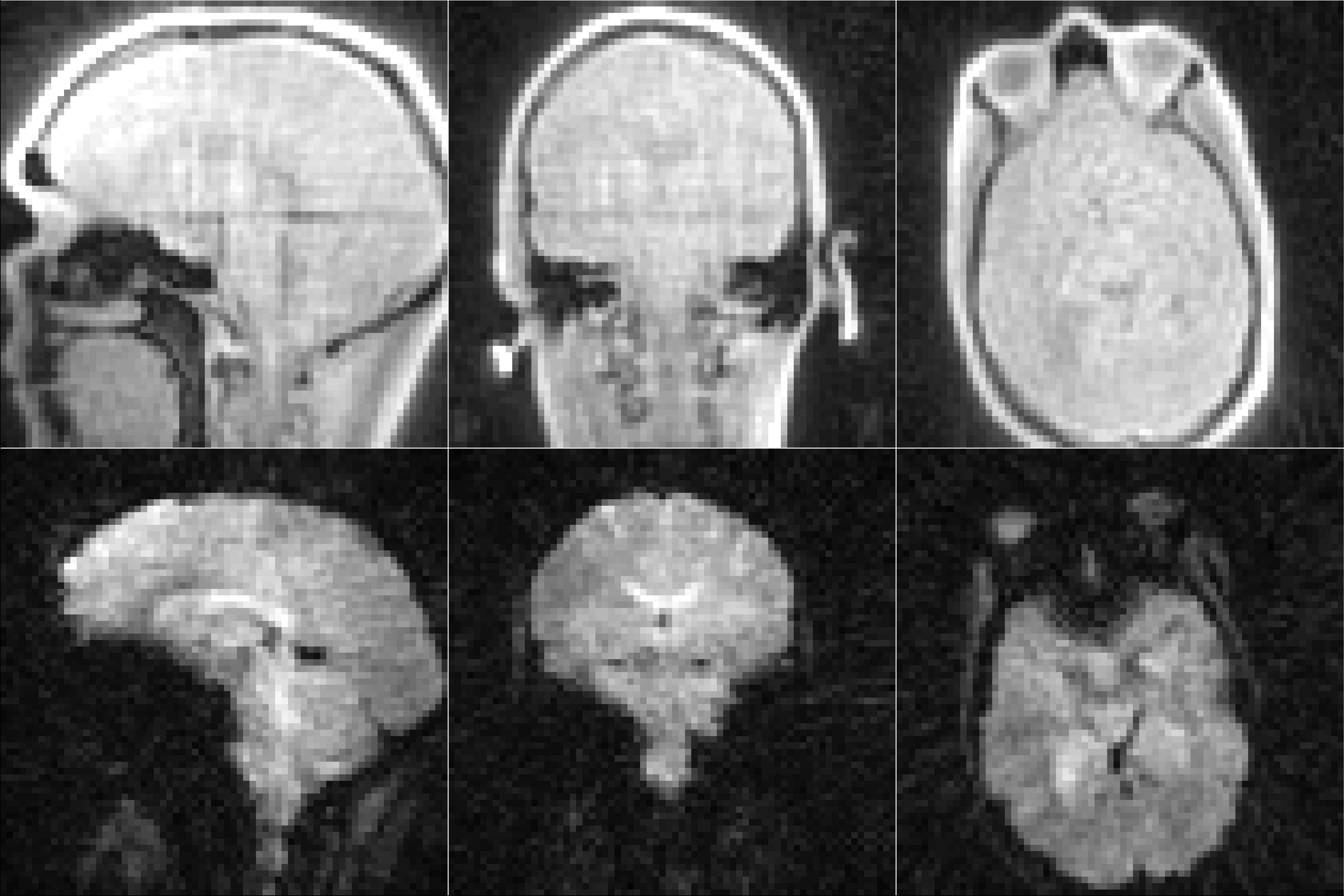
Fig. 5: In vivo 3mm-resolution, with short acquisition duration. From the
three acquisitions, only the ZTE and the second burst echo (TE=23.3ms) are
shown.
(FOV=(21cm)3, resolution=(3mm)3, matrix
size=703, flip angle=3°, 3 acquisition trains, nex=0.5, BW=±31.25kHz,
acquisition duration=11.5sec).