0305
Arterial Spin Labeled Input Function (ASLIF): signal acquisition during pseudo-continuous arterial spin labeling1Fraunhofer MEVIS, Bremen, Germany, 2University Bremen, Bremen, Germany, 3mediri GmbH, Heidelberg, Germany
Synopsis
In ASL pseudo-continuous labeling (pCASL) is often used as a labelling scheme due to its increased SNR compared to pulsed variants. After labeling and a subsequent post-labeling delay, the amount of labeled blood in the organ of interest is acquired. In this abstract, we describe a new approach, which allows to measure the blood signal while it is labeled. Results are presented for a four-phase Hadamard-encoded pCASL sequence. This will ultimately allow for a realtime monitoring of the arterial input function in pCASL.
Introduction
In ASL experiments pseudo-continuous labeling (pCASL) is often used as a labelling scheme due to its increased SNR compared to pulsed variants. However, pCASL is sensitive to local field inhomogeneities within the labelling area. Furthermore, labelling efficiency might also depend on other factors like flow velocity. Therefore, a deficit of labelled blood in different vascular regions as measured with pCASL might not necessarily reflect a perfusion deficit. Several approaches were taken to ensure proper labelling. However, it remains challenging to rely on proper delivery of sufficiently labelled blood to the organ of interest. In this work, we describe an approach, where the effect of pCASL is directly imaged during the labeling process.Methods
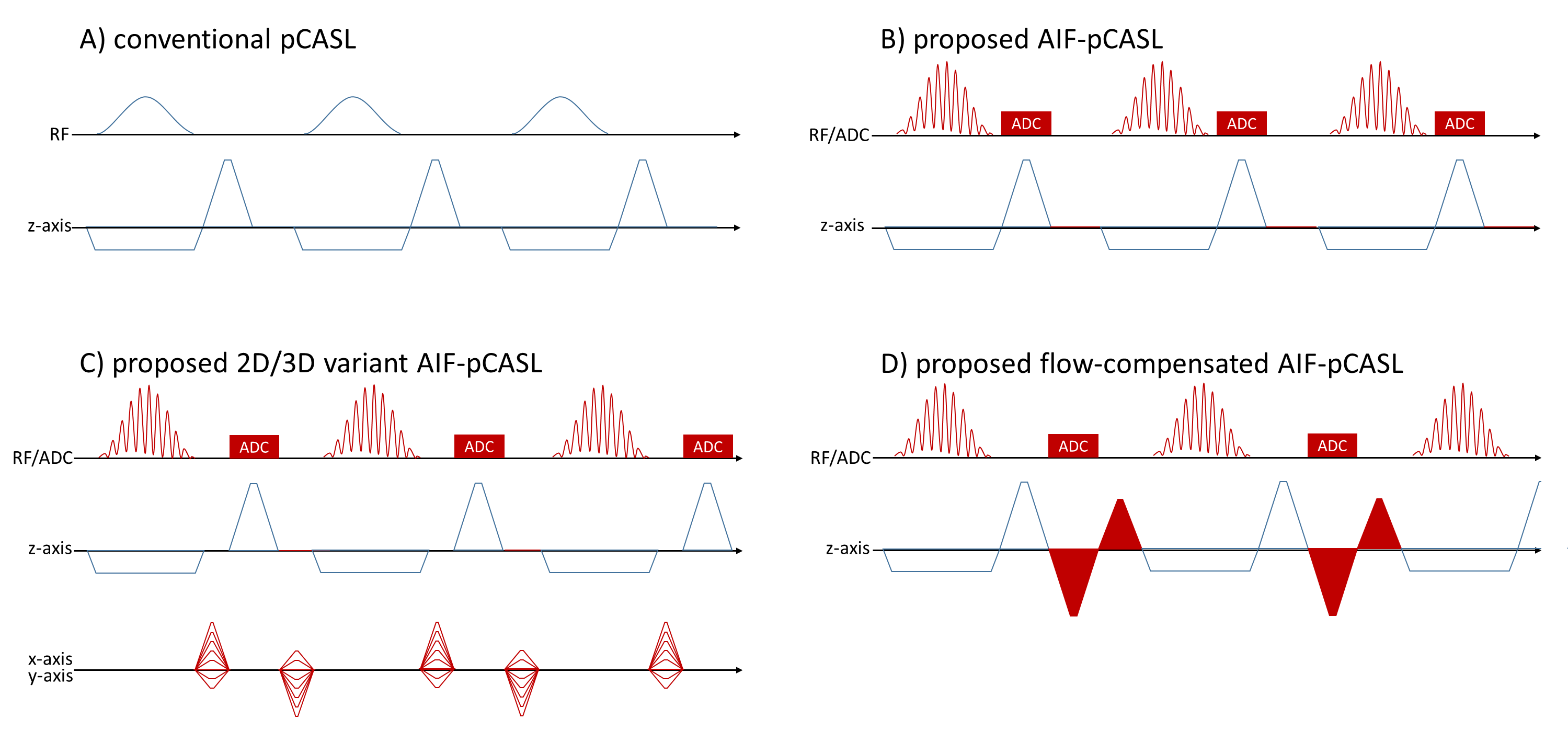
In a typical pCASL preparation, a slice-selective Hanning-shaped RF pulse of 500 us duration is applied about every millisecond. In this work, this RF-RF intervall is called TRpCASL. For labeling the phase of the RF pulse remains the same, while it is changing by 180 degree from pulse to pulse for the control experiment. Essentially, this is identical to a steady state free precession (SSFP) sequence commonly used for imaging1. However, in pCASL a net gradient along the flow direction is applied (i.e. z-axis). This residual gradient moment makes the echo train an unbalanced SSFP with partial dephasing2. In Figure 1A a typical pCASL sequence is depicted. Fig. 1B shows the modifications to enable imaging. In the simplest case, just an ADC readout event can be added during the rephrasing gradient. Due to technical limitation a minimum time of 285us is necessary between end of ADC and start of the next RF pulse. Spatial encoding along x- and/or y-direction for separating individual vessels can be added as in Fig. 1C. Fig. 1D shows a variant with flow-compensated readout echo train.
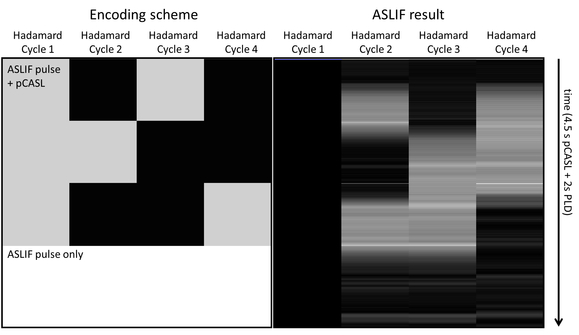
Experiments were performed on a clinical 3 Tesla whole-body MR-scanner (Magnetom Skyra, Siemens Healthineers, Erlangen, Germany). 5 volunteers were examined with a pCASL 3D-GRASE sequence3,4 after signing an informed consent according the ethical standards of the university. ASLIF imaging during 4-phase Hadamard-encoded pCASL5,6 was applied over a period of 4.5s followed by a PLD phase of 2s where only the ASLIF pulse was used.
The ASLIF readout slab was placed 35mm above the labeling slab yielding a 3/2*π shift between ASLIF and labeling pulse. ADC phase was aligned to the phase of the ASLIF pulse. This results in data, where the signal originating from the pCASL pulse will have a π/2 phase shift from pulse to pulse, while the phase shift of the ASLIF pulse is 0 or π. For post-processing, data was added or subtracted from the same data shifted by 2xTRpCASL yielding the ASLIF signal and pCASL signal exclusively. Without additional spatial encoding a minimum TRpCASL of 1070us was possible, with a spatial encoding of 6 or 12 encoding steps TRpCASL was 1140us and 1180us, respectively. This yielded a spatial resolution of about 24 and 12mm, respectively, sufficient to distinguish individual vessels in the neck.
Results
Fig. 2 shows the positions of the labeling and the ASLIF pulse on a low resolution TOF localizer. At the position of the ASLIF pulse the result of the ASLIF with a resolution of 12mm is shown. Fig. 3 and 4 show the labelled blood boli produced by the other three Hadamard-encoded pCASL phases with a temporal resolution of 15ms (6 phase encoding steps). To supress static tissue signal, the first (fully control) Hadamard-phase was subtracted from the other phases.
Discussion
ASLIF allows to measure the ASL signal during the labeling process. The different slice profiles of control and labeling pCASL pulses make it challenging to separate blood from labelled tissue signal. However, the phase shift of an odd multiple of π/2 between ASLIF and pCASL pulse allows for reliable separation of both signal sources. At the edges of Hadamard-subboli the subtraction does not work properly. However, this could easily be removed by a simple median filter. If the ASLIF pulses are played out with a phase shift of pi between RF pulses, it essentially resembles a control experiment ideally not affecting the labelled spins at all (verified by Bloch simulations, data not shown). Nonetheless, in that case, flow-sensitivity is higher and Fig. 1D variant is beneficial to reduce signal fluctuations. However, this comes at a penalty in minimum TRpCASL.
Conclusion
ASLIF is a new approach to directly image the amount of inflowing labelled blood ultimately allowing to estimate (and optimize) labeling efficiency in separate vessels in realtime.Acknowledgements
No acknowledgement found.References
1.Hargreaves BA. Rapid gradient-echo imaging. Journal of magnetic resonance imaging : JMRI. 2012;36(6):1300-13.
2.Han M, Hargreaves BA. Reduction of flow artifacts by using partial saturation in RF-spoiled gradient-echo imaging. Magnetic resonance in medicine. 2011;65(5):1326-34.
3. Gunther M, Oshio K, Feinberg DA. Single-shot 3D imaging techniques improve arterial spin labeling perfusion measurements. Magnetic resonance in medicine. 2005;54(2):491-8.
4. Fernandez-Seara MA, Wang Z, Wang J, Rao HY, Guenther M, Feinberg DA, et al. Continuous arterial spin labeling perfusion measurements using single shot 3D GRASE at 3 T. Magnetic resonance in medicine. 2005;54(5):1241-7.
5. Günther M, Highly efficient accelerated acquisition of perfusion inflow series by cycled arterial spin labeling. . Proceeding of the 15th Annual Meeting of ISMRM; 2007; Berlin, Germany.
6. von Samson-Himmelstjerna F, Madai VI, Sobesky J, Guenther
M. Walsh-ordered hadamard time-encoded pseudocontinuous ASL (WH pCASL).
Magnetic resonance in medicine. 2016;76(6):1814-24.
Figures
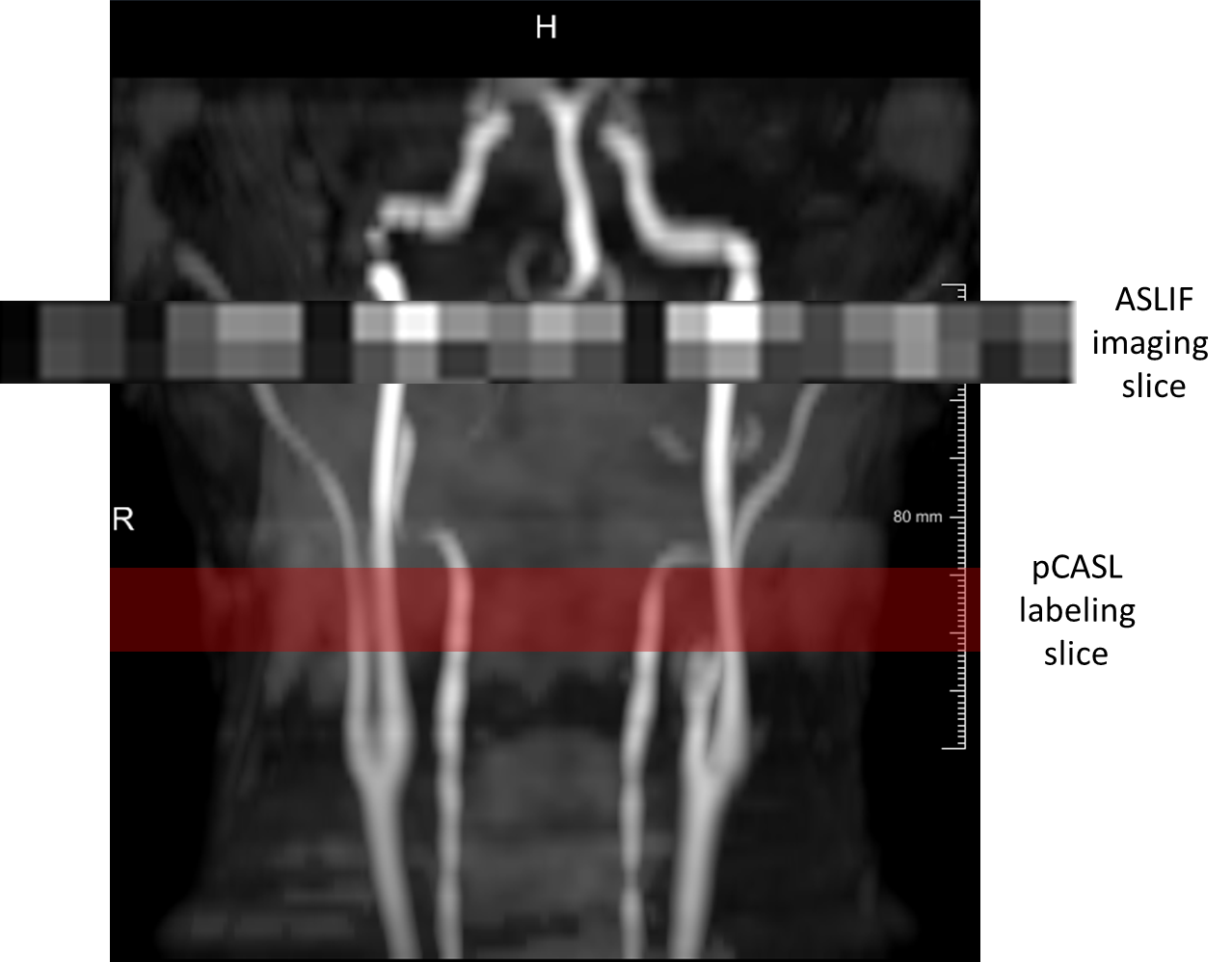
Position of labeling as well as ASLIF slab located 35mm above the pCASL slice. An individual time steps of the spatially resolving ASLIF sequence is shown depicting the acquisition of individual vessel signal.
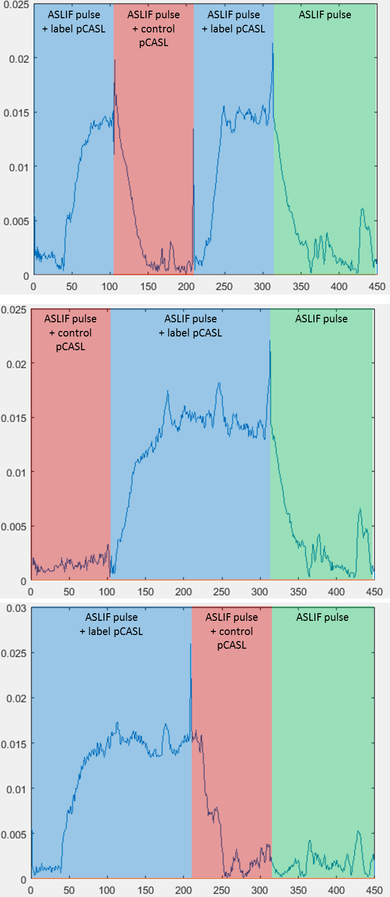
Result of an ASLIF sequence with 6 phase encoding steps along the coronar direction (x-axis). A 4-phase Hadamard pCASL scheme was employed. To suppress the signal from static tissue spins, the first Hadamard phase (employing control phase for all subboli) was subtracted from the other three Hadamard acquisitions. The curves show the signal within the left carotid of a healthy subject, nicely depicting the labeled blood bolus with a temporal resolution of 15 ms. The sharp peaks (not removed for educational purposes) indicate the points where cancellation of the pCASL pulse was not successful due to change of labeling/control paradigm.