0268
Comparison of 3D spoiled-gradient multiple echo with STEAM for proton density fat fraction and fatty acid composition estimation1Univ. Lyon, INSA‐Lyon, Université Claude Bernard Lyon 1, UJM-Saint Etienne, CNRS, Inserm, CREATIS UMR 5220, U1206, F69621, VILLEURBANNE, France, Lyon, France, 2Department of Physiology, Faculty of Biology and Medicine, University of Lausanne, Lausanne, Lausanne, Switzerland, 3Centre de Recherche en Nutrition Humaine Rhône-Alpes (CRNH-RA), Centre Hospitalier Lyon Sud, Pierre-Bénite, Lyon, France, 4Hospices Civils de Lyon, Département d'imagerie digestive, CHU Edouard Herriot, Lyon, Lyon, France
Synopsis
A total of 39 volunteers underwent an imaging and spectroscopy protocol on a 3T Ingenia Philips system with an axial 3D spoiled-gradient multiple echo sequence on abdominal region and a set of three STEAM sequences acquired on subcutaneous adipose tissue, visceral adipose tissue and liver. The quantification of Proton density fat fraction (PDFF), proportion of saturated (SFA), monounsaturated (MUFA) or polyunsaturated (PUFA) fatty acids from both MRI and MRS methods were compared. Good correlation with a little bias was found for the liver PDFF. Values of PUFA, MUFA and SFA from both techniques were poorly correlated.
Introduction
Overweight and obesity are a major worldwide health concern characterized by an abnormal accumulation of fat in adipose tissue and liver1. While 1H MRI is well suited to evaluate subcutaneous (SAT) and visceral (VAT) adipose tissue volumes, Magnetic Resonance Spectroscopy (MRS) is able to provide proportions of saturated (SFA), monounsaturated (MUFA) or polyunsaturated (PUFA) fatty acids. Recent studies demonstrated the feasibility of assessing the composition of fat based on multiple gradient echo imaging2. Some studies3,4 focusing on liver diseases have extensively compared the MRS and MRI methodologies for the quantification of PDFF. To our knowledge, no study has compared in vivo the FA quantification of human adipose tissue from these two techniques. Thus, the goal of this work was to compare FA quantification results estimated from a 3D spoiled-gradient multiple echo with STEAM sequence acquired in human abdominal adipose tissue at 3T. Additionally, estimated PDFF obtained in the liver were also compared.Subjects and methods
Imaging and spectroscopy protocol
A total of 39 healthy male volunteers underwent an imaging and spectroscopy protocol on a 3T Ingenia Philips system constituted by an axial 3D spoiled-gradient multiple echo sequence (MGE) on abdominal region (encompassing lumbar L1 to L5 and the liver) and a set of three STEAM sequences acquired on SAT, VAT and liver (parameters in Table 1 and Figure 1).
Data processing
The segmentation of adipose tissue and the quantification of FA (PUFA, MUFA and SFA) from a MGE has been previously described2,5. On MR images, the liver volume was manually defined on a unique slice, SAT and VAT volumes were automatically segmented on multiple slices (21 ± 2) from lumber L2 to L4. We use the same spectral model function to process the imaging and spectroscopic signal to quantify FA composition. Peak amplitude were directly expressed by the parameters characterizing a triglyceride molecular structure (chain length, number of double bonds and number of methylene interrupted by double bond)6. This fitting model has been described in previous work7 and has given robust results in vitro and in vivo.
Results comparison
PDFF, PUFA, MUFA and SFA quantified in each single MRS volume (for the liver, only PDFF) were compared to the mean value from MRI calculated on the total segmented SAT and VAT volumes. The correlation was evaluated by the coefficient of determination r² and bias was measured by Bland-Altman plot.
Results/Discussion
PDFF in liver
On 39 subjects, 15 MRS measurements were null and were removed from the analyses. Hepatic fat is weak in healthy subjects and difficult to detect. Two subjects presented an abnormal level of PDFF in the liver (PDFF >10%) with MRI measurements. Thus analyses were made with and without these subjects (Figure 2). As demonstrated in the literature, Figures 2-A and C show good correlation between MRI and MRS measurements (figure C, r²=0.76 vs figure A, r²=0.87). However, MRI-based measurements slightly overestimated PDFF compared to MRS measurements (bias=1.9 for the Figure2-B and bias=1.7 D).
Fatty acid composition in SAT and VAT
On 39 subjects, 9 MRS VAT spectra were not analyzable because of poor quality and were removed from the analyses. Regarding MRI results from the whole segmented volume versus the MRS results, the comparison of FA composition of SAT and VAT appeared to be substantially equivalent in average (Figure3). In both cases, paired t-tests showed significant difference between SAT and VAT FA composition (p<0.001). However, MRS and MRI FA composition estimations were poorly correlated (r²=0.43 for PUFA, r²=0.18 for MUFA, r²=0.36 for SFA, n=69). PUFA MRI values were significantly higher than MRS values (bias=+4.1, paired t-test p<0.001). SFA MRI values were significantly higher than MRS values (bias=-4.2, paired t-test p=0.005). No significant difference in MUFA measurements was found. The inter-subject variability was higher in localized MRS measurements than in the MRI measurements which were averaged over the whole adipose tissue volumes.
Conclusion
In the case of a longitudinal study, localized spectroscopy can be difficult to use in particular to reposition the voxel in the same place. Imaging enables to study larger areas while maintaining anatomical landmarks which facilitate registration from one MRI to another. In the comparison of MRI and MRS methods: good correlation was found for the estimation of PDFF in liver as reported in literature with the particularity here to study healthy subjects with small PDFF values (<8%); poor correlation was found in the case of FA composition, but MRS and MRI measurement are consistent in average. At the end of the study, analysis by gas chromatography will be made on biopsies of SAT to confirm the FA composition results.Acknowledgements
LABEX PRIMES (ANR-11-LABX-0063), program "Investissements d'Avenir" (ANR-11-IDEX-0007), IHU Opera and PHRC-IR Visfatir.References
- OMS Infobase globale: www.who.int
- Leporq B, Lambert SA, Ronot M, Vilgrain V, Van Beers BE. Quantification of the triglyceride fatty acid composition with 3.0 T MRI. NMR Biomed (2014). 27(10):1211-21.
- Achmad, E., Yokoo, T., Hamilton, G., et al. (2015). Feasibility of and agreement between MR imaging and spectroscopic estimation of hepatic proton density fat fraction in children with known or suspected nonalcoholic fatty liver disease. Abdominal imaging, 40(8), 3084-3090.
- Zand, K. A., Shah, A., Heba, E., Wolfson, T., et al. (2015). Accuracy of multiecho magnitude‐based MRI (M‐MRI) for estimation of hepatic proton density fat fraction (PDFF) in children. Journal of Magnetic Resonance Imaging, 42(5), 1223-1232.
- Nemeth A, Ratiney H, Leporq B, et al. Quantification of abdominal subcutaneous and visceral fat by magnetic resonance imaging of the proton at 3T: application to an overfeeding protocol. Proceedings of the 25th Annual Meeting ISMRM, Honolulu, Hawaï, USA, 2017; 1331.
- Nemeth A, Ratiney H, Leporq B, et al. Impact of time sample section and model function design on the quantification of fatty acid composition: In vitro and in vivo studies. Proceedings of the 25th Annual Meeting ISMRM, Honolulu, Hawaï, USA, 2017; 4194.
- Hamilton G, Schlein AN, Middleton MS, Hooker CA, Wolfson T, Gamst AC, et al. In vivo triglyceride composition of abdominal adipose tissue measured by 1H MRS at 3T. J Magn Reson Imaging. 2017 mai;45(5):1455–63.
Figures
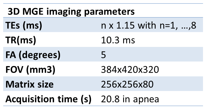
Table 1:
3D spoiled-gradient multiple echo (MGE) sequence parameters
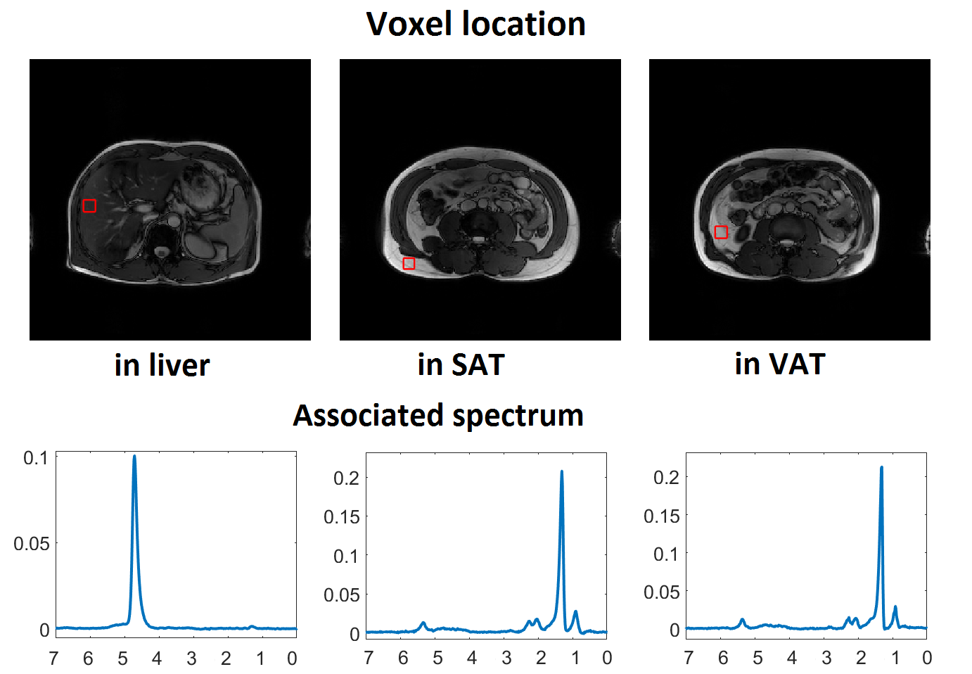
Figure 1:
A STEAM sequence was acquired in liver, subcutaneous adipose tissue (SAT) and visceral adipose tissue (VAT) using the following parameters: TR = 3000 ms, TE= 14 ms, TM= 10 ms, VOI of 20×20×20 mm3, no water suppression, 2048 Hz bandwidth, 1024 data points, 4 signal averages for VAT and SAT and 32 signal averages for the liver. For the SAT acquisitions, the phase of the signal (TE = 14 ms) was corrected with the phase of a second echo (TE = 28 ms) which was less impacted by eddy current effects.
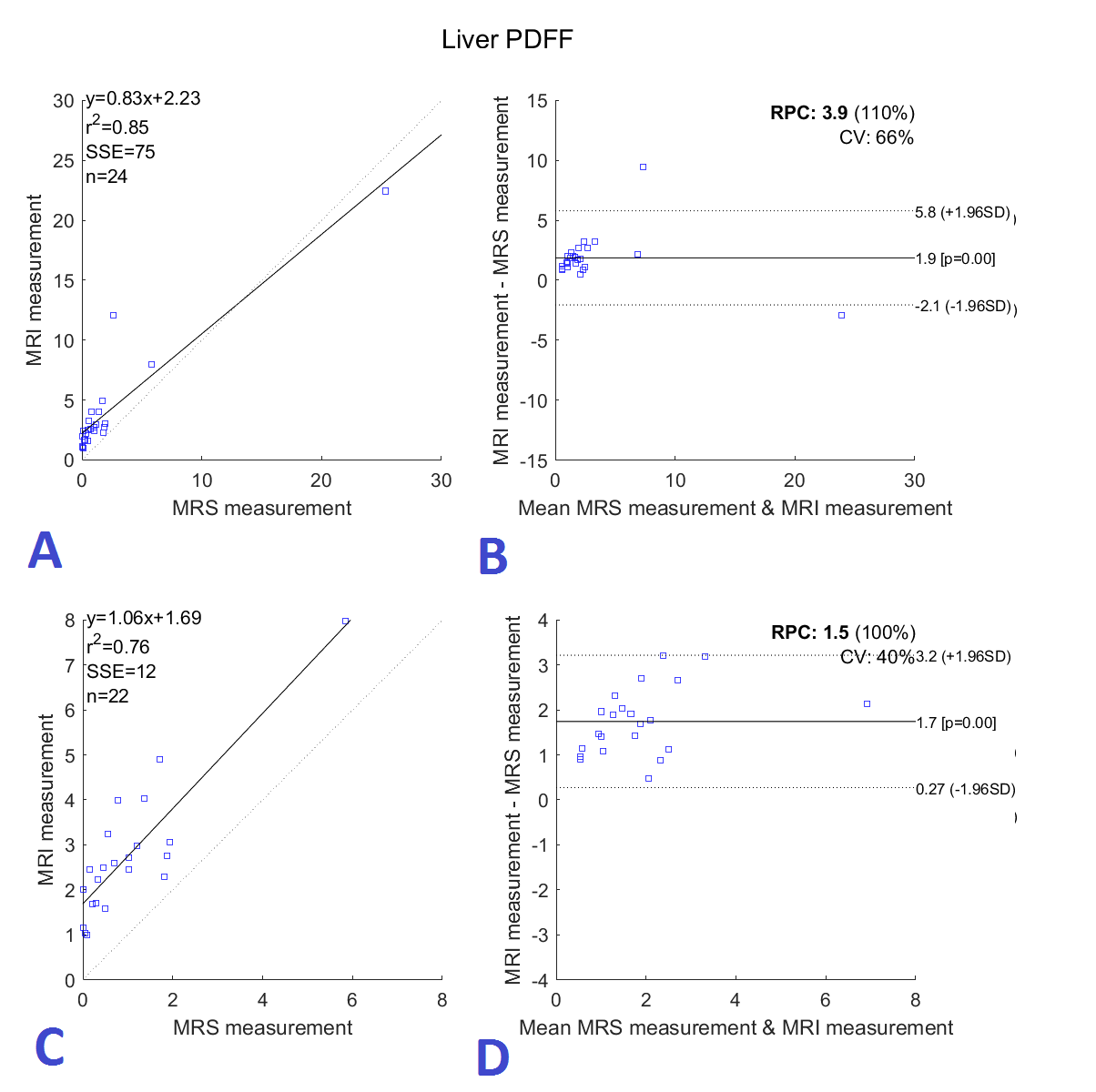
Figure 2:
Figures A and C show the correlation between MRS and MRI PDFF measurements in human liver and Figures B and D show the respective Bland-Altman. PDFF values of MRI were estimated in a manual segmented volume (26.87 ± 7.86 cm3) and MRS in a volume of 8 cm3. Two subjects presented abnormal level of PDFF, analyses were made with (A-B) and without their data (C-D).
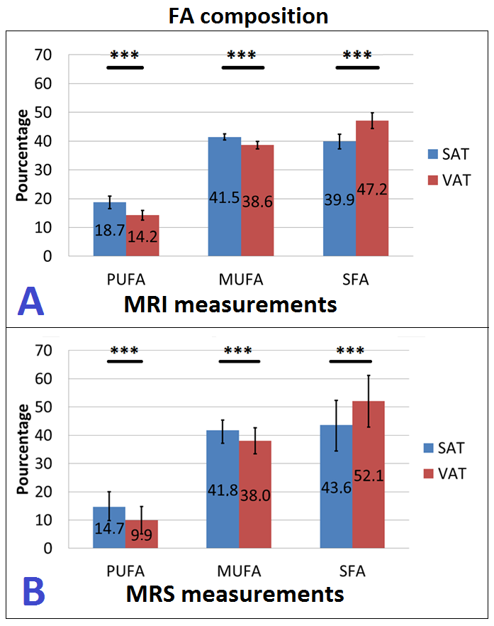
Figure 3:
Figure 3 shows the fatty acid composition of the subcutaneous adipose tissue (SAT) and visceral adipose tissue (VAT) using MRI methodology, n=39 (A) and using spectroscopic methodology, n=30 (B). PUFA = polyunsatured fatty acid, MUFA= mono-unsatured fatty acid and SFA = satured fatty acid. Paired t-tests were made between values from SAT and values from VAT. *** p<0.001