0267
Increased Measurement Efficiency for Fat Quantification Using a Mixed Polarity Bipolar Acquisition Scheme1St Jude Children's Research Hospital, Memphis, TN, United States
Synopsis
A new mixed polarity bipolar GRE sequence is proposed for fat quantification by mixing positive and negative readout polarity in one echo acquisition. Accurate fat fraction maps can be obtained using the proposed method with up to 50% scan time savings compared to a dual shot unipolar GRE method.
Introduction
Multi-gradient echo imaging is widely applied for fat/water separation and quantification. Typically unipolar readout gradient schemes (uniGRE), as opposed to bipolar (biGRE), are used to avoid eddy current induced phase errors between echoes of alternating polarity which severely impede fat quantification. The minimum echo spacing ∆TE in uniGRE is determined by the duration of the required flyback gradients; to realize shorter ∆TE for accurate quantification at 3T, multi-shot uniGRE with interleaved TEs are typically applied at the expense of measurement time. Sufficiently short ∆TEs can only be realized in a single shot scan by eliminating the flyback gradients and using biGRE. This requires correction of phase errors introduced by the bipolar readout gradients, e.g. through additional reference lines1. Another acquisition strategy acquires both polarities in an interleaved fashion across k-space for all TEs, subsequently sorts the data by polarity, applies a parallel imaging type reconstruction to the two undersampled data sets, and then removes phase errors via complex averaging of these two images2. Of note, up to 18% additional reference/calibration lines (161 and 242, respectively) are required for both phase correction methods, and the second approach does not allow for further application of parallel imaging to speed up acquisition time, e.g. for abdominal breath-hold imaging. In this work, a new mixed readout polarity bipolar GRE (mxGRE) method is proposed that uses a single reference line and does not interfere with using parallel imaging for scan time reduction.Method
As shown in Fig. 1, N echoes are acquired in the same manner as a regular bipolar GRE for the first half of k-space and with reversed polarity for the second half. The ky=0 reference line is collected with both positive and negative readout gradients for phase correction. The phase error between negative and positive readout lines is corrected by an approach proposed by Li et al3. Seven cylindrical 1L polystyrene bottles with fat fractions of 0, 10, 20, 30, 40, 50, and 100% were made from peanut oil, agar, and Gadolinium-doped water. The phantom and five healthy volunteer scans were performed on a 3T clinical MRI system. Data were collected using 3D dual shot uniGRE and 3D mxGRE. The phantoms were also scanned with 3D biGRE. All scans used N=6 echoes, a bandwidth of 1.5KHz/px and a flip angle of 5°. The phantom measurements were performed with TE1/∆TE/TR 1.7/1.2/10ms, matrix 256×256×32, FOV 280×280×160mm, TA(mxGRE&biGRE/uniGRE) 82/164s. Liver scans were carried out with TE1/∆TE/TR 1.7/1.2/10ms, matrix 160×104×16, FOV 450×315×80mm, GRAPPA with R=2 along ky. and TA(mxGRE/uniGRE) 13/26s. For knee scans TE1/∆TE/TR 1.7/1.2/20ms, matrix 256×152×30, FOV 300×172×180mm, and TA(mxGRE/uniGRE) 152/303s was used. Fat fraction (FF) maps were calculated with a complex-based fat/water reconstruction technique that incorporates a graphcut algorithm for B0 field estimation4.Results and Discussion
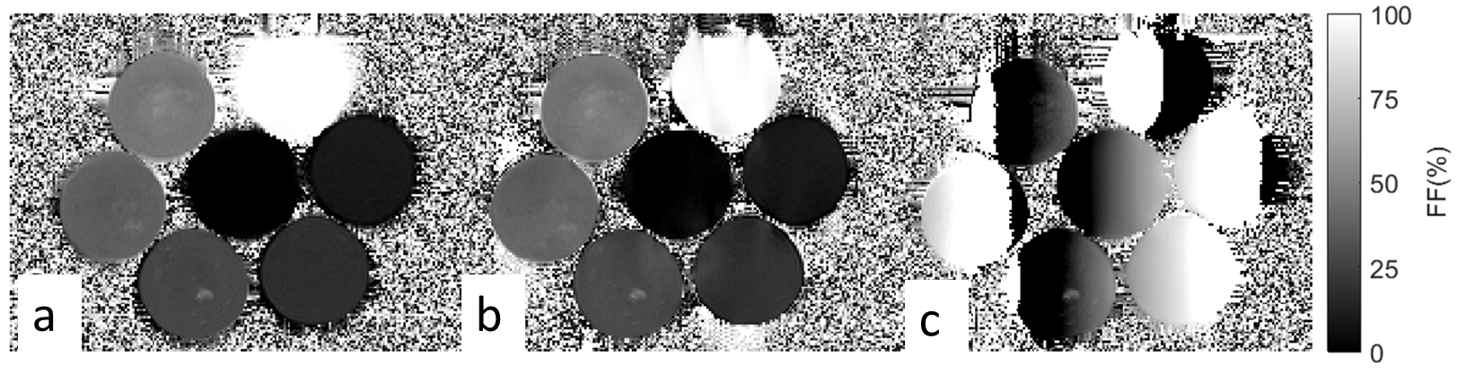
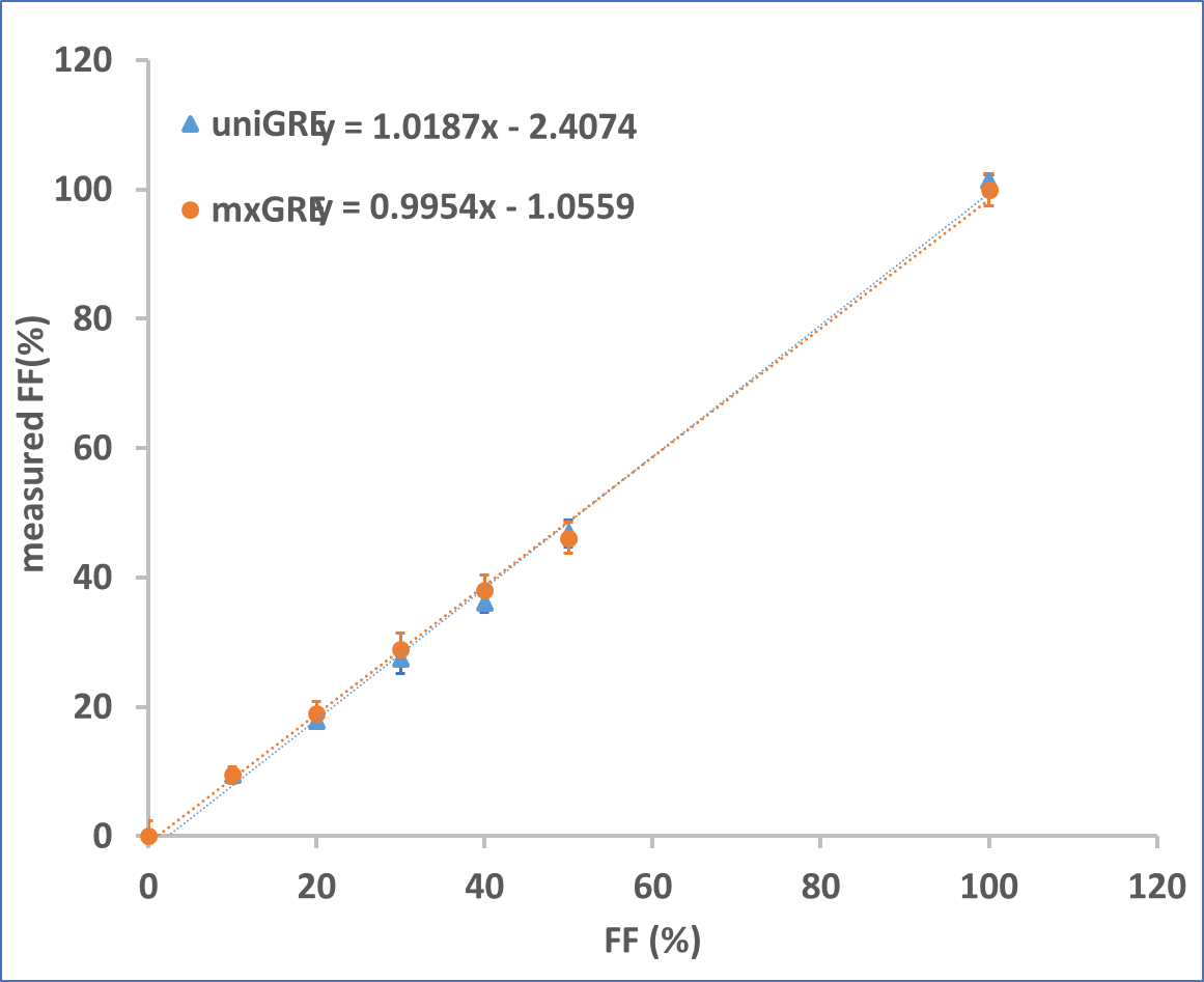
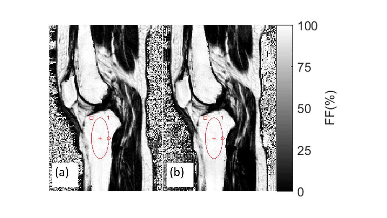
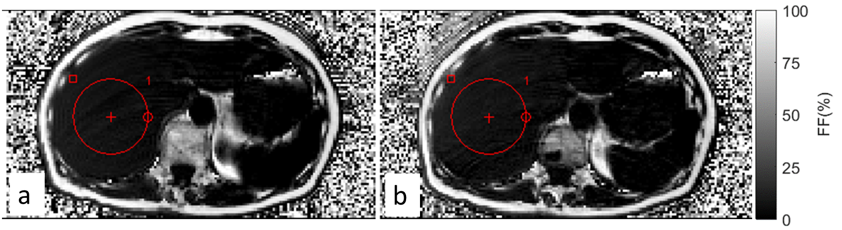
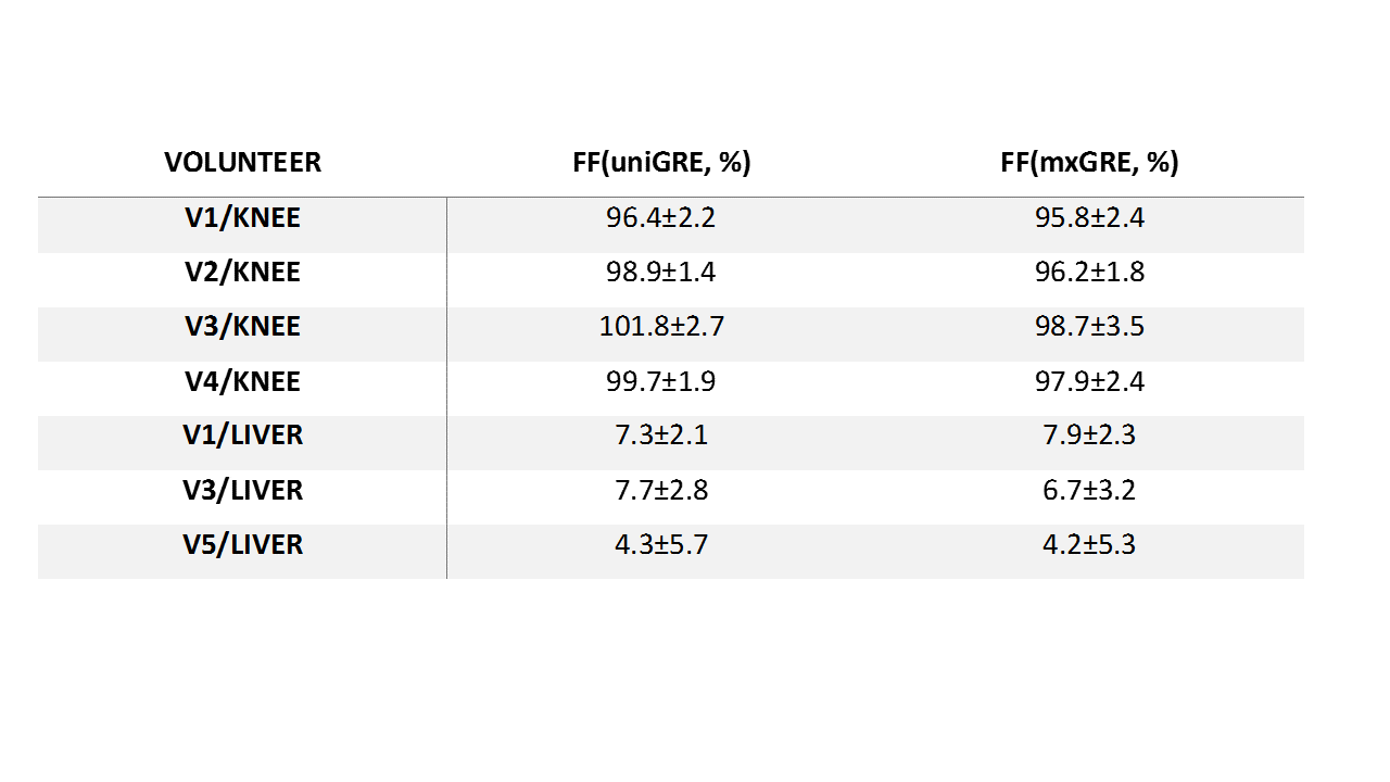
Fig. 2 shows FF maps obtained with uniGRE, mxGRE and biGRE. The biGRE FF values changed sharply in homogenous phantoms, and are incorrect. Therefore, biGRE was excluded in further comparisons. By eliminating the phase error, the mxGRE FF map is similar to the uniGRE FF map. The comparison between the FF values measured with the uniGRE and mxGRE and the nominal FF are presented in Fig. 3. Both methods measured FF satisfactorily with a slight overestimation (slope 1.019) for uniGRE and marginal underestimation (slope=0.995) for mxGRE. Fig. 4 shows a sagittal scan of the knee of a healthy volunteer. The FF values obtained in identically positioned ROIs within the bone marrow of uniGRE and mxGRE FF maps are in good agreement (difference <3.1% across all the knee scans). Abdominal FF maps of a volunteer obtained with uniMGRE (a) and mxMGRE (b) are shown in Fig. 5. An ROI was placed in the liver to compare the FF values as shown in Fig. 5, and they are found to be in good agreement (difference < 1.0% across all the liver scans). The FF comparison between the two methods in the healthy volunteers is listed in Table 1. The good agreement between with the FF maps from uniGRE and mxGRE occurs despite a 50% reduction in scan time for the mxGRE. As demonstrated in the liver scans, the proposed method is compatible with imaging acceleration techniques, e.g. parallel imaging to further reduce scan time.Conclusion
A new mixed polarity bipolar GRE sequence for fat quantification is presented. Accurate FF maps can be achieved using the proposed method with up to 50% scan time savings in comparison to a dual shot unipolar GRE method. This approach uses fewer reference lines than other fat/water bipolar approaches without compromising the ability to accelerate scans using parallel imaging.Acknowledgements
No acknowledgement found.References
1. Yu et al, Phase and amplitude correction for multi-echo water-fat separation with bipolar acquisitions, J. Magn Reson Imaging 2010; 31:1264-1271
2. Soliman et al, Fat quantification using an interleaved bipolar acquisition, Magn Reson Med, 2016; 75:2000-2008
3. Li et al, Fast Decomposition of Water and Lipid Using a GRASE Technique With the IDEAL Algorithm, Magn Reson Med, 2007; 57:1047-1057
4. Hu HH, et al. ISMRM workshop on fat-water separation: insights, applications and progress in MRI. Magn Reson Med 2012; 68:378–388.
Figures