0240
MRI texture features as predictors of histopathologic and genomic characteristics of hepatocellular carcinoma.1Translational and Molecular Imaging Institute, Icahn School of Medicine at Mount Sinai, New York, NY, United States, 2Department of Radiology, Icahn School of Medicine at Mount Sinai, New York, NY, United States, 3Department of Pathology, Icahn School of Medicine at Mount Sinai, New York, NY, United States, 4Department of Medicine/Division of Liver Diseases, Icahn School of Medicine at Mount Sinai, New York, NY, United States
Synopsis
The goal of this study was to assess the diagnostic value of texture features measured with MRI compared to qualitative imaging traits for the prediction of histopathologic and genomic characteristics of hepatocellular carcinoma (HCC) lesions. Texture features exhibited additional, complementary correlations with histopathology and genomics compared to qualitative imaging traits, including association with microvascular invasion and expression of immunotherapy target CTLA4. These promising results warrant further investigation of texture features as predictors of histopathologic and genomics measurements in HCC.
Purpose
Pathological and genomics evaluation of hepatocellular carcinoma (HCC) may play an important role for prediction of prognosis and treatment decision and response. However, these techniques require tissue sampling, which is invasive and rarely clinically indicated in HCC. Predicting HCC pathological and genomic characteristics using MRI would be of major clinical interest, because MRI is noninvasive and covers the whole lesion 1,2. Image texture analysis has shown promising results in terms of correlations between texture features and histopathologic and genomic features in various tumor types 3-5. The goal of our study was to assess the potential additional value of texture features compared to qualitative imaging traits for the prediction of histopathologic and genomic characteristics of HCC lesions.Methods
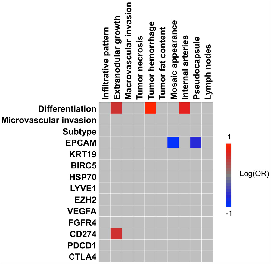
This retrospective study included 53 patients with HCC (M/F 38/15, mean age 60y, range 36-77y) that underwent hepatic resection within 4 months after clinical abdominal MRI at 1.5T (n=39) or 3.0T (n=14). Haralick texture features (Energy, Contrast, Correlation, Variance, Homogeneity, Sum average, Sum variance, Sum entropy, Entropy, Difference Variance, Difference Entropy, Information correlation measures 1 and 2 and Maximal Correlation) were calculated in index HCC lesions on contrast-enhanced images [pre contrast (n=52), early arterial phase (EAP; n=36), late arterial phase (LAP; n=45), portal venous phase (PVP; n=52), late venous phase (LVP; n=50), hepatobiliary phase (HBP; n=40 in patients who received gadoxetic acid)] and ADC maps (n=52). Qualitative imaging traits (infiltrative pattern, extra-nodular growth, macrovascular invasion, tumor necrosis, tumor hemorrhage, tumor fat content, mosaic appearance, internal arteries, pseudocapsule, lymph node involvement) were assessed by two radiologists in consensus. Histologically distinct components of 48 of the analyzed HCC tumors were macro-dissected using H&E staining of serial tissue sections as reference. Isolated total RNA samples were profiled to determine transcriptomic HCC subtypes 6 with digital transcript counting technology using the nearest template prediction algorithm 7. In addition, gene expression levels of key HCC markers and therapeutic targets were determined 2. Binary logistic regression analysis was used to determine the association of the texture and qualitative imaging features with histopathologic (tumor differentiation and microvascular invasion) and genomic features (subtype and gene expression levels) of the lesions. Prior to regression analysis, texture features were standardized to eliminate scaling differences between the features. ROC analysis was performed to test the diagnostic performance of texture features for detection of histopathologic and genomics of HCC.Results
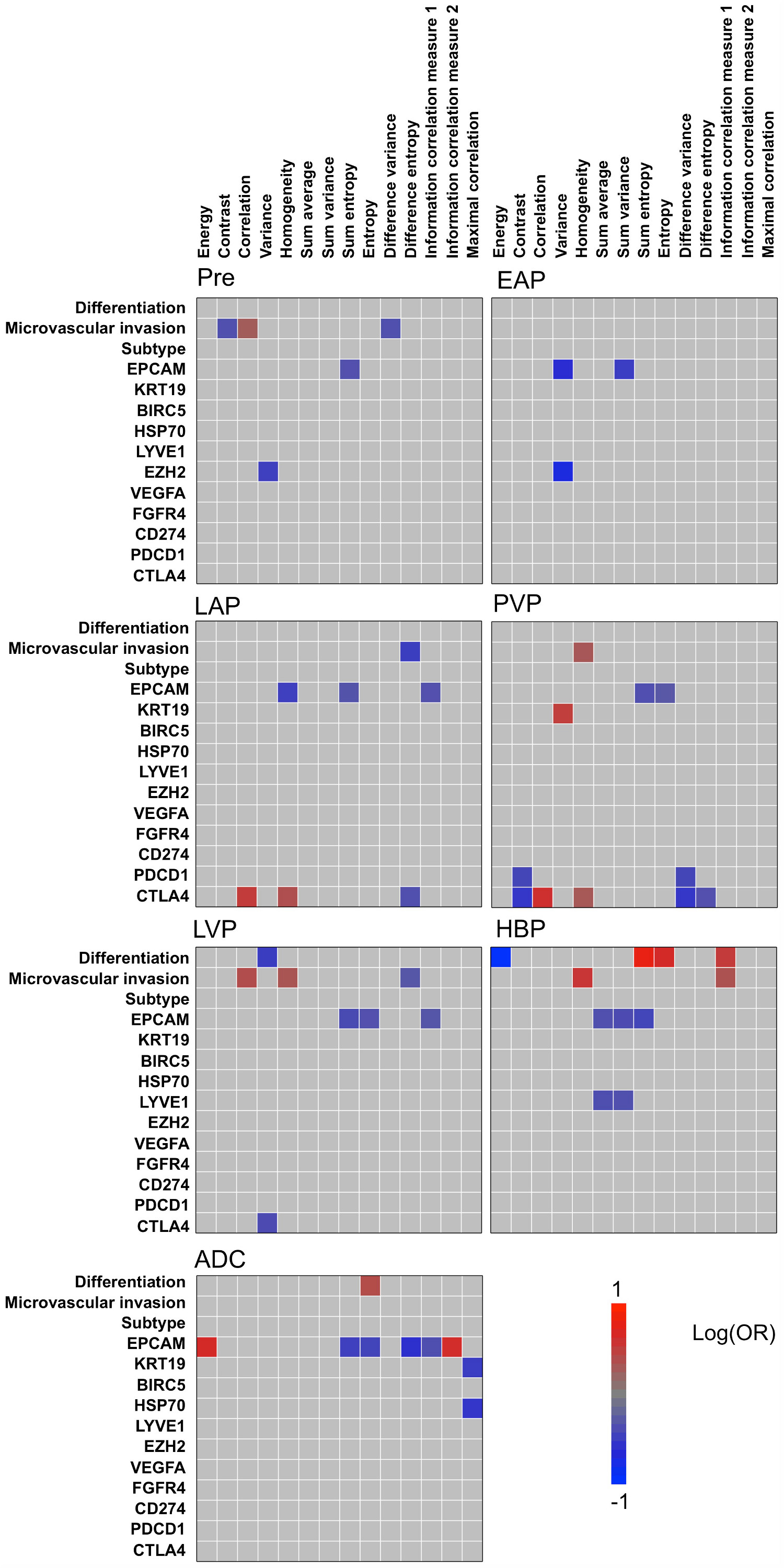
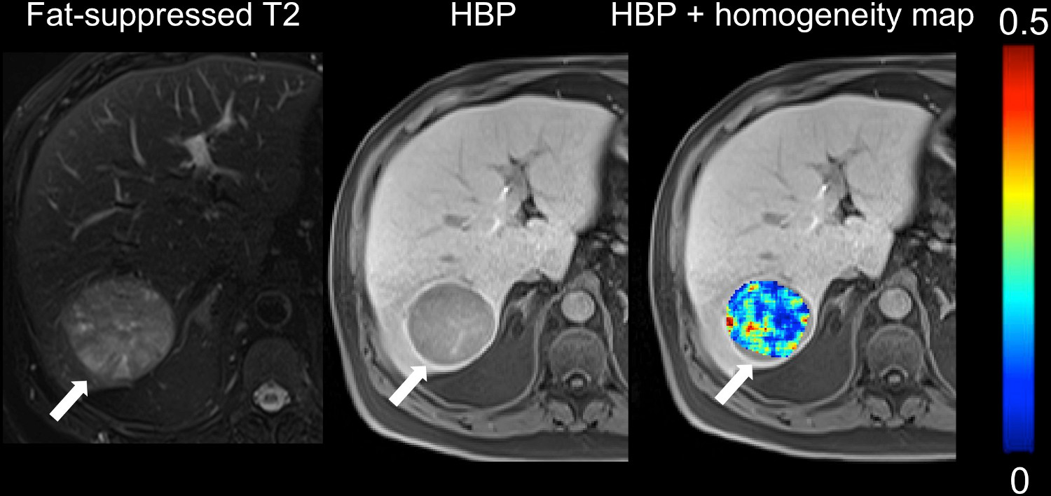
Mean lesion size was 4.1±3.4 cm (range 1.0 – 15.0 cm). Observed significant associations between MRI texture features and histopathological and genomics characteristics of HCC are displayed in the heatmaps in Figure 1. Significant associations of texture with histopathological and genomics features were particularly seen on T1w HBP and PVP images, including a significant strong association between HBP Entropy and tumor differentiation (odd ratio (OR) =4.778, p=0.018, AUC=0.784), between HBP Homogeneity and microvascular invasion (OR=3.799, p=0.016, AUC=0.804; Figure 3) and between PVP correlation and expression of immunotherapy target CTLA4 (OR=4.253, p=0.018, AUC=0.677). Qualitative imaging traits showed fewer correlations with histopathology and genomics (Figure 2). Nevertheless, the observed significant correlations were generally strong, such as strong associations between mosaic appearance and expression of stemness marker EPCAM (OR=0.079, p=0.020; Figure 3) and between hemorrhage and tumor differentiation (OR=17.33, p=0.015). Overall, texture features showed complementary associations compared to qualitative imaging traits, since texture features exhibited significant associations with histopathological microvascular invasion and gene expression levels of stemness marker KRT19, HCC markers HSP70, LYVE1 and EZH2 and immunotherapy targets PDCD1 and CTLA4 that were not seen with qualitative imaging traits.Discussion and Conclusion
Our study demonstrates significant associations between texture features and histopathologic and genomic characteristics of HCC. While qualitative imaging traits also showed significant association with several of the histopathologic and genomics features, texture analysis exhibited complementary correlations, suggesting additional value of texture analysis for noninvasive assessment of HCC. While texture analysis of MRI images has already previously shown promising for noninvasive prediction of HCC differentiation 4, our study describes the first results on the correlation of MRI texture features of HCC with histopathological microvascular invasion and gene expression levels of key HCC markers. The promising results observed in our study warrant further investigation of texture features as surrogate histopathological and genomics measurements in HCC.Acknowledgements
This research was supported by RSNA Research Seed Grant #RSD1608 and U01 CA172320.References
1. Semaan S, Makkar J, Lewis S, Chatterji M, Kim E, Taouli B. Imaging of Hepatocellular Carcinoma Response After 90Y Radioembolization. AJR American journal of roentgenology 2017;209(5):W263-W276.
2. Hectors SJ, Wagner M, Bane O, et al. Quantification of hepatocellular carcinoma heterogeneity with multiparametric magnetic resonance imaging. Scientific reports 2017;7(1):2452.
3. Fehr D, Veeraraghavan H, Wibmer A, et al. Automatic classification of prostate cancer Gleason scores from multiparametric magnetic resonance images. Proceedings of the National Academy of Sciences of the United States of America 2015;112(46):E6265-6273.
4. Zhou W, Zhang L, Wang K, et al. Malignancy characterization of hepatocellular carcinomas based on texture analysis of contrast-enhanced MR images. Journal of magnetic resonance imaging : JMRI 2017;45(5):1476-1484.
5. Li H, Zhu Y, Burnside ES, et al. MR Imaging Radiomics Signatures for Predicting the Risk of Breast Cancer Recurrence as Given by Research Versions of MammaPrint, Oncotype DX, and PAM50 Gene Assays. Radiology 2016;281(2):382-391.
6. Hoshida Y, Nijman SM, Kobayashi M, et al. Integrative transcriptome analysis reveals common molecular subclasses of human hepatocellular carcinoma. Cancer research 2009;69(18):7385-7392.
7. Tan PS, Nakagawa S, Goossens N, et al. Clinicopathological indices to predict hepatocellular carcinoma molecular classification. Liver international : official journal of the International Association for the Study of the Liver 2016;36(1):108-118.
Figures