0235
Elevated brain iron in cocaine addiction as indexed by magnetic field correlation imaging1Neuroscience, Medical University of South Carolina, Charleston, SC, United States, 2Psychiatry and Behavioral Sciences, Medical University of South Carolina, Charleston, SC, United States, 3Radiology and Radiological Science, Medical University of South Carolina, Charleston, SC, United States
Synopsis
Brain iron is critical for neural processes implicated in addiction. Recently, disrupted iron regulation was detected in individuals with cocaine use disorder (CUD) using quantitative susceptibility mapping. Our goal was to replicate these findings using an alternative iron imaging method called magnetic field correlation imaging. Consistent with the only study of brain iron in CUD, we detected elevated brain iron levels in globus pallidus regions and loss of age-related iron accumulation in CUD. Our replication of aberrant brain iron findings in CUD using a different MRI modality lends support for further investigation of iron homeostasis in CUD.
Introduction
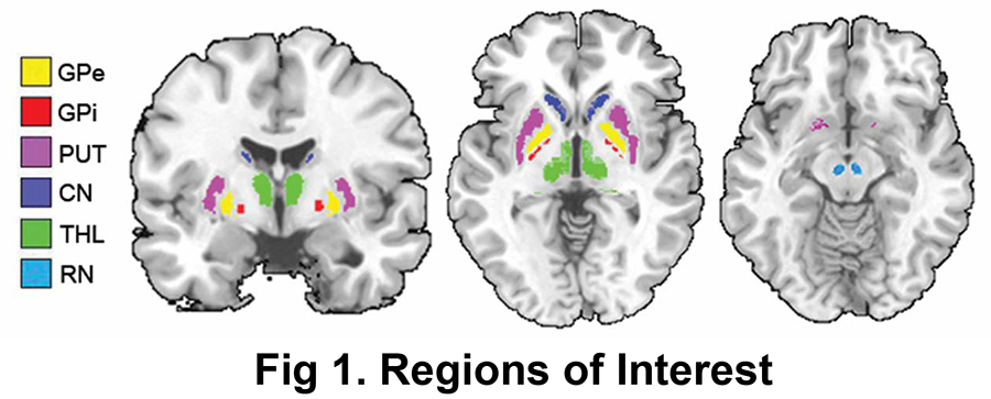
Substantial brain alterations have been detected in cocaine use disorder (CUD).1 However, the neurobiological mechanisms underlying different stages of CUD are not fully understood and may explain why effective medication treatments remain elusive.2 Recently, a quantitative susceptibility mapping (QSM) study demonstrated that iron regulation is also disrupted in CUD.3 Iron plays a vital role in neural processes implicated in addiction including dopamine synthesis, myelin maintenance and blood oxygen transport.4 Our goal is to replicate the QSM findings from the only study of brain iron in CUD3 using an alternative iron imaging method called magnetic field correlation (MFC) imaging.5,6 We hypothesize that aberrant brain iron levels in CUD would also be detectable using MFC imaging which provides similarly sensitive and complementary iron indices to QSM.7 The globus pallidus external segment (GPe), globus pallidus internal segment (GPi), putamen (PUT), caudate nucleus (CN), thalamus (THL) and red nucleus (RN) were chosen as regions of interest (ROIs) as they are targets of cocaine,1 have the highest concentrations of dopamine and iron,4,8 and were previously studied in CUD.3Methods
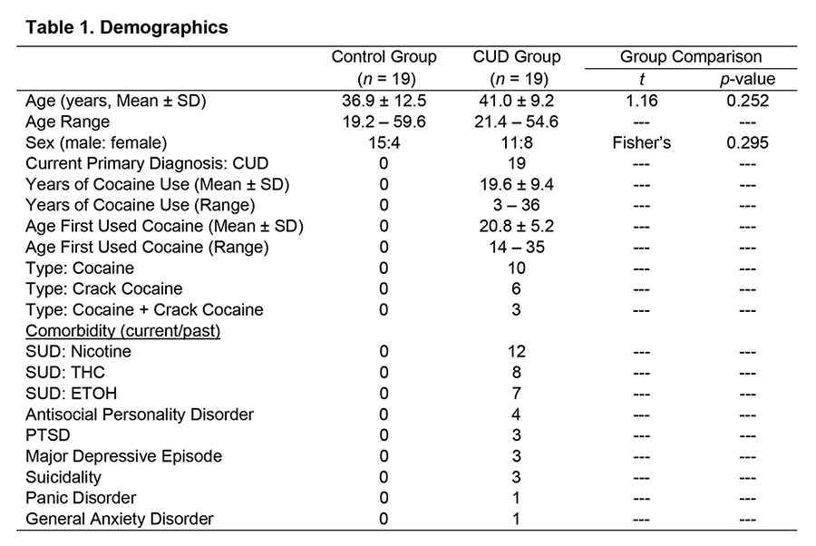
We studied 19 individuals with CUD (41.0 [mean] ± 9.2 [SD] years old, 11 males) and 19 healthy controls (36.9 ± 12.5 years old, 15 males). CUD patients met DSM-IV-TR criteria for cocaine dependence9; controls had no history of DSM-IV-TR diagnoses, including drug, nicotine or alcohol abuse/dependence. CUD patients were abstinent on the scan day (negative urine drug test). Imaging was conducted on a 3T Siemens Trio; MFC asymmetric spin echo (ASE): TR/TE = 5550/40 ms, voxel = 1.7 mm³, matrix = 128 × 128 × 40, averages = 4, flip angle = 90°, EPI factor = 33, no gaps, refocusing pulse time shifts = 0, -4 and -16 ms, acquisition = 6 min, 40 sec. MPRAGE: TR/TE = 1900/2.34 ms, voxel = 0.9 × 0.9 × 1 mm³, matrix = 256 × 256 × 192, acquisition = 5 min, 42 sec. The MFC parametric maps were calculated as previously described.6 Automatic ROI segmentation of each subject’s MPRAGE was performed using Freesurfer (http://surfer.nmr.mgh.harvard.edu). The whole globus pallidus (GP) was manually delineated into GPe and GPi. ROIs and MFC maps were normalized with ART210 to MNI space. To exclude partial volume effects, ROIs were constrained with a consensus mask (ROI voxels with overlap among subjects). The RN ROI was drawn on a normalized group average 0-shift ASE image (automatic segmentation unavailable). ROIs were visually inspected for anatomical accuracy and applied to the MFC map to extract subject means (Figure 1). Group comparisons were conducted (normally distributed measures: two-tailed Student’s t-test, effect size Cohen’s d; non-normally distributed: Mann-Whitney U test, effect size rank biserial correlation [rrb]; nominal: Fisher’s exact test). Within group MFC correlations with age and collinearity tests were conducted (normally distributed: Pearson’s correlation [r]; non-normally distributed: Spearman’s correlation [rs]; control for collinear covariates: partial correlations; all two-tailed). False discovery rate (FDR) correction was used for multiple comparisons.11Results
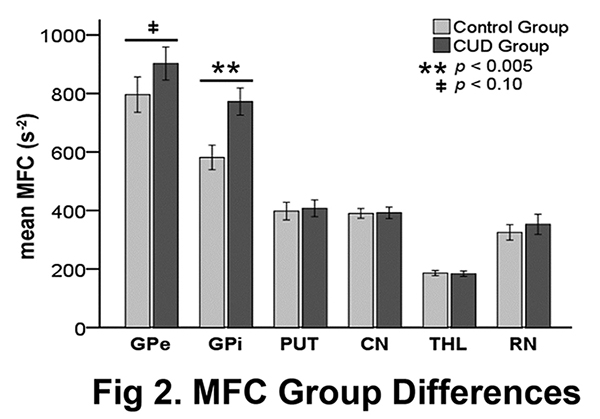
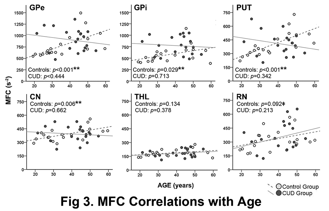
The groups did not significantly differ in age or gender (Table 1). CUD patients had significantly higher MFC in the GPi than controls (32.9% higher, large effect size: rrb = 0.5). There was a similar trend in the GPe (13.4% higher in CUD, medium effect size: rrb = 0.3). There were no significant group differences/trends in the PUT, CN, THL and RN (Figure 2). In controls, MFC significantly increased with age in the GPe, GPi, PUT and CN, with a similar trend in the RN (Figure 3). In CUD patients, MFC did not correlate with age in any of the ROIs (Figure 3) or with years of cocaine use (even when controlling for age or vice versa). All significant findings survived FDR correction.Discussion
Consistent with the only study of brain iron in CUD,3 we found that CUD patients had elevated brain iron in GP regions and lacked age-related brain iron accumulation seen in normal aging.8 The GPe and GPi are key regions involved in the transition from goal-directed to compulsive behavior that is the hallmark of addiction.12 As excess iron causes oxidative stress and cell death,4 over accumulation of iron in GP regions suggests that disruption of brain iron homeostasis may be involved in the development of CUD and implicates neurodegeneration of these key regions in CUD.Conclusion
Iron homeostasis in addiction is an emerging area of research that may shed light on novel therapeutic targets.3,13 Our replication of aberrant brain iron findings in CUD using a different MRI modality lends support for pursing this new line of research. Further investigation of brain iron in different stages of CUD is warranted.Acknowledgements
Funding was provided by The Klingenstein Third Generation Foundation (VA) and The Litwin Foundation (JAH).References
1. Ashok AH, Mizuno Y, Volkow ND, et al. Association of stimulant use with dopaminergic alterations in users of cocaine, amphetamine, or methamphetamine: a systematic review and meta-analysis. JAMA Psychiatry. 2017;74(5):511-519.
2. Czoty PW, Stoops WW, Rush CR. Evaluation of the "pipeline" for development of medications for cocaine use disorder: a review of translational preclinical, human laboratory, and clinical trial research. Pharmacol Rev. 2016;68(3):533-62.
3. Ersche KD, Acosta-Cabronero J, Jones PS, et al. Disrupted iron regulation in the brain and periphery in cocaine addiction. Transl Psychiatry. 2017;7(2):e1040.
4. Beard JL, Connor JR. Iron status and neural functioning. Annu Rev Nutr. 2003;23:41-58.
5. Jensen JH, Chandra R, Ramani A, et al. Magnetic field correlation imaging. Magn Reson Med. 2006;55(6):1350-61.
6. Jensen JH, Szulc K, Hu C, et al. Magnetic field correlation as a measure of iron-generated magnetic field inhomogeneities in the brain. Magn Reson Med. 2009;61(2):481-5.
7. Adisetiyo V, Jensen JH, Lee CY, et al. Comparative analyses of magnetic field correlation imaging, quantitative susceptibility mapping and transverse relaxation rate R2* indices of brain iron in healthy adults. Proceedings of the ISMRM 23rd Annual Meeting & Exhibition in Toronto, Ontario, Canada. 2015;1447.
8. Hallgren B, Sourander P. The effect of age on the non-haemin iron in the human brain. J Neurochem 1958;3(1):41-51.
9. American Psychiatric Association. Diagnostic and statistical manual of mental disorders. 4th ed. Arlington, VA: American Psychiatric Association, 2000.
10. Ardekani BA, Guckemus S, Bachman A, et al. Quantitative comparison of algorithms for inter-subject registration of 3D volumetric brain MRI scans. J Neurosci Methods 2005;142(1):67-76.
11. Benjamini Y, Hochberg Y. Controlling the false discovery rate: a practical and powerful approach to multiple testing. J R Stat Soc Series B Stat Methodol 1995;57(1):289-300.
12. Keeler JF, Pretsell DO, Robbins TW. Functional implications of dopamine D1 vs. D2 receptors: A 'prepare and select' model of the striatal direct vs. indirect pathways. Neuroscience. 2014;282:156-75.
13. Juhás M, Sun H, Brown MR, et al. Deep grey matter iron accumulation in alcohol use disorder. Neuroimage. 2017;148:115-122.
Figures