0231
Reduced Local Segregation in Single-Subject Grey Matter Networks in Adult PTSD1HMRRC, West China Hospital, Chengdu, China, China, 2Department of Psychosis Studies, Institute of Psychiatry, Psychology & Neuroscience, King’s College London, London, United Kingdom, London, United Kingdom, 3uaxi MR Research Center (HMRRC), Department of Radiology, West China Hospital of Sichuan, Chengdu, China
Synopsis
The present study investigated graph properties of single-subject grey matter networks in PTSD using a new method proposed by Tijms and colleagues to statistically describe gray matter networks in individual subjects using T1-weighted MRI scans. Compared with trauma exposed controls, global topology of single-subject grey matter networks of PTSD was characterized by decreased clustering coefficient and local efficiency. The reduced segregation in grey matter network and its negative relation with increased segregation in the functional network may be important for understanding the nature of the brain network disorganization associated with PTSD and facilitate clinical diagnosis in patients with suspected PTSD
INTRODUCTION
Posttraumatic stress disorder (PTSD) is a psychiatric condition that can develop following exposure to extremely traumatic life events. Previous data suggest that dystrophic changes in grey matter are associated with cortical dysfunction in PTSD [1]. However, most previous grey matter morphological studies have been region focused, an approach which can fail to capture the complex connectivity alterations of grey matter that typically supports higher cognitive and affective processes [2]. The present study investigated graph properties of single-subject grey matter networks in PTSD using a new method proposed by Tijms and colleagues to statistically describe gray matter networks in individual subjects using T1-weighted MRI scans [3]. We predicted that grey matter networks in our PTSD sample would be shifted toward a randomized configurationand altered nodal degree and efficiency would be found in our study based on previous findings [4], and there would be a correlation between those network alterations and clinical severity of PTSD
MATERIALS AND METHODS
Eighty-nine PTSD patients and 88 trauma-exposed controls (TEC) underwent a structural T1 magnetic resonance imaging scan. The single-subject brain structural networks were constructed based on grey matter similarity of 90 brain regions starts with defining the network’s nodes as 3×3×3 voxel cubes (27 voxels). These cubes keep the 3D structure of the cortex intact, thereby including spatial information from the MRI scan in addition to gray matter values in voxels. As network properties can vary with network size [5], it is critical to normalize grey matter networks to have the same number of nodes across participants. To achieve this, we followed the methodology proposed by Batalle et al. [6] to normalize single subject grey matter networks based on the unified Automated Anatomical Labeling (AAL) parcellation template available in SPM software corresponding to the 90 nodes of the AAL atlas. Graph theory analyses were applied to investigate the topological properties of brain networks. We used nonparametric permutation tests to identify differences of topological metrics in two groups. Relationship of brain network measures with CAPS scores that reflect psychiatric manifestations of PTSD were analyzed in SPSS.
RESULTS:
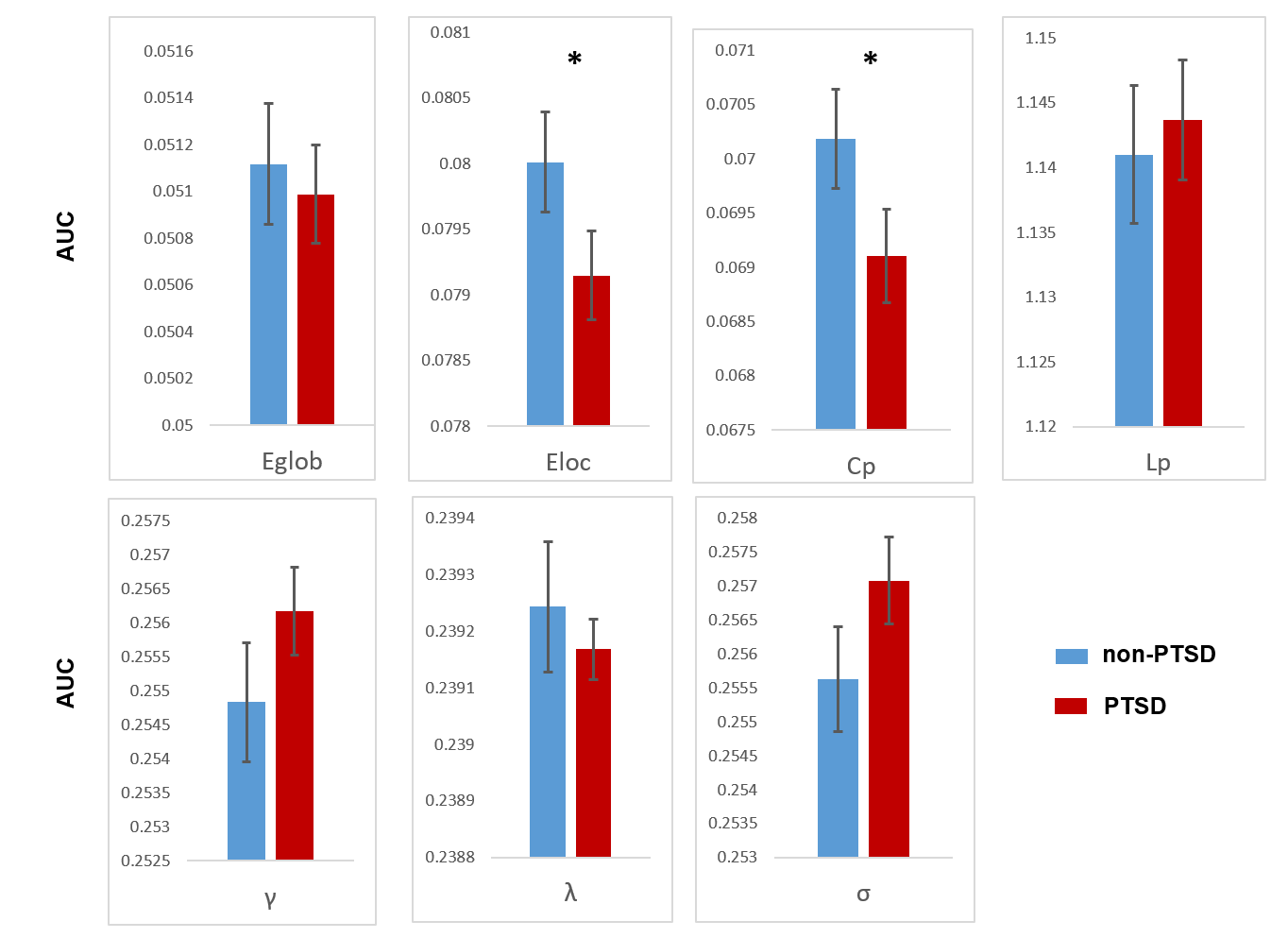
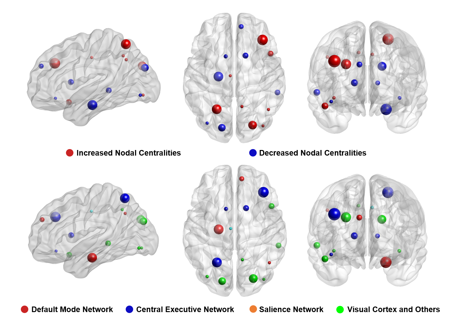
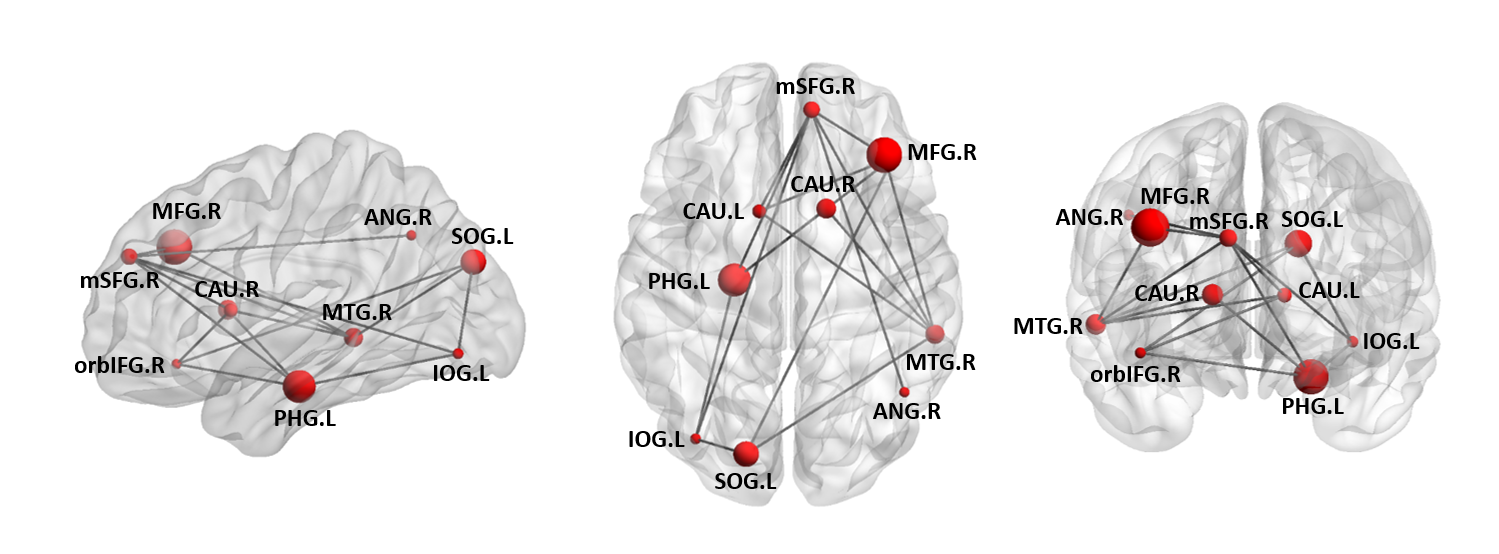
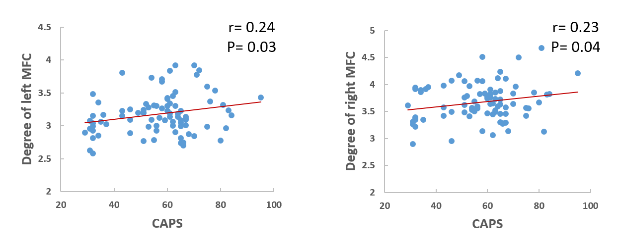
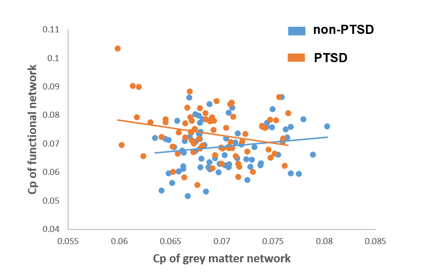
Compared with TEC, brain networks of PTSD patients were characterized by decreased clustering coefficient (Cp) (P=.04) and local efficiency (Eloc) (P=.04). Locally, patients with PTSD exhibited altered centrality involving medial superior frontal (mSFG), inferior orbital frontal (iOFG), superior parietal (SPG), middle frontal (MFG), angular and para-hippocampal gyri (P<0.05, corrected). Nodal Degree of left and right MFG in patients with PTSD were positively correlated with CAPS scores (r=0.24, p=0.03 and r=0.23, p=0.04 respectively). A negative correlation between the segregation (Cp) of grey matter and functional networks was specifically found in the PTSD group.DISCUSSION
The human brain is typically organized as a “small world ” network with two main organizational principles, segregation (reflected by clustering or local efficiency), and integration (reflected by path length and global efficiency) [7]. Our findings of decreased clustering coefficient and local efficiency in PTSD indicate that the grey matter networks of PTSD adults were shifted towards to a “more random” organization, which is a common alteration of grey matter networks as seen in Alzheimer’s disease [8], schizophrenia [9], and major depressive disorder [10]. The reduced segregation of brain networks reflected by lower Cp and Eloc might lead to impairments in cognitive function and emotion processing in patients with PTSD. The alteration of network segregation reflected by reduced Cp and Eloc in the present study was in the opposite direction from that seen in our previous functional study [11]. Previous studies have shown that in the healthy adult brain, volumetric covariance and resting-state functional connectivity are positively correlated [12]. This relationship was corroborated in our control group, in which there was a positive correlation of Cp in grey matter with Cp in functional network (p>0.05). In contrast, our PTSD patients demonstrated an inverse correlation between Cp of structural and functional networks. Because the damages of grey matter interplaying way between two brain regions might lead to compensatory hyperactivated changes in functional connectivity in these regions, the inverse relationship in PTSD might represent a compensatory response to anatomic alteration. In conclusion, our analyses of brain topology based on single-subject grey matter networks indicate a more randomly organized brain network in PTSD. The reduced segregation in grey matter network and its negative relation with increased segregation in the functional network may be important for understanding the nature of the brain network disorganization associated with PTSD and facilitate clinical diagnosis in patients with suspected PTSD.Acknowledgements
No acknowledgement found.References
1. Yehuda, R., et al., Post-traumatic stress disorder. Nat Rev Dis Primers, 2015. 1: p. 15057. 2. Alexander-Bloch, A., J.N. Giedd, and E. Bullmore, Imaging structural co-variance between human brain regions. Nat Rev Neurosci, 2013. 14(5): p. 322-36. 3. Tijms, B.M., et al., Similarity-based extraction of individual networks from gray matter MRI scans. Cereb Cortex, 2012. 22(7): p. 1530-41. 4. Suo, X., et al., Anatomic Insights into Disrupted Small-World Networks in Pediatric Posttraumatic Stress Disorder. Radiology, 2017. 282(3): p. 826-834. 5. van Wijk, B.C.M., C.J. Stam, and A. Daffertshofer, Comparing Brain Networks of Different Size and Connectivity Density Using Graph Theory. Plos One, 2010. 5(10). 6. Batalle, D., et al., Normalization of similarity-based individual brain networks from gray matter MRI and its association with neurodevelopment in infants with intrauterine growth restriction. Neuroimage, 2013. 83: p. 901-11. 7. Bullmore, E. and O. Sporns, The economy of brain network organization. Nat Rev Neurosci, 2012. 13(5): p. 336-49. 8. Tijms, B.M., et al., Single-subject grey matter graphs in Alzheimer's disease. PLoS One, 2013. 8(3): p. e58921. 9. Bassett, D.S., et al., Hierarchical organization of human cortical networks in health and schizophrenia. J Neurosci, 2008. 28(37): p. 9239-48. 10. Singh, M.K., et al., Anomalous gray matter structural networks in major depressive disorder. Biol Psychiatry, 2013. 74(10): p. 777-85. 11. Lei, D., et al., Disrupted Functional Brain Connectome in Patients with Posttraumatic Stress Disorder. Radiology, 2015. 276(3): p. 818-27. 12. Di, X., et al., Do all roads lead to Rome? A comparison of brain networks derived from inter-subject volumetric and metabolic covariance and moment-to-moment hemodynamic correlations in old individuals. Brain Struct Funct, 2017.Figures