0214
Focused, High-Resolution, Distortion-Free Diffusion Imaging1Department of Radiology, Mayo Clinic, Rochester, MN, United States
Synopsis
This study reports focused, high-resolution, distortion-free diffusion imaging using a combination of DIADEM (Distortion-free Imaging: A Double Encoding Method) and reduced field-of-view (rFOV) imaging. DIADEM is a hybrid, multi-shot approach inspired by the point-spread-function mapping technique for distortion-free imaging. The multiple-shots effectively signal average and compensate for the reduced image SNR resulting from the rFOV. The rFOV reduces the number of phase-encoding steps, which shortens the scan time, making it more clinically feasible. The results demonstrate focused distortion-free diffusion images with a high in-plane resolution (0.86 mm2), which could provide improved anatomic depiction of local brain tissue structures.
Introduction
Single-shot, echo-planar imaging (EPI) requires a long readout acquisition time (i.e., echo train length × echo spacing) to acquire high-resolution, diffusion-weighted imaging (DWI). Consequently, it usually suffers from strong off-resonance effects, such as geometric distortions and T2* blurring in the phase-encoding (PE) (i.e., blipped) direction. If the target region of interest (ROI) is a subsection of the brain, it is well-known that reduced field of view (rFOV) in the EPI-PE dimension (y) can provide potential advantages in mitigating the artifacts by shortening the readout acquisition time1. Nevertheless, the off-resonance effects are still not negligible, especially for high-resolution imaging. In addition, the image signal-to-noise (SNR) decreases with the reduced imaging volume. Recently, a hybrid multi-shot approach using spin-warp (SW) and echo-planar encoding strategy inspired by the point-spread-function mapping (PSF) method2 has been proposed for distortion-free high-resolution imaging, which is referred to here as DIADEM (Distortion-free Imaging: A Double Encoding Method). In this study, to address those challenges outlined above, DIADEM is combined with a rFOV approach using a 2D echo-planar spatially-selective RF excitation2.Methods
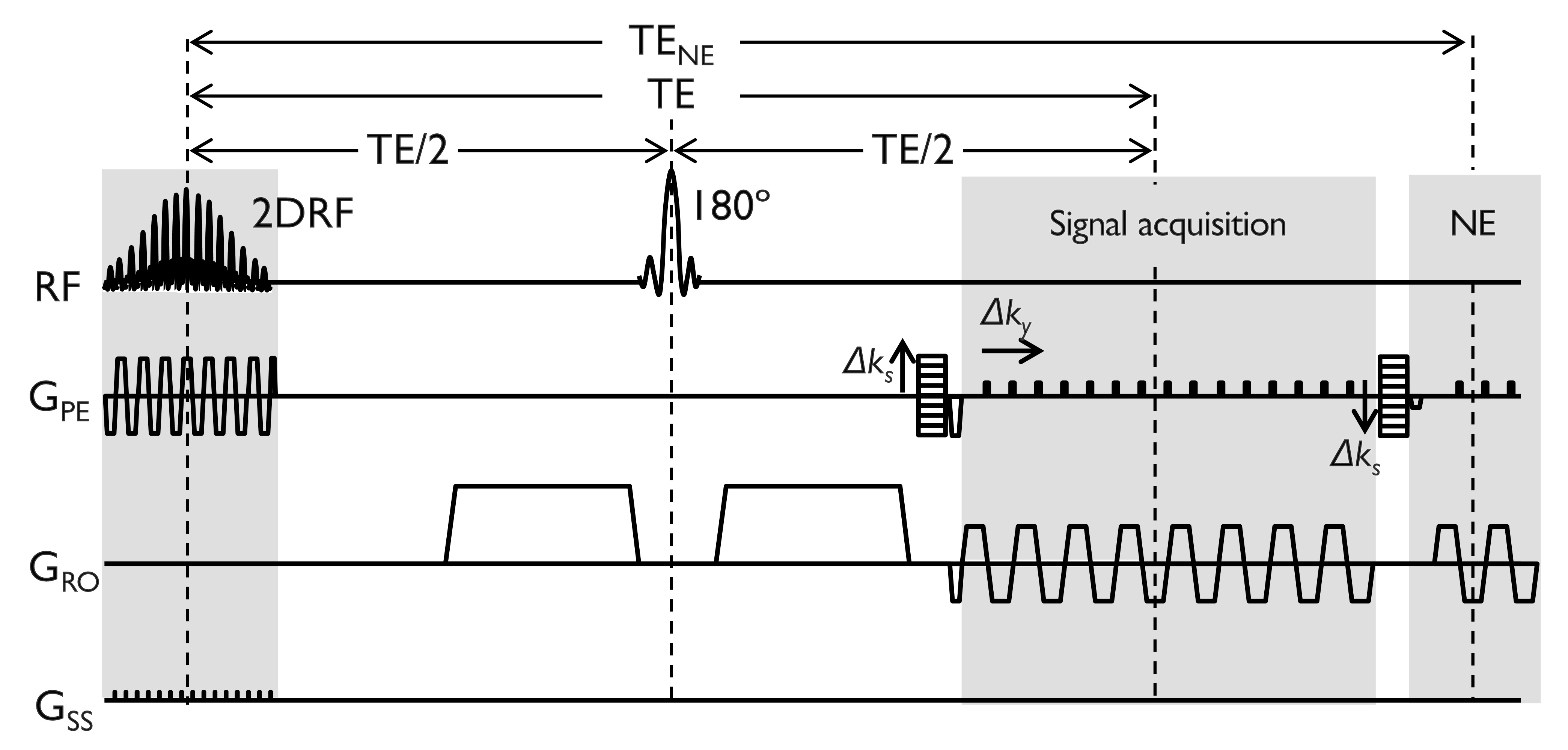
Under an IRB-approved protocol, localized high-resolution DWI was performed in the right cerebral hemisphere and brain stem in two healthy volunteers on a compact 3T3-5 with high-performance gradient system (700 T/m/s and 80 mT/m). The diffusion DIADEM sequence was implemented and optimized on the GE platform6, and was combined with a 2D echo-planar spatially-selective RF excitation1 for localized imaging (Fig. 1). Imaging parameters were: TR/TE(signal acquisition)/TENE(navigator echo)=2000/73/105 ms, no partial Fourier in the EPI-PE dimension, 16 slices, slice thickness=3.0-3.5mm, readout bandwidth=±200 kHz, FOV=220×55 mm2 (i.e. a rFOV factor of 4 in the EPI-PE dimension), matrix size=256×64, echo spacing=784 µs, 7 PSF scans with one b-value=0, and 6 diffusion directions with b-value=800 s/mm2. For DIADEM acceleration, each PSF data set was acquired with an acceleration factor of 2, and partial Fourier of 85% in the SW-PE dimension(s), which resulted in 27 segments (or averages). Cardiac triggering was not applied. The total scan time was 6 minutes and 20 seconds. As a geometrical reference, corresponding T2-weighted anatomical images, which have an identically-matched spatial resolution to the diffusion imaging, were acquired with a standard 2D fast spin echo sequence. Both distorted I(x,y) and distortion-free I(x,s) images were reconstructed from the same 3D PSF data I(x,y,s), as described in a previous study2. Since the geometric distortions presented in the distorted images I(x,y), the need to acquire an additional conventional diffusion images with rFOV is eliminated. The distorted and distorted-free images and their corresponding DTI scalars calculated by FSL7 were directly compared against each other.Results and Discussion
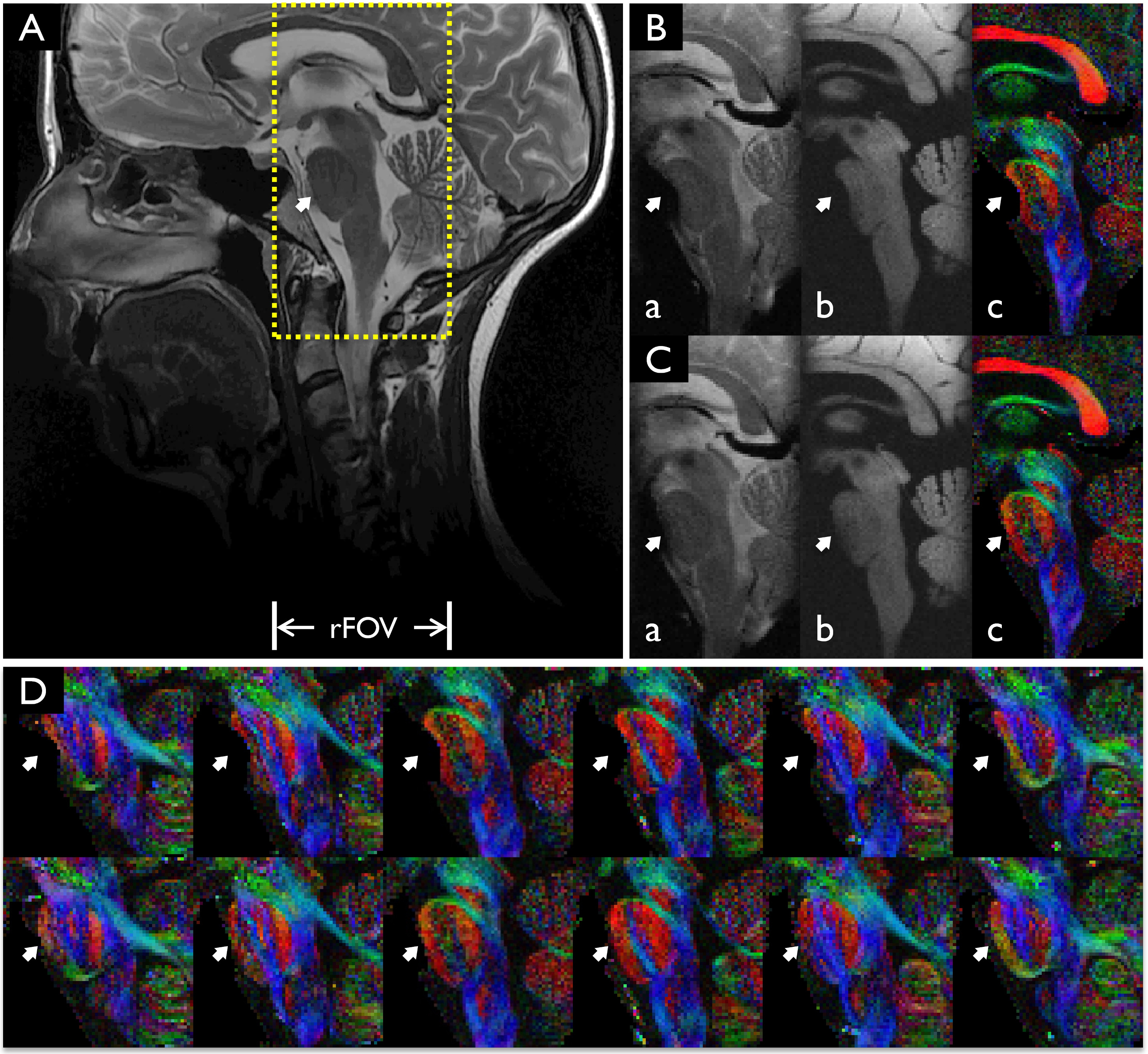
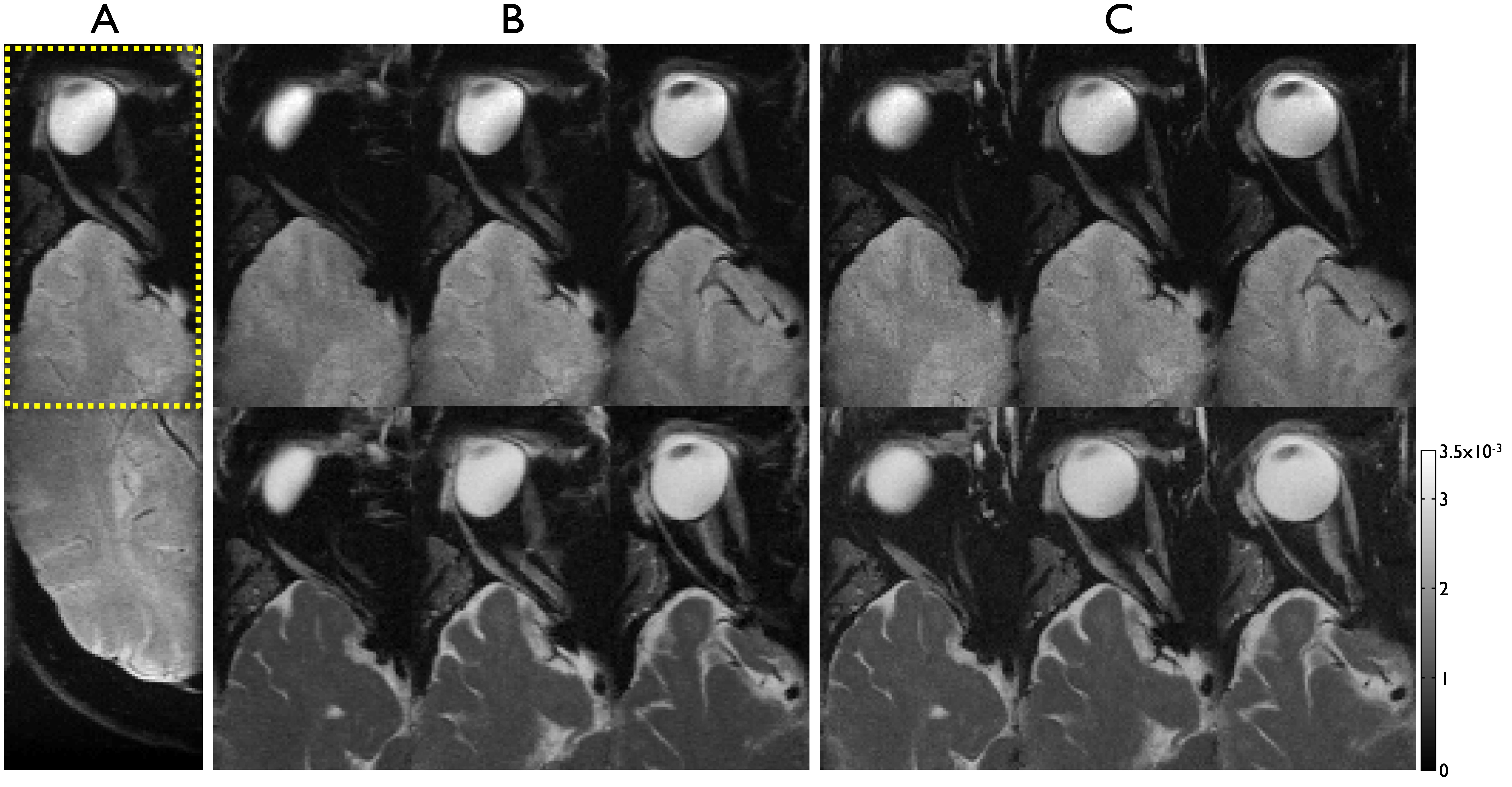
Figure 2 demonstrates that localized DWI with very high in-plane resolution of 0.86 mm2 is possible with the proposed approach. The SNR reduction from using the rFOV is compensated by the multi-shot nature of the proposed approach, which effectively signal averages. With a high rFOV factor of 4 in the EPI-PE dimension and benefit of high-performance gradients on the compact 3T8, the distortion level was substantially reduced in the distorted rFOV-DWI, I(x,y), corresponding to the rFOV-EPI. The echo spacing was 784 µs, which compares to 1440 µs obtainable with a whole-body 3T with standard gradients (50 mT/m, 200 m/T/s). However, local geometric distortion still appeared for the high-resolution imaging, especially in the region of the pons (white arrows in Fig. 2B). Consequently, loss of spatial information occurred in the corresponding areas. However, the lost spatial information was fully recovered in the proposed rFOV-DWI without apparent distortion (Figs. 2C and 2D). Due to the benefits of the high-resolution and distortion-free imaging, this approach is desirable for regions where susceptibility image artifacts are usually severe for high-resolution diffusion imaging9 such as the orbit and optic nerve as demonstrated in Fig. 3. The measured maximum distortion was 8.16 pixels, which corresponds to 7.0 mm.Conclusion
This study demonstrates the feasibility and advantage of combining DIADEM with rFOV approaches. High-resolution distortion-free diffusion imaging was demonstrated in localized areas of the brain, typically subject to severe susceptibility artifacts. The proposed localized diffusion approach may offer improvements for understanding local brain tissue structures and the changes associated with disease pathology.Acknowledgements
This work was supported by NIH U01 EB024450-01.References
1. Saritas EU, Cunningham CH, Lee JH, Han ET, Nishimura DG. DWI of the spinal cord with reduced FOV single-shot EPI. Magn Reson Med. 2008 Aug;60(2):468-73. doi: 10.1002/mrm.21640.
2. In MH, Posnansky O, Speck O, High-resolution distortion-free diffusion imaging using hybrid spin-warp and echo-planar PSF-encoding approach. Neuroimage. 2017 Mar 1;148:20-30. doi: 10.1016/j.neuroimage.2017.01.008.
3. Lee S-K, Mathieu J-B, Graziani D, et al. Peripheral nerve stimulation characteristics of an asymmetric head-only gradient coil compatible with a high-channel-count receiver array. Magn Reson Med 2016;76:1939–1950. doi: 10.1002/mrm.26044.
4. Tao S, Weavers PT, Trzasko JD, Shu Y, Huston J 3rd, Lee SK, Frigo LM, Bernstein MA. Gradient pre-emphasis to counteract first-order concomitant fields on asymmetric MRI gradient systems. Magn Reson Med. 2017 Jun;77(6):2250-2262. doi: 10.1002/mrm.26315.
5. Weavers PT, Tao S, Trzasko JD, Frigo LM, Shu Y, Frick MA, Lee SK, Foo TKF, Bernstein MA. B0 concomitant field compensation for MRI systems employing asymmetric transverse gradient coils. Magn Reson in Med 2017. doi:10.1002/mrm.26790
6. In MH, Gray EM, Tan ET, Trzasko JD, Shu Y, Houston J 3rd, Foo TK, Bernstein MA. Distortion-free high-resolution diffusion imaging on a compact 3T MRI with high-performance gradients within a clinically feasible scan time. ISMRM 2018, (submitted)
7. FSL package, https://fsl.fmrib.ox.ac.uk/fsl/fslwiki/FSL
8. Tan ET, Lee SK, Weavers PT, Graziani D, Piel JE, Shu Y, Huston J 3rd, Bernstein MA, Foo TK. High slew-rate head-only gradient for improving distortion in echo planar imaging: Preliminary experience. J Magn Reson Imaging. 2016 Sep;44(3):653-64. doi: 10.1002/jmri.25210.
9. Jeong HK, Dewey BE, Hirtle JA, Lavin P, Sriram S, Pawate S, Gore JC, Anderson AW, Kang H, Smith SA. Improved diffusion tensor imaging of the optic nerve using multishot two-dimensional navigated acquisitions. Magn Reson Med. 2015 Oct;74(4):953-63. doi: 10.1002/mrm.25469.
Figures